


Epithelial cells in urine are cells that line the urinary tract and sometimes appear in a urine sample; a few squamous or transitional epithelial cells is usually normal, but the presence of many cells — especially renal tubular epithelial cells — can suggest contamination, infection, or kidney injury. According to MedlinePlus (part of the U.S. National Library of Medicine), most labs classify epithelial cells qualitatively (none, few, moderate, many) and interpret them alongside symptoms and other test results. In short: occasional squamous cells often reflect contamination; renal tubular epithelial cells are more likely to indicate a problem with the kidneys and usually prompt further testing (MedlinePlus; MSD Manual).
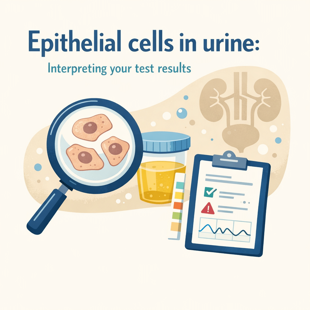
What are epithelial cells in urine?
Epithelial cells are thin, flat or polygonal cells that form the inner lining of body surfaces, including the skin and the urinary tract. When you provide a urine specimen, small numbers of these cells can slough off naturally and appear in the urine sediment. Labs detect them during microscopic examination of urine (urine microscopy or urinalysis). According to the MSD Manual, the clinical significance depends on the cell type and number and on other urine findings such as white blood cells, bacteria, or casts.
Types of epithelial cells and what they indicate
- Squamous epithelial cells: These originate from the urethra and external genitalia (skin). Finding a few squamous cells is common and often reflects contamination of the sample during collection rather than disease. NHS guidance notes that squamous cells often indicate a poorly collected sample.
- Transitional epithelial cells (urothelial cells): These line the bladder, ureters and renal pelvis. A small number can be normal, but increased counts may appear with bladder inflammation, infection, or urine catheterization (MSD Manual).
- Renal tubular epithelial cells (RTECs): These come from the kidney’s tubules. Their presence in more than a rare number can suggest tubular injury, acute tubular necrosis, or other intrinsic renal disease. The MSD Manual and MedlinePlus explain that RTECs are the most clinically significant epithelial cells when present in urine.
How labs measure epithelial cells and typical reference ranges
Urine labs usually report epithelial cells qualitatively (none, few, moderate, many) or quantitatively as cells per high-power field (cells/HPF) under a microscope. Reference ranges vary between laboratories and by method, but commonly used general guidance includes:
- Squamous epithelial cells: –5 cells/HPF (few is typically considered normal)
- Transitional epithelial cells: –5 cells/HPF (small numbers may be acceptable)
- Renal tubular epithelial cells: cells/HPF (any consistent presence may be abnormal)
MedlinePlus and the MSD Manual emphasize that laboratories differ in reporting formats and that results must be interpreted with clinical context. Always check your lab’s reference information on the report.
Common causes of increased epithelial cells in urine
- Sample contamination: Poor collection technique (not cleaning the genital area, not using midstream catch) commonly increases squamous epithelial cells (NHS).
- Urinary tract infection (UTI): Infections in the bladder or urethra can shed transitional epithelial cells; the CDC describes infection as a common reason to perform urinalysis when symptoms suggest UTI.
- Instrumentation or catheterization: Catheters or recent cystoscopy can dislodge epithelial cells from the urinary tract (MSD Manual).
- Kidney tubular injury: Conditions that injure the renal tubules (certain toxins, ischemia, severe infection) can release renal tubular epithelial cells into urine; this finding often leads clinicians to check kidney function tests (creatinine, BUN) and consider renal imaging or further tests (MSD Manual; MedlinePlus).
- Inflammation or cancer: Chronic inflammation or urothelial carcinoma can raise transitional epithelial cells, but these causes are less common and require specific clinical evaluation.
Symptoms and related test findings that change interpretation
Epithelial cells alone provide limited information; doctors interpret them alongside symptoms and other lab results:
- With fever, dysuria (painful urination), frequency, or urgency and bacteria/white cells in urine, epithelial cells likely point to infection (CDC).
- If epithelial cells appear together with rising serum creatinine, reduced urine output, or pigmented granular casts, clinicians may suspect tubular damage (MSD Manual).
- Isolated squamous cells with no other abnormalities usually indicate sample contamination and may prompt a repeat, clean-catch specimen (NHS).
How doctors interpret epithelial cells in urine
Clinicians use a stepwise approach:
- Review the urine collection method and note whether the sample was clean-catch or catheterized (NHS).
- Check the type and quantity of epithelial cells on microscopy and compare with white blood cells, bacteria, red blood cells, and casts (MedlinePlus).
- Correlate with symptoms (fever, pain, urinary symptoms), and with blood tests such as creatinine and electrolytes if kidney injury is suspected (MSD Manual).
- Order follow-up tests when indicated: repeat urinalysis with a properly collected sample, urine culture for suspected infection, urine cytology or imaging for persistent transitional cell abnormalities, or renal function tests and nephrology referral for suspected tubular injury.
Common mistakes and how to avoid them
- Assuming all epithelial cells mean infection: Many epithelial cells are contamination-related; interpret alongside symptoms and other markers (NHS).
- Ignoring renal tubular epithelial cells: Even a small but consistent number of RTECs may merit further evaluation of kidney function (MSD Manual).
- Not repeating the test with correct collection: If squamous cells dominate and clinical suspicion for infection is low, repeating with a midstream clean-catch sample usually clarifies the result (MedlinePlus).
How the urine sample collection affects results
Proper collection reduces contamination:
- For adults, the midstream clean-catch method (clean genital area, start urinating, collect midstream) reduces squamous epithelial contamination.
- For infants or when accurate sterile samples are needed, catheterization or suprapubic aspiration may be used in clinical settings, but these have different risks and are performed by professionals (NHS).
- If you collect at home, follow your lab’s instructions and tell your clinician if you used a catheter or had recent procedures.
Treatment and follow-up based on epithelial cell findings
There is no treatment for epithelial cells per se; clinicians treat the underlying cause:
- If contamination is suspected: repeat the urinalysis with proper collection.
- If infection is suspected: clinicians confirm with urine culture and treat with antibiotics as guided by culture results and local guidelines (CDC).
- If renal tubular injury is suspected: clinicians address the cause (remove offending drugs, treat hypotension/infection) and monitor kidney function; some cases require hospitalization or nephrology consultation (MSD Manual).
Decisions depend on clinical context, and your doctor can assess whether further testing or treatment is appropriate.
When to see a doctor
See your doctor promptly if you have any of the following in combination with a urine result that reports many epithelial cells or specifically notes renal tubular epithelial cells:
- Fever plus painful, frequent, or burning urination (possible infection).
- New or worsening flank pain or side pain (possible kidney involvement).
- Decreased urine output or sudden swelling in the legs or face and a reported rise in serum creatinine (possible kidney injury).
- Blood visible in the urine (visible hematuria) with persistent abnormal epithelial findings.
- A lab report that explicitly states “renal tubular epithelial cells present” or “many epithelial cells” together with abnormal kidney function tests.
If your lab report shows only a few squamous epithelial cells and you have no symptoms, you may only need a repeat clean-catch specimen. Always follow your clinician’s advice and seek urgent care when symptoms are severe or rapidly worsening.
Frequently asked questions
What does “few epithelial cells” on my urine report mean?
Most labs use qualitative language; “few epithelial cells” usually indicates a small, often clinically insignificant number, commonly from the urethra or external genitalia and possibly from contamination (NHS; MedlinePlus).Can epithelial cells mean I have cancer?
Increased transitional epithelial cells can be a red flag in some settings, but they do not prove cancer. Your clinician may order urine cytology or imaging if there is persistent, unexplained increase, especially with risk factors or other concerning signs (MSD Manual).Do epithelial cells always mean a urinary tract infection?
No. Epithelial cells can appear because of contamination, instrumentation, or kidney injury. Infection is more likely when epithelial cells appear with white blood cells, bacteria, and urinary symptoms (CDC; MSD Manual).Should I repeat the test if squamous cells are reported?
Often yes: if the report shows mostly squamous epithelial cells and you have no other symptoms, clinicians commonly request a repeat midstream clean-catch sample to rule out contamination (NHS).What are renal tubular epithelial cells and why are they important?
Renal tubular epithelial cells line the kidney tubules. Their presence in urine, particularly in larger numbers, may indicate tubular injury and prompt further evaluation of kidney function (MedlinePlus; MSD Manual).How quickly will abnormal epithelial findings be followed up?
That depends on the overall clinical picture. If you have symptoms like fever or decreased urine output, follow-up is often immediate. If the abnormality is isolated and you feel well, your clinician may repeat the test or monitor over days to weeks.
Glossary of key terms
- Urinalysis: A set of tests on urine that includes dipstick chemistry and microscopic examination.
- High-power field (HPF): The area visible under a microscope at high magnification; used to count cells.
- Squamous epithelial cells: Flat cells from external genitalia or lower urethra; often indicate contamination.
- Transitional epithelial cells: Cells that line the bladder and ureters; may increase with bladder inflammation or catheterization.
- Renal tubular epithelial cells (RTECs): Cells from the kidney tubules; their presence can suggest tubular injury.
- Urine culture: A microbiology test to grow and identify bacteria from urine, used to confirm infection.
Sources
Understand your lab results with AI DiagMe
Understanding lab findings like epithelial cells in urine often requires combining the test result with symptoms, medical history, and other tests. AI DiagMe helps you interpret lab reports in minutes and provides clear, evidence-aware explanations you can discuss with your clinician.

