


A urine cytology test examines cells shed into urine to look for abnormal or cancerous cells; in most clinical settings it is used to help detect and monitor cancers of the urinary tract (especially bladder cancer) and to flag suspicious cellular changes that need further investigation. Urine cytology does not give a numeric result but reports categories such as negative (no malignant cells seen), atypical, suspicious, or positive for malignant cells; interpretation depends on the lab and the clinical context. According to the MSD Manual and diagnostic guidelines, urine cytology is most useful when paired with cystoscopy (a visual exam of the bladder) and other tests because it is specific but less sensitive for certain tumors.
What is cytology urine and why is it done?
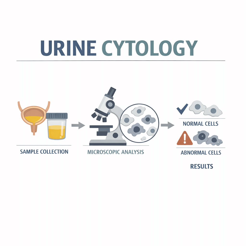
Urine cytology (often called cytology urine) inspects urine under a microscope to find abnormal cells from the lining of the urinary tract. Clinicians commonly order it when they suspect bladder cancer, when patients have visible blood in the urine (hematuria), or when monitoring people with known urothelial (urinary tract lining) cancers. The test can help detect high-grade (more aggressive) cancers reliably but misses many low-grade (less aggressive) tumors, so doctors usually combine it with imaging or direct visualization techniques such as cystoscopy (Mayo Clinic; MSD Manual).
How the test works
A urine sample is collected and processed in the laboratory so a cytotechnologist or cytopathologist can view individual cells. Technicians may concentrate cells by centrifugation or use special slides and stains to highlight nuclear and cellular features. The trained specialist looks for changes in cell size, nuclear shape, staining patterns, and other features that suggest malignancy. When available, additional techniques such as fluorescent in situ hybridization (FISH) can look for genetic changes in cells to improve detection (NHS; MSD Manual).
What results mean: categories and common interpretations
- Negative/no malignant cells: No cancer cells were identified. This result reduces the likelihood of a high-grade tumor but does not completely rule out cancer, particularly low-grade disease (MSD Manual).
- Atypical urothelial cells: Cells look abnormal but not clearly cancerous. This finding often prompts repeat testing, closer monitoring, or cystoscopy.
- Suspicious for malignancy: Many features suggest cancer but the sample is not definitive. This generally leads to urgent further evaluation.
- Positive for malignant cells: Cells characteristic of cancer are present; clinicians will usually proceed with diagnostic imaging and cystoscopy with biopsy.
Reference ranges do not apply because cytology reports categories rather than numeric values. Laboratories may use different reporting systems and words, so your doctor will explain what the lab’s categories mean in your specific situation (NHS).
When clinicians order urine cytology
Clinicians commonly request urine cytology when:
- A patient has unexplained visible blood in the urine (macroscopic hematuria) or persistent microscopic hematuria detected on urinalysis.
- A patient has risk factors for bladder cancer (e.g., age over 50, smoking history, occupational exposure to certain chemicals).
- A patient with a prior urothelial cancer requires surveillance for recurrence.
- Symptoms such as persistent urinary urgency, pain with urination, or unexplained weight loss raise concern for urinary tract malignancy (NHS; Mayo Clinic).
How to prepare and how the sample is collected
Most urine cytology tests use a voided (midstream) urine sample collected at the clinic or at home following instructions. For better yield, labs sometimes request a first-morning specimen or a specimen collected after bladder irrigation or catheterization, especially if previous tests were inconclusive. The NHS and lab-specific instructions often emphasize:
- Follow the kit directions exactly.
- Deliver the sample promptly or use preservatives if the lab requests them.
- Inform the laboratory if you have recent instrumentations (like catheterization), infections, or recent urologic procedures, as these can change the appearance of cells.
Limitations: sensitivity, specificity and false results
Urine cytology is specific (a positive result commonly indicates real disease) but has limited sensitivity for low-grade tumors—many low-grade bladder cancers shed few abnormal cells and escape detection (MSD Manual; Mayo Clinic). False negatives occur when samples lack sufficient abnormal cells or when low-grade tumors have subtle changes. False positives can arise from inflammation, infection, recent instrumentation, or stones that cause reactive cellular changes. Because of these limitations, clinicians interpret cytology alongside imaging, cystoscopy, and biomarker tests.
Complementary tests and newer urine-based markers
Because urine cytology alone can miss some cancers, clinicians may use additional tests:
- Cystoscopy: Direct visualization and biopsy remain the diagnostic standard for bladder tumors (Mayo Clinic).
- Urine-based molecular tests: Tests such as UroVysion FISH (detects chromosomal abnormalities) and protein-marker assays (for example, NMP22) can improve detection in some settings but vary in performance and cost (NHS; PubMed literature).
- Urine culture and urinalysis: These check for infection or blood that may explain symptoms and influence cytology interpretation.
Current evidence suggests some molecular assays help in surveillance and specific clinical scenarios, but no single urine biomarker fully replaces cystoscopy at present (NHS; MSD Manual).
How doctors interpret results and decide next steps
Clinicians integrate cytology results with symptoms, risk factors, imaging, and cystoscopy findings. For example:
- Negative cytology with ongoing hematuria will usually prompt cystoscopy and imaging because a negative cytology does not exclude low-grade tumors.
- Atypical results often lead to repeat cytology and cystoscopy within weeks to months.
- Positive or suspicious cytology typically leads to urgent cystoscopy with biopsy to confirm diagnosis and stage the disease.
Decisions depend on patient age, prior cancer history, and risk factors; your urologist can assess which path fits your case (Mayo Clinic; MSD Manual).
Practical tips for patients
- Provide the sample as instructed and notify the lab about recent infections, catheter use, or urologic procedures.
- Ask your clinician whether your lab uses additional urine biomarkers or FISH testing; these may be more helpful in surveillance than initial diagnosis depending on your case (NHS).
- Understand that one test result rarely gives a final answer; expect a plan for follow-up if results are atypical or if symptoms continue.
Common causes of abnormal cytology results besides cancer
- Urinary tract infection or inflammation (can cause reactive atypia).
- Recent urologic instrumentation (catheterization, cystoscopy) that disturbs cells.
- Kidney stones or other sources of bleeding that change cell appearance.
- Radiation or certain medications that alter cell morphology.
Clinicians consider these factors when assessing whether abnormal cells likely represent malignancy (MSD Manual).
Red flags and what increases concern
Findings that raise higher concern include:
- Positive or suspicious cytology.
- Repeated atypical results without an obvious benign explanation.
- Cytology findings in someone with gross hematuria, weight loss, or known risk factors for bladder cancer.
In these situations, prompt cystoscopy and imaging are typically recommended (Mayo Clinic; NHS).
When to see a doctor
See your doctor promptly if you have any of the following related to urine cytology or urinary symptoms:
- Visible blood in your urine (bright red or dark brown) even if it happens once.
- A urine cytology report of suspicious or positive for malignant cells.
- New or worsening urinary symptoms (painful urination, strong persistent urgency, difficulty passing urine).
- Repeated atypical cytology results without a clear benign explanation.
- A history of bladder or urothelial cancer and new urinary symptoms or a change in routine surveillance findings.
If you have a cytology result that concerns you, your clinician can explain urgency based on your history and may arrange cystoscopy within days to weeks depending on risk.
Frequently asked questions
Q: Is urine cytology the same as a regular urine test?
A: No. A routine urinalysis checks for infection, blood, protein, or other chemical markers in the urine, while urine cytology inspects individual cells under a microscope to look for abnormal or cancerous cells (Mayo Clinic).
Q: Can urine cytology detect all bladder cancers?
A: No. Urine cytology detects many high-grade tumors reliably but misses a substantial number of low-grade tumors. Clinicians therefore use it alongside cystoscopy and imaging for a complete evaluation (MSD Manual).
Q: How long does it take to get results?
A: Turnaround varies by laboratory but typically takes a few days to a week. If additional testing (for example, FISH) is ordered, expect a longer wait. Your clinic should give a timeline when they order the test.
Q: Will an infection affect the cytology result?
A: Yes. Infection or inflammation can cause reactive changes in cells that may appear atypical, which can complicate interpretation and sometimes lead to repeat testing after treating the infection (MSD Manual).
Q: If my cytology is atypical, what happens next?
A: Your doctor may repeat the test, perform cystoscopy, order imaging, or use urine molecular tests depending on your risk factors and symptoms. The exact plan depends on your clinical context (NHS).
Q: Can urine cytology replace cystoscopy for surveillance?
A: Not by itself. While cytology helps monitor for recurrence, cystoscopy remains the standard for direct visualization and biopsy; some guidelines use cytology and molecular tests to complement cystoscopy in surveillance (Mayo Clinic; MSD Manual).
Glossary of key terms
- Cytopathologist: A doctor who examines cells under a microscope to diagnose disease.
- Cystoscopy: A procedure where a urologist uses a thin camera to look inside the bladder.
- Hematuria: Blood in the urine; can be visible (macroscopic) or only on lab tests (microscopic).
- Sensitivity: The ability of a test to correctly identify those who have a disease (true positive rate).
- Specificity: The ability of a test to correctly identify those who do not have a disease (true negative rate).
- FISH (fluorescent in situ hybridization): A lab method that detects genetic abnormalities in cells.
Sources
- Urinalysis (Mayo Clinic)
- Urine cytology (Urology Care Foundation)
Understand your lab results with AI DiagMe
Understanding lab tests can feel overwhelming because results depend on context, prior health, and test methods. AI DiagMe can help interpret lab reports quickly and clearly so you and your clinician can discuss the best next steps based on current evidence and your individual risk factors.
➡️ Get your results interpreted in minutes

