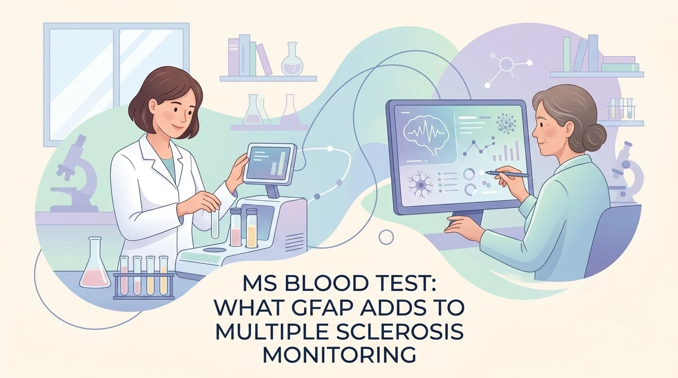
MS 혈액 검사는 아직 다발성 경화증을 진단하는 방법은 아니지만, 질환의 경과를 추적하는 방법으로 빠르게 자리잡고 있습니다. 2026년 8월 JAMA Neurology에 발표된 연구에서는 2,300명 이상의 다발성 경화증 환자를 대상으로 GFAP라는 단백질을 추적 관찰한 결과, 혈중 GFAP 수치가 재발 사이에 나타나는 느리고 눈에 띄지 않는 악화와 관련이 있음을 발견했습니다. 이 글에서는 GFAP가 무엇을 측정하는지, 이미 연구에서 사용되고 있는 신경필라멘트 표지자와 어떻게 다른지, 이번 연구 결과가 증명하는 것과 그렇지 않은 것은 무엇인지, 그리고 다음 신경과 진료 시 담당 의사에게 물어볼 만한 질문은 무엇인지 알아봅니다.
2026년 8월 연구에서 실제로 측정한 것
바젤 대학교와 캘리포니아 대학교 샌프란시스코 캠퍼스의 연구진은 두 개의 장기 환자 코호트, 즉 스위스 MS 코호트와 샌프란시스코의 EPIC 연구를 통합했습니다. 두 코호트를 합치면 총 2,329명, 18,600건 이상의 혈액 측정 데이터가 포함되며, 일부 참가자는 10년 이상 추적 관찰되었습니다. 6개월에서 12개월마다 일반 채혈을 통해 두 가지 단백질을 측정했습니다: GFAP(신경교 섬유성 산성 단백질)와 NfL(신경필라멘트 경쇄)입니다.
연구의 핵심 질문은 명확하고 실용적이었습니다. 두 단백질 중 하나가 재발 활동과 무관한 진행, 즉 뚜렷한 발작 없이 장애가 서서히 악화되는 현상을 미리 예측할 수 있을까요? GFAP는 그럴 수 있었습니다. 연령 대비 GFAP 수치가 높은 사람은 다음 정기 방문 전에 상태가 악화될 가능성이 약 40% 더 높았습니다. 또한 치료 첫 2년 동안 GFAP가 감소한 경우, 이후 악화 위험이 상당히 낮아졌습니다.
연구진은 연구 활용 측면에서도 주목할 만한 점을 언급했습니다. GFAP를 기준으로 임상시험 참가자를 선별하면 진행형 질환을 대상으로 한 임상시험 규모를 약 5분의 1 정도 줄일 수 있으며, 이는 진행형 다발성 경화증이 연구하기 가장 어려운 형태라는 점에서 중요한 의미를 가집니다.
GFAP와 NfL은 서로 다른 것을 반영합니다
두 단백질은 흔히 함께 언급되지만, 서로 다른 세포에서 유래하며 서로 다른 정보를 전달합니다. NfL은 손상된 신경 섬유에서 누출되므로 재발 중이나 재발 전후에 수치가 상승합니다. GFAP는 뇌와 척수의 지지 세포인 성상세포에서 유래하며, 급성 악화보다는 느리게 진행되는 배경 과정을 반영합니다.
| 항목 | NfL (신경필라멘트 경쇄) | GFAP (신경교 섬유성 산성 단백질) |
|---|---|---|
| 기원 세포 | 신경 섬유 (축삭) | 성상세포, 뇌의 지지 세포 |
| 수치 상승이 의미하는 것 | 급성 염증, 새로운 병변, 재발 | 재발 사이의 느린 진행 |
| 신호의 시간적 범위 | 수 주에서 수 개월 | 수개월에서 수년 |
| 나이의 영향 | 예, 결과는 연령 보정됩니다 | 예, 성별 및 체중에 의해서도 영향을 받습니다 |
| 2026년 출시 예정 | 전문 진료 및 연구 환경 | 주로 연구 코호트 |
이러한 상호 보완성이 실질적인 핵심입니다. 두 가지 마커를 함께 사용하면 같은 질환의 서로 다른 두 측면을 파악할 수 있으며, 이것이 바로 현재 연구에서 하나만 선택하지 않고 두 마커를 모두 측정하는 이유입니다.
이것이 여러분에게 무엇을 바꾸는지, 그리고 바꾸지 않는 것은 무엇인지
세 가지 사항을 명확히 짚어 둘 필요가 있습니다.
첫째, 이것은 선별 검사가 아닌 모니터링 마커입니다. GFAP는 뇌에 손상을 주는 다양한 질환에서 상승할 수 있으며, 나이가 들면서 자연스럽게 증가하기도 합니다. 단독으로 측정한 단일 수치는 큰 의미가 없으며, 이 때문에 연구자들은 단순 수치 대신 연령 보정 점수를 사용했습니다.
둘째, 다발성 경화증의 진단 방식은 작년과 동일합니다. 저희 개요에서는 다발성 경화증의 증상, 원인 및 진단에 대해 설명합니다. 진단은 임상 소견, MRI 결과, 그리고 필요한 경우 뇌척수액 분석을 바탕으로 이루어집니다. 어떤 혈액 단백질도 이를 대체할 수 없습니다.
셋째, GFAP는 대부분의 국가에서 아직 일상적인 진료에 포함되어 있지 않습니다. 이 코호트 연구에서 사용된 검사법은 초고감도 연구용 플랫폼이며, 현재 검사실에서 검사 결과지에 기재할 수 있는 합의된 기준치가 존재하지 않습니다. 담당 신경과 전문의가 GFAP를 언급한다면, 대개 연구 또는 전문 센터의 맥락에서일 것입니다.
달라지는 것은 대화의 방식입니다. 지금까지 무증상 진행은 대부분 기능이 이미 저하된 후에야 사후적으로 인식되었습니다. 보행 검사보다 먼저 변화를 보이는 마커가 있다면, 임상의가 더 일찍 대응할 수 있는 근거가 생깁니다.
최신 과학적 발전
지난 3년간 발표된 네 편의 논문은 GFAP가 단순한 실험실 연구 대상에서 신뢰할 수 있는 모니터링 도구로 어떻게 발전했는지를 설명합니다.
2023년, 스위스 연구팀이 355명을 대상으로 GFAP와 NfL을 직접 비교했습니다. 연구 결과, 진행성 질환이 악화되는 환자들은 NfL 수치를 보정한 후에도 안정적인 환자들보다 GFAP가 뚜렷하게 높았으며, GFAP가 높을수록 MRI에서 회백질이 더 빠르게 감소하는 것으로 나타났습니다. 이것이 의미하는 바: 두 마커는 이미 서로 다른 정보를 전달하고 있었으며, GFAP는 재발이 아닌 꾸준한 진행과 연관된 마커였습니다.
2024년, 같은 연구팀이 B세포 억제 치료(다발성 경화증에 사용되는 강력한 면역 치료 중 하나)를 시작한 362명을 추적 관찰했습니다. 치료 시작 1년 후에도 GFAP가 높게 유지된 환자들은 이후 증상이 악화될 가능성이 더 높았습니다. 이것이 의미하는 바: 이 마커는 질환 자체뿐만 아니라 치료에도 반응하는 것으로 보이며, 바로 그 점이 추적 관찰에 유용한 이유입니다.
2025년 《The Lancet Neurology》에 발표된 리뷰 논문은 이러한 혈액 마커들의 현재 위치를 정리했습니다. NfL은 활성 염증 시 신경 섬유 손상을 반영하고, GFAP는 재발과 무관한 진행의 새로운 마커로 설명되며, 세 번째 단백질인 CHI3L1은 현재 연구 중입니다. 이것이 의미하는 바: 이 분야는 하나의 완벽한 검사가 아닌, 여러 마커를 조합한 패널 방식으로 수렴되고 있습니다.
2026년 8월 연구는 네 연구 중 가장 규모가 크며, 별도의 독립적인 두 번째 코호트에서 검증을 거친 첫 번째 연구입니다. 이 점이 중요한 이유는, 다른 나라에서 다른 환자들과 다른 임상의들을 대상으로도 동일한 결과가 재현된다면 훨씬 더 신뢰할 수 있기 때문입니다. 그럼에도 네 연구 모두 관찰 연구로, 인과관계를 증명하는 것이 아니라 연관성을 보여주는 것이며, 현재 임상에서 개별 환자에게 적용할 수 있는 기준값을 확립한 연구는 아직 없습니다.
신경과 전문의와 상담해야 할 때
혈액 마커는 연구 단계의 주제이지만, 진료를 받아야 하는 이유는 변함없이 명확합니다:
- 24시간 이상 지속되는 새로운 또는 악화되는 근력 저하, 감각 이상, 시력 저하, 균형 장애
- 뚜렷한 발작 없이도 수개월에 걸쳐 보행 거리, 손의 민첩성 또는 피로감이 서서히 변화하는 경우
- 질환 조절 치료 중 부작용이나 혈구 수치 변화가 나타나는 경우
- 현재 치료가 여전히 효과가 있는지 불확실한 경우
대부분의 다발성 경화증 치료에는 정기적인 혈액 검사 모니터링이 동반되며, 이를 이해하는 것이 도움이 됩니다. 별도의 가이드에서 혈액 검사 결과 읽는 법 가이드, 또 다른 가이드에서는 비정상 혈액 검사 결과에 대해 설명합니다. 검사가 처음이시라면, 다음 내용도 참고하세요. 혈액 검사 중 어떤 일이 일어나는가.
다발성 경화증은 면역 매개 질환이므로 관련 페이지도 도움이 될 수 있습니다. 한 글에서는 자가면역 질환의 증상과 치료를 다루고, 다른 글에서는 자가면역 패널 검사, 그리고 세 번째 항목에서는 CRP 수치 상승.
용어 설명
| 용어 | 정의 |
|---|---|
| GFAP | 신경교 섬유산성 단백질. 성상세포의 구조 단백질로, 이 세포가 활성화되거나 손상될 때 혈액에서 측정할 수 있습니다. |
| NfL | 신경필라멘트 경쇄. 신경 섬유가 손상될 때 방출되는 단백질로, 활성 염증의 지표로 사용됩니다. |
| 성상세포 | 뇌와 척수에 있는 별 모양의 지지 세포로, 신경세포에 영양을 공급하고 염증 조절을 돕습니다. |
| PIRA | 재발과 무관한 진행. 뚜렷한 발작 없이 장애가 서서히 악화되는 상태입니다. |
| 재발 | 새로운 신경학적 증상이 나타나거나 기존 증상이 재발하여 최소 24시간 이상 지속된 후 부분적 또는 완전한 회복이 이루어지는 상태. |
| 코호트 연구 | 동일한 집단을 일정 기간 추적 관찰하며 변화를 기록하는 연구. |
| 연령 보정 점수 | 원시 농도 수치가 아닌, 같은 연령대의 일반적인 수치와의 차이로 표현한 결과. |
| 질병 조절 치료 | 재발을 줄이고 다발성 경화증의 진행을 늦추는 것을 목표로 하는 장기 치료. |
자주 묻는 질문
혈액 검사로 다발성 경화증을 진단할 수 있나요?
아닙니다. 다발성 경화증을 확진하는 단일 혈액 검사는 존재하지 않습니다. 진단은 임상 병력, 신경학적 검진, 뇌와 척수의 MRI, 그리고 경우에 따라 요추 천자를 통한 뇌척수액 분석을 종합하여 이루어집니다. 혈액 검사는 주로 특정 감염, 비타민 결핍, 기타 자가면역 질환 등 다발성 경화증과 유사한 증상을 보이는 질환을 배제하는 데 활용됩니다.
의사에게 GFAP 검사를 요청할 수 있나요?
현재 대부분의 국가에서는 그렇지 않습니다. 이 검사는 연구 환경 외에서는 일반적으로 제공되지 않으며, 개인적인 판단에 활용할 수 있는 검증된 기준치도 아직 없습니다. 연구에 참여 중인 다발성 경화증 전문 센터에서는 측정이 가능할 수 있습니다. 담당 신경과 전문의에게 해당 센터가 바이오마커 연구에 참여하고 있는지 문의해 보는 것이 좋습니다.
GFAP 수치가 높으면 다발성 경화증이 악화되고 있다는 의미인가요?
그것만으로는 판단할 수 없습니다. 2026년 연구는 대규모 집단에서 평균적인 위험도가 높다는 것을 보고한 것이지, 특정 개인에게 반드시 그렇다는 의미가 아닙니다. GFAP 수치는 나이가 들수록 높아지고 성별에 따라 차이가 있으며, 뇌졸중이나 두부 외상 같은 다른 뇌 손상 후에도 증가합니다. 이는 여러 신호 중 하나일 뿐이며, 증상 및 영상 검사 결과와 함께 종합적으로 해석해야 합니다.
이것이 새로운 알츠하이머 혈액 검사와 같은 건가요?
아닙니다. 두 검사 모두 초고감도 혈액 분석이라는 같은 계열에 속하지만, 알츠하이머 검사는 아밀로이드 및 타우 병리와 관련된 p-tau217 같은 단백질을 측정합니다. GFAP와 NfL은 뇌 및 신경 손상의 일반적인 지표로, 여러 신경계 질환에 걸쳐 연구되고 있습니다.
MS 치료에는 정기적인 혈액 검사가 필요한가요?
네, 대부분의 질병 조절 치료제는 주기적인 모니터링을 필요로 합니다. 일반적으로 복용하는 약에 따라 정해진 간격으로 전혈구 검사, 간 효소 수치, 경우에 따라 신장 기능 검사를 받게 됩니다. 이는 바이오마커 연구와는 별개로, 표준 안전성 추적 관찰의 일환입니다.
이 검사 결과는 얼마나 걸리나요?
일반적인 안전성 검사 결과는 보통 영업일 기준 1~3일 이내에 확인할 수 있습니다. GFAP와 같은 연구용 분석 검사는 일괄 처리 방식으로 진행되어 몇 주가 걸릴 수 있으며, 이것이 현재 즉각적인 임상 결정에 활용되지 않는 또 다른 이유입니다.
출처
- National Library of Medicine — Multiple Sclerosis, MedlinePlus, 2026 — medlineplus.gov
- National Institute of Neurological Disorders and Stroke — Multiple Sclerosis (MS), NIH, 2025 — ninds.nih.gov
- Mayo Clinic — Multiple sclerosis: diagnosis and treatment, 2026 — mayoclinic.org
- Einsiedler M, Sandgren S, Schaedelin S, et al. — Serum Glial Fibrillary Acidic Protein Dynamics, Disease Progression, and Therapy Response in Multiple Sclerosis — JAMA Neurology, 2026 — doi.org/10.1001/jamaneurol.2026.2500
- Meier S, Willemse EAJ, Schaedelin S, et al. — Serum Glial Fibrillary Acidic Protein Compared With Neurofilament Light Chain as a Biomarker for Disease Progression in Multiple Sclerosis — JAMA Neurology, 2023 — doi.org/10.1001/jamaneurol.2022.5250
- Benkert P, et al. — Serum GFAP and Neurofilament Light Chain Levels Reflect Different Mechanisms of Disease Progression under B-Cell Depleting Treatment in Multiple Sclerosis — Annals of Neurology, 2024 — consensus.app
- Chitnis T, Magliozzi R, Abdelhak A, et al. — Blood and CSF biomarkers for multiple sclerosis: emerging clinical applications — The Lancet Neurology, 2025 — doi.org/10.1016/S1474-4422(25)00249-2
추가 읽을거리
AI DiagMe로 내 검사 결과 이해하기
신경계 질환을 안고 생활한다는 것은 혈구 수치, 간 효소, 신장 기능, 비타민 D 등 반복적인 혈액 검사 모니터링을 의미하는 경우가 많습니다. 이 수치들은 쉽게 측정할 수 있지만, 혼자서 해석하기는 쉽지 않습니다. AI DiagMe는 검사 결과를 분석하여 각 수치가 무엇을 의미하는지 쉬운 언어로 설명하고, 정상 범위와 비교해 드립니다. 검사 결과를 이해하고 의사에게 질문을 준비하는 데 도움을 드리며, 진단을 내리거나 의사를 대신하지 않습니다.