


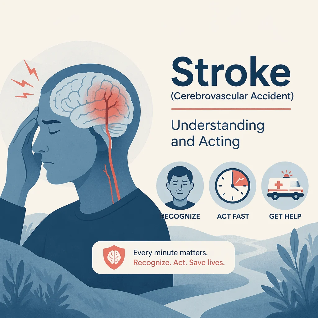
A stroke, also called a cerebrovascular accident, happens when the blood supply to part of the brain is suddenly interrupted, either by a clot that blocks an artery or by a vessel that bursts and bleeds. Brain cells starved of oxygen begin to die within minutes, which is why a stroke is always a medical emergency where every minute counts. The encouraging news is that fast treatment can limit the damage and improve recovery, and many strokes can be prevented by managing everyday risk factors. In this article you will learn what a stroke is, how to recognise the warning signs with BE-FAST, the main types and causes, the tests used to diagnose it, how it is treated, and what recent research is changing for patients.
Call 999 now: recognise a stroke with BE-FAST
A stroke is a life-threatening emergency. If you notice even one of the signs below, in yourself or in someone else, call 999 (or your local emergency number) right away. Do not wait to see whether it passes, do not drive yourself to the hospital, and try to note the exact time the symptoms started, because that timing guides treatment. The letters BE-FAST make the main warning signs easy to remember.
| Sign | What to check right now |
|---|---|
| B — Balance | Sudden loss of balance, dizziness, or trouble walking |
| E — Eyes | Sudden trouble seeing in one or both eyes, or double vision |
| F — Face | One side of the face droops or feels numb; ask the person to smile |
| A — Arms | One arm drifts down or feels weak; ask the person to raise both arms |
| S — Speech | Speech is slurred or hard to understand; ask for a simple sentence |
| T — Time | If you see any of these signs, call 999 now and note the start time |
Act quickly even if the signs seem mild or fade after a few minutes. A brief episode can be a warning of a much larger stroke to come, and only a hospital can tell the difference. Other sudden symptoms can also signal a stroke, including a severe headache with no known cause, numbness on one side of the body, or confusion.
What is a stroke (cerebrovascular accident)?
The brain needs a constant flow of oxygen-rich blood to work. When that flow stops, the affected area of the brain cannot function, and nerve cells start to die. Because different parts of the brain control different abilities, the symptoms depend on which region is affected: movement, speech, vision, balance, or memory can all be disrupted.
A stroke is not the same as a heart attack, although both involve blocked blood flow. A heart attack affects the heart muscle, whilst a stroke affects the brain. The two share many of the same risk factors, however, which is why protecting your heart also protects your brain.
The three types of stroke: ischaemic, haemorrhagic, and TIA
Strokes fall into three main categories. Telling them apart matters because the treatments are very different, and only brain imaging can confirm which type has occurred.
| Type of stroke | What happens in the brain | Share of cases | Key point |
|---|---|---|---|
| Ischaemic stroke | A clot blocks an artery that supplies the brain | About 85% | The most common type; may be treated by dissolving or removing the clot if caught early |
| Haemorrhagic stroke | A blood vessel in or around the brain bursts and bleeds | About 15% | Often linked to high blood pressure; managed very differently from an ischaemic stroke |
| Transient ischaemic attack (TIA) | A brief blockage that clears on its own, usually within minutes | Warning event | Symptoms disappear but signal a high short-term risk of a full stroke; treat it as an emergency |
Ischaemic stroke
In an ischaemic stroke, a clot forms in a brain artery or travels there from elsewhere, often from the heart or from a narrowed neck artery. This is by far the most common type, and it is the one that can sometimes be reversed with clot-busting drugs or clot removal when treatment starts quickly.
Haemorrhagic stroke
A haemorrhagic stroke is caused by bleeding, when a weakened vessel ruptures inside or on the surface of the brain. Long-standing high blood pressure and, less often, a bulge in a vessel wall called an aneurysm are frequent causes. Because there is bleeding rather than a clot, clot-busting drugs must never be used, which is why imaging comes first.
Transient ischaemic attack (TIA), the mini-stroke
A transient ischaemic attack, often called a mini-stroke, produces stroke-like symptoms that resolve on their own. It causes no lasting visible damage, but it is a serious red flag: it means a larger stroke could follow within hours or days. Anyone who experiences one should be assessed urgently so that preventive treatment can begin.
What causes a stroke? Risk factors you can and cannot change
Most strokes result from a mix of factors that build up over years. Some cannot be changed, but many can, and this is where prevention makes the biggest difference.
Risk factors you can change
You lower your risk the most by treating high blood pressure, a condition that strains and damages artery walls throughout the body and brain. Smoking, physical inactivity, an unhealthy diet, and excess weight all add to the risk. Plaque forms and arteries narrow faster in people who also manage high cholesterol, and blood-vessel damage is common in people who develop diabetes. An irregular heart rhythm called atrial fibrillation deserves special attention, because it lets clots form in the heart and travel to the brain.
Risk factors you cannot change
Age is the strongest fixed risk factor, as stroke becomes more likely after 55. A family history of stroke, your sex, and certain ethnic backgrounds also play a role. These cannot be altered, but knowing about them is a good reason to be stricter with the factors you can influence.
How is a stroke diagnosed?
Diagnosis is a race against the clock. When a stroke is suspected, brain imaging comes first, because it is the only way to tell an ischaemic stroke from a haemorrhagic one and to choose a safe treatment.
Imaging tests come first
A CT scan is usually done immediately; it quickly shows bleeding and helps rule out a haemorrhagic stroke. A brain MRI gives a more detailed picture and can reveal small or early ischaemic damage. Imaging of the blood vessels, called angiography, maps where a clot is lodged and helps the team plan clot removal. Doctors may also use a carotid ultrasound to check the neck arteries and an electrocardiogram (ECG) to look for an irregular heartbeat.
Blood tests support the workup and target risk factors
Blood tests do not diagnose a stroke on their own, but they guide safe treatment and uncover the risk factors behind it. In the emergency setting, teams check blood sugar and clotting before giving certain drugs. To look at how your blood clots, doctors run a coagulation panel, and in selected cases they also order a D-dimer blood test to investigate abnormal clotting. Afterwards, the search for causes continues, and the table below shows the everyday tests used to manage the main risk factors.
| Risk factor | Test or measure | Why it matters for stroke |
|---|---|---|
| High blood pressure | Blood pressure reading | The single biggest modifiable risk factor for both stroke types |
| High LDL cholesterol | LDL cholesterol, part of a lipid panel | Feeds the artery-narrowing plaque that can trigger a clot |
| High blood sugar and diabetes | A1C and fasting glucose | Long-term high sugar damages blood vessels and raises stroke risk |
| Atrial fibrillation | Heart rhythm check (ECG); cardiac markers when heart strain is suspected | An irregular heartbeat lets clots form and travel to the brain |
| Clotting tendency and inflammation | Coagulation panel, D-dimer, and other markers | Help investigate unusual causes in selected patients |
To measure long-term blood sugar, clinicians use the A1C blood test, and they may also check your fasting glucose levels. When the heart is under strain, they can measure the BNP cardiac marker to look for heart problems that raise stroke risk. Some workups also check homocysteine levels, an amino acid linked to blood-vessel damage.
How is a stroke treated? Why time is brain
Treatment depends entirely on the type of stroke, and it works best when it starts fast. The phrase clinicians use is simple: time is brain, because millions of nerve cells are lost for every hour a large stroke goes untreated.
Treating an ischaemic stroke
For an ischaemic stroke, doctors try to restore blood flow. Clot-busting medicine, known as thrombolysis, can dissolve the clot when given generally within about 4.5 hours of the first symptoms. For larger clots, a procedure called thrombectomy threads a thin catheter into the artery to physically pull the clot out, and in carefully selected patients it can be done up to 24 hours after symptoms begin. The sooner either treatment starts, the better the outcome, which is why calling 999 immediately matters so much.
Treating a haemorrhagic stroke
A haemorrhagic stroke is managed by controlling the bleeding and the pressure inside the skull. Doctors work to lower very high blood pressure, reverse any blood-thinning medication, and reduce swelling. Surgery is sometimes needed to repair a damaged vessel or to drain pooled blood. Clot-busting drugs are never used here, which is the reason imaging must confirm the type first.
Rehabilitation and recovery
After the emergency is over, rehabilitation helps the brain relearn lost abilities. Physical therapy rebuilds strength and movement, occupational therapy restores daily skills, and speech-language therapy addresses difficulties with talking or swallowing. Recovery can continue for months, and starting early makes a real difference.
Preventing a stroke and recovering afterwards
Up to four in five strokes are considered preventable, so prevention is powerful. The core steps are steady rather than dramatic: keep blood pressure in a healthy range, treat high cholesterol, control blood sugar if you have diabetes, stop smoking, stay active, eat a balanced diet low in salt, and limit alcohol. If you have atrial fibrillation, taking prescribed medication as directed sharply lowers the risk of a clot reaching the brain.
After a first stroke or a TIA, this same prevention becomes even more important, because the risk of a second event is higher. Regular check-ups let your care team track your risk factors over time and adjust treatment. Support from family, friends, and stroke support groups also plays a major role in emotional recovery and motivation.
Latest scientific advances in stroke care
Stroke treatment has changed quickly over the past few years. The advances below come from recent clinical trials and reviews. They are shared here to inform, not to guide your own treatment, which always belongs to your medical team.
A simpler clot-buster: tenecteplase
Tenecteplase is a clot-dissolving drug that can be given as a single quick injection, unlike the older drug alteplase, which needs a slow one-hour drip. Large trials published in 2023 and 2024, including TRACE-2 and ATTEST-2, found that tenecteplase works about as well as alteplase for eligible ischaemic strokes, and professional societies in Europe have moved to endorse its use. What this means for you: a faster, simpler treatment can save precious minutes, especially when a patient needs to be transferred between hospitals.
Clot removal now helps more people
Thrombectomy, the catheter procedure that pulls out a clot, used to be offered mainly to people with a small area of damage. Two 2023 trials published in a leading medical journal, SELECT2 and ANGEL-ASPECT, together with a systematic review that pooled several studies (a way of combining trials to see the overall picture), showed that many patients with a larger area of early damage also benefit. What this means for you: people who were once considered beyond help may now be eligible for clot removal, so reaching a stroke-ready hospital quickly is more valuable than ever.
Treatment that starts in the ambulance
A mobile stroke unit is a specially equipped ambulance that carries a small CT scanner, so imaging and clot-busting treatment can start before the patient reaches hospital. A 2024 analysis found that this head start helps more patients get treated within the crucial early window. What this means for you: in cities where these units operate, calling 999 at the first sign can put expert care at your door sooner.
Glossary of stroke terms
| Term | Definition |
|---|---|
| Ischaemic stroke | A stroke caused by a clot blocking a brain artery; the most common type. |
| Haemorrhagic stroke | A stroke caused by bleeding when a brain blood vessel bursts. |
| Transient ischaemic attack (TIA) | A mini-stroke with temporary symptoms that warns of a possible future stroke. |
| Thrombolysis | Treatment that uses a drug to dissolve a clot and restore blood flow. |
| Thrombectomy | A procedure that removes a clot from an artery using a thin catheter. |
| Atrial fibrillation | An irregular heart rhythm that can let clots form and travel to the brain. |
| Atherosclerosis | Gradual narrowing of arteries by fatty plaque that can lead to a clot. |
| Aphasia | Difficulty speaking or understanding language after brain injury. |
| Aneurysm | A weak, balloon-like bulge in a vessel wall that can burst and bleed. |
Frequently asked questions about stroke
What are the first warning signs of a stroke?
The first signs usually appear suddenly. Using BE-FAST, watch for loss of balance, trouble seeing, a drooping face, arm weakness, and slurred speech. Other sudden signs include numbness on one side of the body, confusion, or a severe headache with no clear cause. If any of these appear, call 999 immediately, even if they seem to ease.
What is the difference between a stroke and a mini-stroke (TIA)?
A mini-stroke, or transient ischaemic attack, causes the same kind of symptoms as a stroke, but they clear on their own, usually within minutes. It leaves no lasting visible damage, yet it is a strong warning that a full stroke may follow soon. A TIA should be treated as an emergency and assessed by a doctor without delay.
What are the symptoms of a stroke in women?
Women have the same core warning signs as men, described by BE-FAST. Women may also report less typical symptoms such as sudden fatigue, nausea, general weakness, or confusion. Because these can be mistaken for other conditions, it is important not to dismiss them. When in doubt, call 999 and note the time symptoms began.
What causes a stroke?
Most strokes come from a build-up of risk factors over time. High blood pressure is the leading cause, alongside smoking, high cholesterol, diabetes, obesity, physical inactivity, and atrial fibrillation. A clot or a burst vessel is the immediate trigger, but these underlying factors set the stage. Managing them is the best way to lower your risk.
Can you recover from a stroke?
Many people recover well, especially when treatment starts quickly and rehabilitation begins early. The degree of recovery depends on the type of stroke, how much of the brain was affected, and how fast blood flow was restored. Physical, occupational, and speech therapy can help the brain relearn lost skills over weeks and months.
Is a stroke the same as a heat stroke?
No, they are completely different conditions that share only a name. A stroke involves the brain losing its blood supply, while a heat stroke is dangerous overheating of the body, usually in hot weather. Both are emergencies, but their causes and treatments are unrelated. This article is about the brain condition, the cerebrovascular accident.
Sources
- Centers for Disease Control and Prevention (CDC) — About Stroke — cdc.gov
- American Stroke Association — Stroke Symptoms and Warning Signs (B.E. F.A.S.T.) — stroke.org
- Mayo Clinic — Stroke: Symptoms and Causes — mayoclinic.org
- Wang Y, et al. — Tenecteplase versus alteplase in acute ischaemic cerebrovascular events (TRACE-2) — The Lancet, 2023 — doi.org/10.1016/S0140-6736(22)02600-9
- Muir KW, et al. — Tenecteplase versus alteplase for acute stroke within 4.5 h (ATTEST-2) — The Lancet Neurology, 2024 — doi.org/10.1016/S1474-4422(24)00377-6
- Alamowitch S, et al. — European Stroke Organisation expedited recommendation on tenecteplase — European Stroke Journal, 2023 — doi.org/10.1177/23969873221150022
- Sarraj A, et al. — Trial of Endovascular Thrombectomy for Large Ischaemic Strokes (SELECT2) — New England Journal of Medicine, 2023 — doi.org/10.1056/NEJMoa2214403
- Huo X, et al. — Trial of Endovascular Therapy for Acute Ischaemic Stroke with Large Infarct (ANGEL-ASPECT) — New England Journal of Medicine, 2023 — doi.org/10.1056/NEJMoa2213379
- Li C, et al. — Mechanical Thrombectomy for Large Ischaemic Stroke: A Systematic Review and Meta-analysis — Neurology, 2023 — doi.org/10.1212/WNL.0000000000207536
- Mac Grory B, et al. — Mobile Stroke Unit Management in Patients Eligible for Intravenous Thrombolysis — JAMA Neurology, 2024 — doi.org/10.1001/jamaneurol.2024.3659
Further reading
- High Blood Pressure: Understanding, Treating, and Preventing
- Lipid Panel Explained: Cholesterol, LDL, HDL, and Triglycerides
- LDL Cholesterol: Understanding and Reading This Blood Marker
- D-dimer Blood Test: A Comprehensive Guide to Your Results
- Understanding Homocysteine Levels and Risks
Understand your lab results with AI DiagMe
A stroke is diagnosed with brain imaging, not with a blood test, but blood tests are central to managing the risk factors behind it. Cholesterol, blood sugar, and clotting results all help you and your doctor see where your stroke risk stands and what to work on. AI DiagMe helps you understand these lab results in plain language; it does not provide a diagnosis and does not replace your doctor.
Get your results interpreted in minutes