





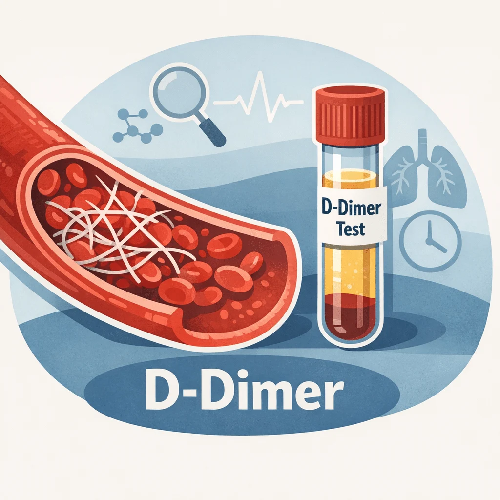
A D-dimer test measures a small protein fragment that appears in your blood when a clot breaks down. Doctors order it most often to help rule out a dangerous clot, such as a deep vein thrombosis (DVT) or a pulmonary embolism (PE), when combined with a clinical risk assessment. This guide explains what the test measures, how to read your number against the reference range, why age-adjusted cutoffs matter, and which other conditions can raise this marker even without a clot. You will also find a plain-language summary of the latest research, a glossary, and clear guidance on when a result deserves prompt medical attention.
What is a D-dimer test?
D-dimer is a fragment left over after your body breaks down a blood clot. It does not exist in meaningful amounts in healthy blood unless clotting and clot breakdown are actively happening somewhere in your body. When a clot forms, the protein fibrin builds a mesh that holds it together. Once the injury heals, an enzyme called plasmin dissolves that mesh, and the debris includes D-dimer fragments that circulate until your kidneys and liver clear them.
A D-dimer test picks up those fragments in a standard blood sample. Because the fragment only appears when a clot is being made and broken down, a raised level tells a clinician that clotting activity is happening somewhere. It does not say where, and it does not say why — those answers require more testing.
Why the test matters in everyday practice
The main strength of this marker is what a normal result rules out, not what a high result confirms. When a doctor already judges a clot unlikely based on your symptoms and history, a normal D-dimer helps exclude a DVT or PE without further imaging. This combination — clinical judgement plus a blood test — is what makes the D-dimer so useful in emergency and outpatient settings, sparing many people an unnecessary CT scan or ultrasound.
How to read your D-dimer test results
Your report lists a number, a unit, and a reference range. A typical result looks like this: D-dimer 320 ng/mL FEU (reference range: below 500 ng/mL FEU). FEU stands for fibrinogen equivalent units, one of the two common ways labs report this test; some labs instead use D-dimer units (DDU), which carry a different numeric cutoff. If you are comparing a result to information you find elsewhere, always check which unit your own lab used.
Most adult laboratories set a standard cutoff around 500 ng/mL FEU. A result below that threshold is usually called negative, meaning clot activity was not detected at a meaningful level. A result at or above the threshold is called positive, which prompts your doctor to look further rather than to diagnose a clot outright.
Why age changes the picture
D-dimer naturally climbs with age, even in people who feel completely well. Using the same flat cutoff for a 30-year-old and an 80-year-old means older adults are flagged "positive" far more often without actually having a clot. To correct for this, many labs and emergency departments now use an age-adjusted cutoff for patients over 50: multiply your age in years by 10 to get your personal cutoff in ng/mL FEU. A 70-year-old, for example, would have a cutoff around 700 ng/mL instead of the standard 500.
This adjustment has been studied in large groups of patients and is now supported by several professional societies, though it has not been formally approved by the FDA as a labelling change. Its purpose is narrow and specific: it improves how well a normal result rules out a clot in older adults, without meaningfully missing real clots. It is not a general "normal range for your age" chart — it only applies to interpreting a result that is being used to investigate a possible DVT or PE.
A simple decision flow for reading your number
Use this sequence as a starting point, not a substitute for medical advice.
| Step | What to check |
|---|---|
| 1. Find your unit | Confirm whether your lab reports in ng/mL FEU or DDU, printed next to your result |
| 2. Compare to the printed range | Use the reference range on your own report, not a number from another source |
| 3. Check your age | If you are over 50, ask whether an age-adjusted cutoff (age x 10 ng/mL) applies |
| 4. Note recent context | Recent surgery, pregnancy, infection, or a long illness can raise the result on their own |
| 5. Discuss with your doctor | Only a clinician can combine this result with your symptoms and risk factors |
Why D-dimer is sensitive but not specific
A test is sensitive when it reliably picks up a condition when present, and specific when it reliably stays normal when the condition is absent. D-dimer is a textbook example of a highly sensitive but poorly specific marker: it almost always rises when a significant clot is forming or dissolving, but plenty of other situations raise it too, so a positive result alone proves little.
Conditions and situations known to raise D-dimer without any dangerous clot include pregnancy, recent surgery or trauma, active infection or sepsis, cancer, liver disease, and simply being older. Intense physical exertion, such as running a marathon, can also cause a temporary rise that settles within a day or two. This is precisely why doctors combine the test with a structured risk assessment — such as the Wells score, which weighs factors like recent immobility, prior clots, and specific symptoms — rather than acting on the number in isolation.
Conditions linked to a high D-dimer
A raised D-dimer is a signal to investigate further, not a diagnosis. The sections below cover the situations doctors consider most often.
Deep vein thrombosis (DVT)
DVT is a clot that forms in a deep vein, most often in the calf or thigh. As your body attempts to break the clot down, D-dimer fragments enter your bloodstream and raise your test result. Typical symptoms include swelling, pain, warmth, or redness in one leg. A leg ultrasound remains the standard way to confirm or rule out DVT once suspicion is raised.
Pulmonary embolism (PE)
A pulmonary embolism happens when a clot — usually one that started as a DVT — breaks loose and lodges in an artery in the lung. Sudden shortness of breath, sharp chest pain that worsens with breathing, and a fast heart rate are common warning signs. Because a PE can be life-threatening, doctors often move quickly from a positive D-dimer to imaging with a CT pulmonary angiogram. Our detailed guide on pulmonary embolism symptoms and treatment covers the full diagnostic and treatment pathway.
Disseminated intravascular coagulation (DIC)
DIC is a rare, serious condition in which the clotting system activates throughout the body at once, forming many small clots that consume clotting factors faster than the body can replace them. This paradoxically raises the risk of both clotting and bleeding at the same time. D-dimer levels in DIC are usually very high, and the test is used alongside a platelet count and other clotting values to confirm the diagnosis and track treatment response.
Infection, inflammation, and cancer
Any process that activates inflammation tends to nudge clotting activity upward, which is why severe infections, chronic inflammatory conditions, and some cancers can all produce an elevated D-dimer without a clot ever forming somewhere dangerous. In these situations, a doctor typically reviews the D-dimer together with other inflammation markers, checking your C-reactive protein levels to help separate ordinary inflammation from a genuine clotting problem.
What a normal D-dimer means
A normal D-dimer is a genuinely reassuring finding when a doctor already judges your risk of a clot to be low or intermediate. In that setting, the test has a very high negative predictive value, meaning it is excellent at confirming that a DVT or PE is unlikely, often sparing you a CT scan or ultrasound. This is the single most valuable use of the test in everyday medicine.
A few caveats are worth knowing. The test can occasionally read normal very early in a clot, before enough breakdown has occurred to raise the level. It also does not exclude every type of vascular problem, and if your doctor has a strong clinical suspicion despite a normal result, further testing may still be arranged. Blood thinner medications taken before the test can also produce a falsely normal reading, so it matters to tell your care team about any anticoagulant use.
The coagulation panel: where D-dimer fits
D-dimer rarely appears alone on a lab report. It is usually part of a broader coagulation panel that also reports PT, PTT, and INR, each of which measures a different part of the clotting process. The table below shows how D-dimer compares with the other common clotting tests.
| Test | What it measures | Common use |
|---|---|---|
| D-dimer | Fragment left when a clot dissolves | Helps rule out DVT or PE when risk is low |
| PT / INR | Speed of the extrinsic clotting pathway | Monitors warfarin, checks liver function |
| PTT / aPTT | Speed of the intrinsic clotting pathway | Monitors heparin, investigates bleeding disorders |
| Fibrinogen | The protein that forms the clot’s fibrin mesh | Assessed alongside D-dimer in DIC |
Two of the natural anticoagulants that keep clotting in check are measured separately when a hereditary clotting disorder is suspected: Protein C and Protein S. Doctors also check the aPTT results in more detail when a bleeding or clotting tendency needs a closer look, and unexplained clots sometimes prompt testing for antithrombin III deficiency.
Special situations that raise D-dimer
Pregnancy
Pregnancy naturally increases clotting activity to prepare the body for delivery, so D-dimer levels rise steadily through each trimester and are typically well above the standard adult cutoff by the third trimester. Because of this, doctors do not use the general reference range to judge a pregnant patient’s result; a suspected clot in pregnancy is usually investigated with imaging rather than D-dimer alone. If you want a broader picture of prenatal labs, our guide covers the full set of blood tests during pregnancy, including which markers shift and why.
Recent surgery or hospitalisation
Any operation triggers tissue repair and clotting activity, so it is common and expected for D-dimer to rise for one to several weeks after surgery. Surgical teams already factor this expected pattern into how they read your pre-surgical blood work, so an elevated result soon after an operation rarely triggers alarm on its own.
Older age
As covered above, ageing alone raises baseline D-dimer, which is exactly why the age-adjusted cutoff exists for patients over 50.
When to see a doctor
Most abnormal D-dimer results are followed up calmly with your regular doctor rather than as an emergency. However, certain combinations of symptoms need prompt attention regardless of your lab number.
Seek urgent or emergency care if you have:
- Sudden shortness of breath, chest pain that worsens with breathing, or coughing up blood.
- Swelling, pain, warmth, or redness in one leg or arm that appears suddenly.
- A fast heart rate with dizziness or fainting alongside any of the symptoms above.
Contact your doctor to discuss your result, without needing emergency care, if you have:
- A mildly elevated D-dimer with no symptoms and a known recent cause, such as surgery or pregnancy.
- A moderately elevated result that your doctor wants to monitor with a repeat test in a few weeks.
- Questions about how a medication or supplement might be affecting your result.
Latest scientific advances
Research on D-dimer interpretation has moved quickly in the past few years, largely refining how clinicians use adjusted cutoffs rather than changing what the test itself measures. A 2023 systematic review and meta-analysis pooling 68 studies and more than 140,000 patients compared several ways of adjusting the D-dimer cutoff — by age, by pretest clinical probability, and using a combined algorithm called YEARS. All of the adjustment strategies kept the test’s ability to catch real clots essentially unchanged while meaningfully improving how well a normal result ruled out a clot, though the strategies varied in how consistent their performance was across different patient groups. What this means for you: age-adjusted and probability-adjusted cutoffs appear safe and are not just a theoretical idea — they are backed by very large combined patient data (Gerber et al., Journal of Internal Medicine, 2023; DOI).
A 2024 narrative review focused specifically on comparing the age-adjusted cutoff against the clinical-probability-adapted cutoff, the two most widely used refinements. The review found that the age-adjusted approach tends to be the more cautious of the two, missing very few real clots but ruling out somewhat fewer people as clot-free, while the clinical-probability approach rules out more people but carries a slightly higher chance of missing a case. What this means for you: neither method is universally “better” — your emergency department’s choice of cutoff reflects a considered trade-off between safety and avoiding unnecessary scans, not an arbitrary shortcut (Righini et al., Journal of Clinical Medicine, 2024).
A 2025 secondary analysis of the landmark ADJUST-PE study tested whether the age-adjusted cutoff works consistently across different D-dimer laboratory assays, since hospitals do not all use the same testing equipment. Repeating the original test on stored samples with several alternative assays found that two of the four alternative assays performed similarly safely to the original method, while two others classified more patients as low-risk in a way that would have missed a small number of real clots. What this means for you: the specific assay your lab uses matters, and this is an early finding, still being confirmed, that explains why hospitals validate their own equipment before adopting an age-adjusted cutoff rather than assuming all D-dimer tests behave identically (Robert-Ebadi et al., Journal of Thrombosis and Haemostasis, 2025).
Beyond cutoff refinement, a 2024 review in the journal Haematologica examined how haematologists should approach otherwise healthy outpatients found to have an unexplained high D-dimer with no signs of a clot — an increasingly common scenario as the test is ordered more widely. The review emphasised that an isolated high D-dimer in someone without symptoms rarely needs aggressive investigation and should prompt a structured but measured work-up rather than alarm, taking into account age, medications, and any subtle risk factors for cancer or an underlying clotting tendency. What this means for you: if you are told your D-dimer is “a bit high” with no other findings, current expert guidance leans towards calm, stepwise follow-up rather than immediate invasive testing (Franchini et al., Haematologica, 2024).
Glossary
| Term | Definition |
|---|---|
| Age-adjusted cutoff | A personalised D-dimer threshold for patients over 50, calculated as age multiplied by 10 ng/mL |
| D-dimer | A protein fragment released into the blood when a clot is broken down |
| Deep vein thrombosis (DVT) | A blood clot that forms in a deep vein, most often in the leg |
| Disseminated intravascular coagulation (DIC) | A serious condition where clotting and bleeding happen throughout the body at once |
| Fibrin | A protein that forms the mesh structure of a blood clot |
| FEU (fibrinogen equivalent units) | One of two common reporting units for D-dimer results |
| Negative predictive value | How reliably a normal test result rules out the condition being investigated |
| Pulmonary embolism (PE) | A blood clot that blocks an artery in the lung, usually after travelling from a leg vein |
| Sensitivity and specificity | How reliably a test detects a condition when present (sensitivity) and stays normal when absent (specificity) |
| Wells score | A clinical scoring tool that estimates the probability of a DVT or PE before testing |
Frequently asked questions about the D-dimer test
Can stress or intense exercise raise D-dimer levels?
Ordinary psychological stress does not typically raise D-dimer to a meaningful degree. Very intense physical exertion, such as running a marathon or another endurance event, can cause a temporary rise due to increased clotting and breakdown activity in the muscles. This effect usually settles back to baseline within 24 to 48 hours, so a recent hard workout is worth mentioning to your doctor if your test was scheduled soon afterwards.
Does hormonal birth control affect D-dimer levels?
Yes. Combined hormonal contraceptives containing both oestrogen and progestin can produce a modest rise in D-dimer because oestrogen influences the clotting system. The effect is usually mild rather than dramatic. Progestin-only methods generally have little to no measurable impact. If you take hormonal contraception, mention it to whoever orders or interprets your test.
Which medications can influence D-dimer test results?
Anticoagulant medications, commonly called blood thinners, are often prescribed precisely because of a high D-dimer finding, but they can also cause a falsely normal result if the test is repeated whilst you are already being treated. Fibrinolytic drugs, which actively dissolve clots in emergency settings, cause a sharp temporary surge as they work. Always give your care team a complete list of medications and supplements before this test.
Why would a doctor order a D-dimer test for chest pain?
Symptoms of a pulmonary embolism, including sudden breathlessness and chest pain, can closely resemble a heart problem. Ordering a D-dimer test alongside cardiac markers helps a doctor quickly distinguish between the two possibilities. A normal result, combined with a low clinical probability, makes a PE less likely and allows the workup to focus on other causes of chest discomfort more efficiently.
Can you have a pulmonary embolism with a normal D-dimer?
This is uncommon but possible, particularly with a very small or long-standing (chronic) clot that is not actively breaking down at the time of testing. It can also occur if you are already taking an anticoagulant that suppresses the result. This is exactly why a doctor’s clinical judgement always accompanies the number — if suspicion remains high despite a normal test, imaging will typically still be arranged.
Can D-dimer results vary between different laboratories?
Yes, meaningfully so. Different labs use different testing methods and may report in different units, which means both the number and the reference range printed on your report can differ from one facility to another. Always interpret your result against the range provided by the specific lab that ran your test, and mention the assay or unit if you are being compared across visits at different locations.
Sources
- D-Dimer Test — MedlinePlus, U.S. National Library of Medicine
- D-Dimer Test: What It Is, What It Is Used For, Risks and Results — Cleveland Clinic
- Deep Vein Thrombosis (DVT): Diagnosis and Treatment, including the D-dimer blood test — Mayo Clinic
- Gerber et al., Utility and limitations of patient-adjusted D-dimer cut-off levels for diagnosis of venous thromboembolism — a systematic review and meta-analysis, Journal of Internal Medicine, 2023 (PubMed PMID 37143392) — DOI
- Righini, Robert-Ebadi and Le Gal, Age-Adjusted and Clinical Probability Adapted D-Dimer Cutoffs to Rule Out Pulmonary Embolism: A Narrative Review of Clinical Trials, Journal of Clinical Medicine, 2024 (PubMed PMID 38929970) — DOI
- Robert-Ebadi et al., Different D-dimer assays with age-adjusted cutoffs to exclude pulmonary embolism: secondary analysis of ADJUST-PE study, Journal of Thrombosis and Haemostasis, 2025 (PubMed PMID 40252844) — DOI
- Franchini, Focosi, Pezzo and Mannucci, How we manage a high D-dimer, Haematologica, 2024 (PubMed PMID 37881856) — DOI
Further reading
- Coagulation panel: PT, PTT, INR and D-dimer explained
- Pulmonary embolism: symptoms, diagnosis, and treatment
- aPTT blood test: understanding your levels
- How to read your blood test results
- Blood markers: a comprehensive glossary
Coagulation results rarely tell the full story on their own, and a D-dimer is easiest to understand alongside the rest of your report. AI DiagMe can help you make sense of related values such as your platelet count, fibrinogen, and PT/INR in plain language, showing how they fit together with your D-dimer result. It is built to help you understand your labs, not to diagnose you or replace your doctor.
Understand your lab results with AI DiagMe
Get your results interpreted in minutes