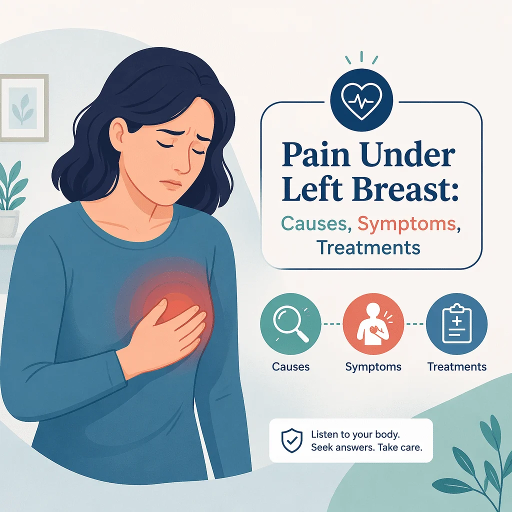
Durerea sub sânul stâng este unul dintre cele mai căutate simptome online și unul dintre cele mai greu de interpretat. Acea mică zonă a pieptului se află deasupra stomacului, lângă splină, sub partea inferioară a plămânului stâng, peste coastele inferioare și cartilajele lor — și direct în fața inimii. Zeci de probleme pot provoca disconfort în același loc, iar mai multe se simt remarcabil de similar.
În acest articol vei afla ce se află sub sânul stâng, cum se simte fiecare grup de cauze, de ce concluzia „pare muscular" nu este niciodată sigură dacă o tragi singur și cum lucrează medicii pentru a clarifica situația. Este scris pentru adulți cu durere nouă, în schimbare sau persistentă în partea inferioară stângă a pieptului. Verifică imediat caseta cu semnele de alarmă de mai jos înainte de a continua lectura.
Sună la 112 acum dacă oricare dintre acestea se aplică
Nu conduce singur și nu aștepta să treacă. Sună la 112, sau la numărul local de urgență, dacă durerea de sub sânul stâng este însoțită de oricare dintre acestea:
- Presiune, strângere, apăsare sau greutate în piept
- Durere care iradiază în braț, maxilar, gât, umăr sau spate
- Dificultăți de respirație în repaus sau la efort ușor
- Transpirații reci sau senzație de răceală a pielii
- Greață sau vărsături însoțite de durere
- Amețeală sau leșin
- Sentimentul puternic că ceva este grav în neregulă, o senzație de rău iminent
- Durere bruscă și ascuțită însoțită de lipsă de aer sau palpitații, care poate indica o embolie pulmonară (un cheag de sânge în plămân) sau un plămân colabat
- Tuse cu sânge
- Durere după o căzătură, o lovitură la piept sau un accident
Sună la 112 și dacă durerea este severă, diferită de orice ai mai simțit, sau durează mai mult de câteva minute. Echipajele de ambulanță pot înregistra un ECG și pot începe tratamentul pe drum, iar persoanele care ajung cu ambulanța primesc adesea îngrijiri mai rapide.
Ce se află sub sânul stâng și de ce poate durea această zonă
Partea stângă inferioară a toracelui este aglomerată. Adăpostește porțiunea inferioară a inimii și sacul pericardic din jurul ei, lobul inferior al plămânului stâng și pleura sa, partea superioară a stomacului, flexura splenică — curbura pronunțată unde colonul se îndreaptă în jos sub coastele stângi — și splina. În jurul tuturor acestora se află coastele, cartilajele care le unesc cu sternul și mușchii intercostali dintre ele.
Aceste structuri împart căi nervoase comune. Semnalele provenite din peretele toracic, inimă, diafragmă și tractul digestiv superior ajung la creier pe trasee suprapuse, motiv pentru care o problemă gastrică poate fi resimțită ca o durere cardiacă, iar o problemă cardiacă poate părea o simplă indigestie. Această suprapunere este o caracteristică anatomică normală și reprezintă principalul motiv pentru care autodiagnosticarea în această zonă eșuează atât de des.
Cauze la nivelul peretelui toracic: coaste, cartilaje și mușchi
Peretele toracic este o explicație frecventă pentru durerea de sub sânul stâng, dar de obicei doar după ce un medic a exclus cauzele grave.
Costocondrită și sindromul Tietze
Costocondrită este inflamația locului unde cartilajul costal se unește cu sternul. Provoacă durere ascuțită sau surdă într-un punct precis, adesea mai intensă la respirații adânci, tuse, răsuciri sau apăsare. Sindromul Tietze este o afecțiune înrudită, mai puțin frecventă, în care o articulație costală este vizibil umflată și sensibilă. Ambele pot dura săptămâni și se pot agrava cu activitatea fizică.
Contractură musculară, traumatism costal și durere nervoasă
Mușchii intercostali dintre coaste pot fi întinși prin ridicarea de greutăți, exerciții fizice neobișnuite, o tuse prelungită sau o poziție incomodă în timpul somnului. Coastele vânătăite sau fracturate provoacă o durere ascuțită, localizată, care se agravează cu fiecare respirație. Spasmul acestor mușchi dă un tablou similar, descris în ghidul nostru despre crampe la nivelul coastelor. Zona zoster poate provoca durere arsură, în bandă, de-a lungul unui spațiu intercostal, timp de câteva zile înainte de apariția oricărei erupții cutanate, după cum este explicat în articolul nostru despre simptomele și testele pentru herpes zoster.
Sensibilitatea reproductibilă — durerea care apare în mod constant atunci când apeși același punct — face mai probabilă o cauză la nivelul peretelui toracic. Totuși, nu exclude nimic. Persoanele care fac un infarct pot avea și peretele toracic sensibil la palpare, iar o întindere musculară poate coexista cu o problemă cardiacă la aceeași persoană. Tratează acest indiciu ca pe o informație pentru medicul tău, nu ca pe un verdict.
Cauze digestive, inclusiv gaz blocat la nivelul flexurii splenice
Gazele intestinale și sindromul flexurii splenice
Flexura splenică este curbura ascuțită din stânga colonului, situată sus, sub coaste. Gazele se pot acumula acolo și pot destinde peretele intestinal, provocând crampe, senzație de plenitudine sau o durere surprinzător de ascuțită sub sânul stâng — un tablou numit uneori sindromul flexurii splenice. Durerea își schimbă adesea locul, se ameliorează după eructații sau după scaun și apare și dispare în decurs de ore. Este o afecțiune frecventă, rar explicată pe înțelesul tuturor, dar este o concluzie la care se ajunge după ce au fost excluse cauzele grave, nu o presupunere de la bun început.
Refluxul, gastrita, ulcerul și pancreasul
Boala de reflux gastroesofagian (BRGE) împinge acidul gastric în esofag și provoacă o senzație de arsură care urcă spre gât, mai intensă după mese copioase, aplecarea în față sau culcatul. Gastrita și ulcerul peptic cauzează o durere surdă în partea superioară a abdomenului, simțită sub coastele stângi. Pancreatita provoacă o durere puternică și persistentă în etajul abdominal superior, care iradiază spre spate, de obicei însoțită de vărsături.
Refluxul este cauza cel mai des identificată atunci când durerea în piept se dovedește a nu fi cardiacă — tocmai de aceea este riscant să presupui că este. Lectură suplimentară: reflux acid și tuse, durere în piept după ce bei suc carbogazos, și pancreatita.
Splina
Splina se află chiar sub coastele stângi, în spatele stomacului. Când se mărește — din cauza unor infecții precum mononucleoza infecțioasă, a unor boli hematologice sau a bolilor hepatice — poate provoca o durere surdă sau o senzație de plenitudine, uneori însoțită de sațietate precoce, adică te simți sătul după foarte puțină mâncare. Un infarct splenic, în care o parte a splinei rămâne fără aport de sânge, cauzează o durere mai ascuțită și mai persistentă. O splină mărită poate sângera periculos și în urma unui traumatism minor, ceea ce reprezintă încă un motiv pentru care durerea din stânga apărută după orice impact necesită o evaluare urgentă, nu odihnă și gheață.
Cauze cardiace ale durerii sub sânul stâng
Angina pectorală și infarctul miocardic
Inima se află în spatele sternului și se extinde spre stânga. Angina pectorală este disconfortul toracic cauzat de reducerea fluxului de sânge către mușchiul inimii. Se manifestă clasic ca o presiune, strângere, apăsare sau greutate, nu ca o înțepătură localizată, poate iradia în braț, maxilar, gât, umăr sau spate, iar angina stabilă apare la efort și dispare în câteva minute de repaus. Un infarct miocardic nu cedează la repaus și este adesea însoțit de transpirații, greață, dificultăți de respirație sau amețeală. NHLBI precizează că infarctul miocardic nu debutează întotdeauna cu o durere violentă și bruscă: simptomele pot apărea treptat, pot rămâne ușoare sau pot veni și trece în decurs de ore.
Pericardita și miocardita
Pericardita este inflamația sacului care înconjoară inima și oferă unul dintre puținele indicii poziționale cu adevărat utile în durerea toracică: o durere ascuțită care se agravează când ești culcat pe spate și se ameliorează când stai în șezut și te apleci ușor înainte, adesea mai intensă la o respirație profundă. Miocardita, inflamația mușchiului cardiac, poate cauza durere în piept însoțită de lipsă de aer, palpitații sau oboseală neobișnuită, apărând adesea după o boală virală. Ambele necesită evaluare medicală; niciuna nu se tratează acasă.
Femeile și persoanele cu diabet sunt mai des trecute cu vederea
Acest lucru contează în mod special pe o pagină despre durerea “sub sânul stâng”, o expresie căutată mult mai frecvent de femei. Femeile care fac un infarct miocardic au mai multe șanse decât bărbații să prezinte durere în umăr, spate sau braț, dificultăți de respirație, oboseală prelungită, tulburări digestive sau anxietate — cu sau fără durere în piept. NHLBI afirmă că femeile pot minimiza aceste simptome și pot întârzia să meargă la spital și că screeningul inițial în spital poate să nu detecteze un infarct precoce sau atipic, lăsând femeile cu un risc mai mare ulterior.
Persoanele cu diabet au, de asemenea, o probabilitate mai mare de a prezenta dureri toracice atenuate sau absente, deoarece leziunile nervoase cauzate de un nivel ridicat al glicemiei pe termen lung diminuează semnalul de avertizare. NHLBI descrie acestea drept infarcte silențioase, mai frecvente la adulții în vârstă și la persoanele cu glicemie crescută sau diabet. Dacă ești femeie sau ai diabet, absența durerii clasice în piept, de tip presiune intensă, nu este un motiv de liniște.
Cauze pulmonare
Pleurezia, inflamația membranei care căptușește plămânul, provoacă o durere ascuțită care se agravează la inspirație, tuse sau strănut și se ameliorează când îți ții respirația. Pneumonia adaugă febră, tuse și lipsă de aer și poate iradia durerea spre spate — vezi articolul nostru despre pneumonie și durerea de spate. Pneumotoraxul, sau plămânul colabat, provoacă o durere bruscă, ascuțită, pe o singură parte, însoțită de lipsă de aer, uneori la persoane tinere, înalte și slabe. Embolia pulmonară, un cheag în circulația pulmonară, provoacă durere bruscă, lipsă de aer și bătăi rapide ale inimii, uneori cu tuse cu sânge. Ultimele două sunt urgențe medicale, iar riscul crește după imobilizare prelungită, intervenție chirurgicală recentă, sarcină, cancer sau un episod anterior de tromboză.
Anxietatea și atacul de panică: reale, dar niciodată un autodiagnostic
Atacurile de panică provoacă cu adevărat dureri în piept, palpitații, lipsă de aer, furnicături și o senzație de catastrofă iminentă, și reprezintă un motiv frecvent pentru vizitele la urgențe. Durerea este reală, nu imaginară. Dar anxietatea este un diagnostic de excludere, iar excluderea este treaba medicului: necesită o examinare și, de obicei, un ECG și analize de sânge. Mulți oameni au atât o tulburare de anxietate, cât și o afecțiune cardiacă. Dacă observi un tipar recurent, ghidul nostru despre anxietate te poate ajuta să îl descrii — după ce un medic a evaluat episodul, nu în locul acestuia.
De ce nu poți diferenția singur aceste cauze în mod sigur
Aceasta este cea mai importantă secțiune a acestui articol. Nicio combinație de senzații nu poate diferenția în mod sigur durerea toracică cardiacă de cea non-cardiacă fără investigații. Medicii folosesc scoruri de risc, ECG și analize de sânge tocmai pentru că simptomele singure nu sunt suficient de precise — și chiar și acestea au limitele lor reale.
Trei convingeri greșite provoacă cel mai mult rău. „Trebuie să fie musculară, pentru că pot apăsa pe ea." „Sunt prea tânăr pentru o problemă cardiacă." „Vine și trece, deci nu poate fi ceva grav." Niciuna nu se confirmă. Pereții toracici sensibili la palpare apar și la persoane care fac infarct, infarctele se întâmplă și la persoane de treizeci de ani, iar durerea cardiacă poate să apară și să dispară în decurs de ore. Simptome precum furnicături în piept sau dificultăți de respirație după masă pot apărea atât din cauze inofensive, cât și din cauze grave.
Tabelul de mai jos este un ghid despre ce să faci în continuare, nu un instrument de diagnostic. Niciun rând nu exclude o cauză.
| Cum este durerea | Ce poate sugera | Ce să faci |
|---|---|---|
| Durere ascuțită, localizată precis, reprodusă când apeși pe același punct | Peretele toracic: condrită costală, sindromul Tietze, contractură musculară, traumatism costal | Mai probabil o cauză la nivelul peretelui toracic, dar nu exclude nimic. Solicită o consultație medicală; sună la 112 dacă apare orice semn de alarmă |
| Se agravează în poziție culcată, se ameliorează stând în șezut și aplecat înainte | Pericardită (inflamația sacului din jurul inimii) | Consultație medicală în aceeași zi, nu îngrijire la domiciliu |
| Presiune sau strângere care iradiază în braț, maxilar, gât, umăr sau spate, însoțită de transpirații, greață sau dificultăți de respirație | Posibil infarct miocardic sau angină pectorală | Sună la 112 acum. Nu conduce singur |
| Durere ascuțită bruscă cu dificultăți de respirație sau tuse cu sânge | Posibilă embolie pulmonară sau pneumotorax | Sună la 112 acum |
| Senzație de arsură care urcă spre gât, mai intensă după mese sau în poziție culcată | Reflux, gastrită sau ulcer | Refluxul și durerea cardiacă se pot suprapune. Consultați un medic în loc să presupuneți cauza |
| Senzație de plenitudine cu crampe, ameliorată de eructații, gaze sau scaun | Gaz blocat la nivelul flexurii splenice a colonului | De obicei benign, dar numai după excluderea cauzelor grave |
| Durere după o căzătură, o lovitură la piept sau un accident | Fractură de coastă, contuzie pulmonară sau leziune splenică | Evaluare urgentă; sună la 112 în caz de durere severă, dificultăți de respirație sau leșin |
Ce face efectiv un clinician
Evaluarea începe cu povestea ta: cum se simte durerea, unde iradiază, ce o declanșează și ce o ameliorează, și care sunt factorii tăi de risc. Medicul ascultă inima și plămânii, palpează pieptul și abdomenul superior, verifică tensiunea arterială, pulsul și nivelul de oxigen și examinează picioarele pentru semne de cheag de sânge.
O electrocardiogramă (ECG), o înregistrare rapidă a activității electrice a inimii, se efectuează de obicei devreme, deoarece este rapidă și poate schimba totul. Urmează analizele de sânge: troponina, o proteină eliberată atunci când mușchiul cardiac este lezat, adesea repetată la câteva ore distanță; D-dimer când se ia în considerare un tromb; markeri inflamatori precum CRP; și uneori creatinfosfokinaza (CPK) ca marker muscular. Pot urma o radiografie toracică, o ecografie sau o tomografie computerizată, iar acolo unde persistă îndoieli, angiografia CT coronariană vizualizează direct arterele coronare.
Odată confirmată o cauză benignă, medicii pot recomanda măsuri adaptate acelui diagnostic — stretching ușor și căldură locală pentru o contractură confirmată a peretelui toracic, sau modificări ale dietei și posturii pentru reflux confirmat. Aceste recomandări vin după un diagnostic, nu înainte, și trebuie să provină de la medicul care te-a examinat.
Cele mai recente progrese științifice în evaluarea durerii toracice
Cercetările din 2023 până în 2025 arată cât de serios trebuie luată durerea toracică și unde instrumentele actuale au lipsuri. Toate studiile de mai jos sunt indexate în PubMed.
Scorurile de risc nu sunt suficient de sigure pentru a exclude un infarct miocardic
Un studiu din 2025 publicat în Open Heart a aplicat scorurile de risc preHEAR și preHEART — liste de verificare bazate pe anamneză, ECG, vârstă, factori de risc și nivelul troponinei — la două cohorte europene de urgențe din asistența medicală primară. Niciunul nu a exclus un infarct miocardic cu suficientă siguranță pentru a fi utilizat singur, iar fiecare infarct ratat în grupul cu risc scăzut a fost la o femeie.
Ce înseamnă asta pentru tine: chiar și un medic care folosește o listă de verificare validată nu poate exclude întotdeauna un infarct fără investigații suplimentare. Un cititor care se bazează pe un articol cu atât mai puțin.
Femeile cu sindrom coronarian acut primesc mai puțin tratament și au rezultate mai proaste
O analiză sistematică și meta-analiză din 2024, publicată în Indian Heart Journal, a adunat studii despre sindromul coronarian acut — atacuri de cord și angină instabilă — și a constatat că femeile aveau mai puține șanse să primească angiografie diagnostică, proceduri de deschidere a arterelor și medicamente standard, și că aveau rezultate mai proaste decât bărbații.
Ce înseamnă asta pentru tine: subtratamentul femeilor este demonstrabil, nu teoretic. Spune clar că ești îngrijorată de inima ta și întreabă ce s-a făcut pentru a exclude o cauză cardiacă.
Durerea peretelui toracic implică mai mult decât articulațiile costale
O analiză din 2025, publicată în Clinical Anatomy, a examinat continuum-ul miofascial — rețeaua de fascii care leagă gâtul, toracele și membrele superioare — și a susținut că acesta este o sursă insuficient recunoscută de durere toracică non-cardiacă, alături de sindromul Tietze, condrocostită și coasta alunecătoare.
Ce înseamnă asta pentru tine: durerea peretelui toracic poate fi mai difuză decât o singură articulație sensibilă, astfel că o durere care migrează nu o face mai puțin probabilă — și tot nu exclude o cauză cardiacă.
Refluxul și bolile cardiace se suprapun
O analiză narativă din 2023, publicată în Journal of Clinical Medicine, a descris boala de reflux gastroesofagian atât ca cel mai frecvent imitator al durerii toracice de origine cardiacă, cât și ca o afecțiune care coexistă adesea cu bolile cardiovasculare.
Ce înseamnă asta pentru tine: refluxul nu te protejează de o problemă cardiacă. „E doar refluxul meu" este una dintre cele mai periculoase concluzii pe care le poți trage în această situație.
Departamentele de urgență se bazează pe imagistică pentru a evita diagnosticele greșite
O analiză din 2024, publicată în International Journal of Cardiovascular Imaging, a descris modul în care durerea toracică acută este evaluată în departamentele de urgență, subliniind că diagnosticele ratate costă vieți și descriind rolul tot mai important al angiografiei coronariene CT în cazurile ambigue.
Ce înseamnă asta pentru tine: diferențierea durerii toracice grave de cea inofensivă este atât de dificilă, încât spitalele investesc în imagistică mai performantă — un semn al complexității acestei probleme și un motiv pentru a nu încerca să faci același raționament acasă.
Glosar de termeni cheie
| Termen | Definiție |
|---|---|
| Angina pectorală | Disconfort toracic cauzat de reducerea fluxului de sânge către mușchiul inimii, declanșat adesea de efort fizic. |
| Sindrom coronarian acut | Termen general care acoperă infarctul miocardic și angina instabilă, situații în care fluxul de sânge către inimă este brusc redus. |
| Condrită costală | Inflamația cartilajului care unește coastele de stern, provocând durere toracică localizată și sensibilă la atingere. |
| Sindromul Tietze | O formă mai puțin frecventă a condrocostită, în care o articulație costală este vizibil umflată, pe lângă sensibilitatea la atingere. |
| Mușchii intercostali | Mușchii situați între coaste, care mișcă peretele toracic în timpul respirației. |
| Flexura splenică | Curbura pronunțată în care colonul se îndoaie în jos, sus sub coastele stângi — un loc frecvent unde gazele rămân blocate. |
| Pericardita | Inflamația sacului din jurul inimii; durerea se agravează de obicei în poziție culcată și se ameliorează când te apleci înainte. |
| Pleurezie | Inflamația membranei care învelește plămânul, provocând o durere ascuțită care se agravează la inspirație. |
| Embolie pulmonară | Un cheag de sânge blocat în circulația pulmonară, care provoacă durere toracică bruscă și dificultăți de respirație. Urgență medicală. |
| Troponina | O proteină eliberată în sânge atunci când mușchiul inimii este lezat, măsurată pentru a ajuta la detectarea unui atac de cord. |
Întrebări frecvente
Poate gazul prins cu adevărat să provoace durere sub sânul stâng?
Da. Gazele acumulate la nivelul flexurii splenice, curbura superioară stângă a colonului, pot întinde peretele intestinal și pot provoca crampe sau chiar dureri ascuțite exact în acea zonă. De obicei, durerea se mișcă, se ameliorează după eructații, eliminarea gazelor sau scaun și se modifică în funcție de mese. Totuși, gazele sunt o explicație comodă, iar explicațiile comode sunt cele care îi pun pe oameni în pericol atunci când vine vorba de durerea toracică stângă. Dacă durerea este nouă, intensă sau este însoțită de oricare dintre semnele de alarmă menționate la începutul acestui articol, tratează situația ca pe o urgență și abia apoi ia în considerare ipoteza gazelor.
De ce simt o durere ascuțită sub sânul stâng când respir?
O durere care se agravează clar la inspirație indică spre peretele toracic sau membrana care învelește plămânul. Condrocostita, o entorsă intercostală, o leziune costală și pleurezia se comportă astfel, la fel și pericardita. Însă dificultatea de respirație care apare alături de acea durere ascuțită schimbă complet tabloul clinic: o embolie pulmonară sau un pneumotorax se manifestă ambele prin durere bruscă pe o singură parte, însoțită de dificultăți de respirație. Dacă îți lipsește aerul, inima îți bate repede sau te simți pe punctul de a leșina, sună la 112 în loc să aștepți să vezi cum evoluează situația.
De ce apare și dispare durerea?
Durerea intermitentă pare liniștitoare, dar nu ar trebui să fie. Durerea peretelui toracic se intensifică la mișcare și se ameliorează cu repausul. Durerea cauzată de gaze apare și dispare în decurs de câteva ore. Refluxul vine și pleacă în funcție de mese și de postură. Dar și durerea cardiacă apare și dispare: NHLBI notează că simptomele unui infarct pot debuta lent și pot apărea și dispărea pe parcursul mai multor ore. Un tipar intermitent nu îți spune, prin el însuși, mare lucru și nu este un motiv pentru a amâna consultul medical.
Cum știu dacă este inima?
Nu poți ști singur, și acesta este răspunsul sincer. O senzație de presiune sau strângere care se extinde spre braț, maxilar, gât, umăr sau spate, însoțită de transpirații, greață sau dificultăți de respirație, reprezintă tabloul clasic de alarmă și necesită apelarea serviciului de urgență. Însă infarctul miocardic se poate manifesta și ca oboseală, tulburări digestive sau dificultăți de respirație, cu durere toracică minimă sau chiar absentă — mai ales la femei și la persoanele cu diabet. Doar un examen clinic, o electrocardiogramă și analize de sânge precum troponina pot diferenția cu certitudine durerea cardiacă de cea non-cardiacă.
Sunt tânără, de sex feminin și altfel sănătoasă. Tot trebuie să mă îngrijorez?
Faptul că ești tânără și sănătoasă îți reduce riscul. Nu face imposibilă o cauză cardiacă și nu ar trebui să schimbe ce faci atunci când apar semne de alarmă. Cercetările privind evaluarea riscului în urgențele de medicină primară au arătat că infarctele ratate la pacienții cu risc scăzut au apărut exclusiv la femei. Afecțiuni precum disecția spontană de arteră coronară, miocardita și embolia pulmonară afectează și persoanele tinere, inclusiv în timpul sarcinii și după naștere. Ia simptomele în serios, descrie-le clar și întreabă ce s-a făcut pentru a exclude o cauză cardiacă.
Ce înseamnă durerea sub sânul stâng când stai întins?
Două tipare frecvente se agravează când stai întins. Refluxul se agravează deoarece acidul urcă mai ușor înapoi în esofag; de obicei provoacă o senzație de arsură ascendentă și apare după mese. Pericardita se agravează și ea în această poziție, dar se ameliorează caracteristic când te așezi și te apleci ușor înainte — unul dintre cele mai utile indicii în evaluarea durerii toracice. O a treia posibilitate este pur și simplu presiunea exercitată pe un perete toracic sensibil. Deoarece pericardita necesită evaluare medicală în aceeași zi, durerea toracică pozițională este un motiv să consulți un medic, nu să schimbi pernele.
Surse
- Simptomele infarctului miocardic — National Heart, Lung, and Blood Institute (NHLBI), NIH.
- Infarctul miocardic la femei — National Heart, Lung, and Blood Institute (NHLBI), NIH.
- Durere în piept — MedlinePlus, U.S. National Library of Medicine.
- Despre simptomele, riscul și recuperarea după infarct miocardic — Centers for Disease Control and Prevention (CDC).
- Johannessen TR, Melessen IMB, Vallersnes OM, Manten A, Halvorsen S, Atar D, Harskamp RE. Adequately identifying low-risk chest pain in emergency primary care: evaluating the performance of preHEAR(T) based on two European cohorts. Open Heart. 2025. Retrieved from PubMed. DOI: 10.1136/openhrt-2025-003362
- Dutta D, Mahajan K, Verma L, Gupta G, Sharma M. Gender differences in the management and outcomes of acute coronary syndrome in Indians: A systematic review and meta-analysis. Indian Heart Journal. 2024. Retrieved from PubMed. DOI: 10.1016/j.ihj.2024.10.002
- Raja G P, Punja R, Cruz AM, Prabhu A. The Myofascial Continuum: Anatomical Insights Into Noncardiac Chest Pain. Clinical Anatomy. 2025. Preluat din PubMed. DOI: 10.1002/ca.70004
- Gries JJ, Chen B, Virani SS, Virk HUH, Jneid H, Krittanawong C. Heartburn’s Hidden Impact: A Narrative Review Exploring Gastroesophageal Reflux Disease (GERD) as a Cardiovascular Disease Risk Factor. Journal of Clinical Medicine. 2023. Retrieved from PubMed. DOI: 10.3390/jcm12237400
- Sarto G, Simeone B, Spadafora L, Bernardi M, Rocco E, Pelle G, et al. Management of acute chest pain in the Emergency Department: benefits of coronary computed tomography angiography. The International Journal of Cardiovascular Imaging. 2024. Preluat din PubMed. DOI: 10.1007/s10554-024-03274-w
Lectură suplimentară
- Crampe la coaste: cauze, semne de alarmă și ce trebuie să faci
- BNP: markerul cardiac din sânge explicat
- Creatinkinaza-MB (CK-MB): un marker cardiac
- Greață nocturnă: cauze, simptome și tratamente
- Respirație dificilă după masă: cauze și tratamente
Înțelege-ți rezultatele analizelor cu AI DiagMe
Dacă un medic investighează durerea de sub sânul stâng, poate recomanda o electrocardiogramă și analize de sânge precum troponina, D-dimerul sau markerii inflamatori. Aceste rezultate ajung adesea sub formă de cifre fără prea multe explicații. AI DiagMe te ajută să înțelegi ce înseamnă un buletin de analize complet, în limbaj simplu, astfel încât să poți purta o discuție mai bună la consultul de urmărire.
Pentru a fi clar: AI DiagMe nu pune diagnostice, nu triază simptome și nu îți poate spune dacă durerea ta în piept este de natură cardiacă. Nu este destinat urgențelor. Dacă ai durere în piept însoțită de oricare dintre semnalele de alarmă descrise mai sus, sună la 112.
Obține interpretarea rezultatelor în câteva minute