A thyroid nodule blood test is almost always the first thing ordered after a lump is found in the neck, and it is almost never the test that answers the question people actually have. A practical guide on thyroid nodule risk assessment, published for clinicians on 19 August 2026, walks back through that sequence. The logic is worth borrowing, because it explains why a perfectly normal blood result still ends with an ultrasound appointment rather than a reassuring phone call.
Thyroid nodules are extraordinarily common. By age 60, about half of all people have one that can be found on examination or imaging, and more than 90% of those nodules are benign, according to the American Thyroid Association. The work-up exists to sort a very large and mostly harmless population efficiently. It is a triage system, not a cancer test.
What the thyroid nodule blood test actually measures
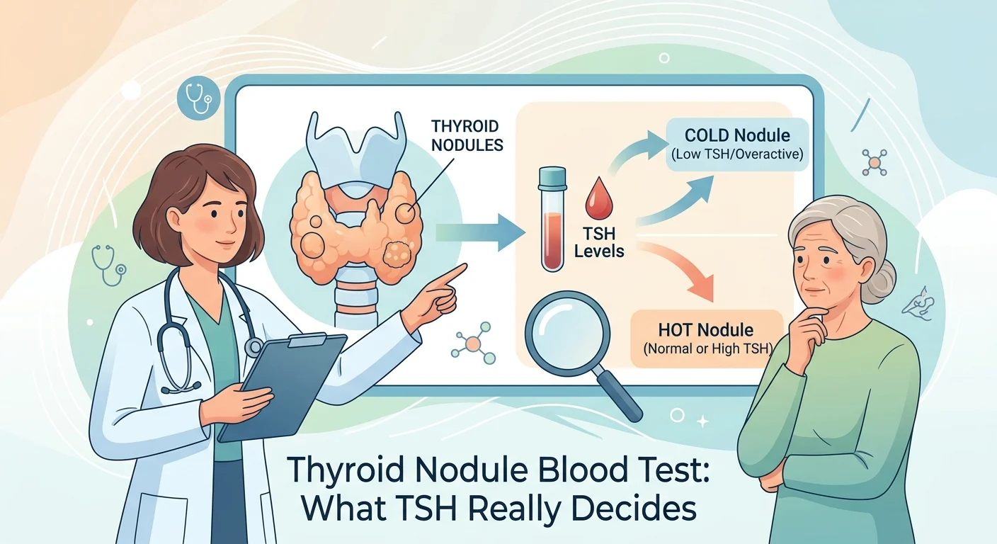
The initial laboratory test is TSH, thyroid-stimulating hormone. TSH is not made by the thyroid at all. It comes from the pituitary gland, and it rises when the body senses too little thyroid hormone and falls when it senses too much. So it measures how hard the thyroid is being pushed overall. It says nothing about what any single lump inside the gland is made of. Our guide to normal thyroid levels and their reference ranges covers what a full panel reports.
Free T4 is usually added when TSH comes back abnormal, to gauge how far off the balance has drifted. Neither value looks at the nodule itself. That is the part worth sitting with before the result arrives.
Why a normal TSH does not rule anything out
The American Thyroid Association is blunt about this: most thyroid nodules, including those that are cancerous, are non-functioning, meaning tests like TSH come back normal. Thyroid function tests are typically normal even when cancer is present in a nodule.
That single sentence is the most useful thing to know before opening the envelope. A normal TSH is not a clean bill of health for the lump. It tells the doctor which branch of the pathway to take, and the branch it points to happens to be the one that needs an ultrasound. If your report reads the other way, our pages explain what sits behind a high TSH result and behind a low TSH result.
What each TSH result sets in motion
| TSH result | What it suggests about the nodule | Usual next step |
|---|---|---|
| Low or suppressed | The nodule may be making hormone on its own. Hyperfunctioning nodules are very rarely cancerous | Nuclear thyroid scan to confirm a hot nodule; biopsy usually avoided |
| Normal | The nodule is non-functioning. This is the commonest situation, including for cancers | Ultrasound risk score plus nodule size decide whether a needle biopsy is needed |
| High | The gland is underactive overall, often from Hashimoto thyroiditis | Same ultrasound pathway, plus assessment of the underactivity itself |
Ultrasound is where the real risk assessment happens. Features such as a solid and darker appearance, microcalcifications, irregular margins and a shape taller than it is wide are scored into a category, and that category, combined with size, sets the threshold for a fine needle biopsy. Nuclear scanning is no longer a first-line method for evaluating nodules, and it keeps its role mainly for the rare nodule causing an overactive thyroid. If your report has already described the gland as uneven, our page explains what a heterogeneous thyroid gland means, and a high TSH result often traces back to hypothyroidism and its causes.
The needle, and what it can and cannot settle
Fine needle aspiration is the test that actually looks at cells. In American Thyroid Association figures, up to 80% of biopsies come back benign, about 5% come back malignant, and up to 20% land in an indeterminate group where the cells cannot reliably be called one or the other. Fewer than 5% are non-diagnostic when ultrasound is used to guide the needle.
That indeterminate fifth is where most of the anxiety and most of the recent progress live. Molecular tests, run on the cells already collected during the biopsy, look at the genes inside them and can often push an ambiguous result back toward benign. They are most useful precisely when the pathologist cannot decide.
The other blood tests, and when they matter
Two more blood values appear in thyroid nodule conversations, and both are frequently misunderstood.
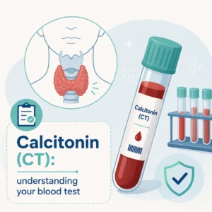
Calcitonin is used to look for medullary thyroid carcinoma, an uncommon type. Practice differs by country: in the United States it is generally reserved for people with a family history of that cancer, for suspicious biopsy findings, or before thyroid surgery, whereas several European groups measure it more systematically. Note that calcitonin is not the same thing as procalcitonin, which is an infection marker. Thyroglobulin is a protein made by thyroid tissue; it is a follow-up tool after treatment for thyroid cancer rather than a screening test for a new nodule, because benign glands make it too.
Hormone results from the neck also travel with the rest of the endocrine picture, which is why thyroid values often sit on the same request form as a female hormone panel or a male hormone panel.
Latest scientific advances
Three strands of recent research help place that TSH number in proportion, without a single statistic being needed to follow them.
- TSH does carry a signal, but a weak one. A 2024 meta-analysis pooling 23 diagnostic studies found that TSH on its own separates cancerous from benign nodules only moderately well, and a 2023 study of people with entirely normal thyroid function found the malignant nodules had slightly higher TSH values than the benign ones. What this means for you: a result sitting in the upper part of the normal range is not an alarm, and it is not a reason to skip the ultrasound either.
- Benign nodules grow, and growth is usually not the story. A 10-year follow-up of 732 nodules judged benign found that growth over a decade was common, while cancer appeared in well under 1% of them. What this means for you: being told a nodule is a little bigger at the next scan is an expected finding, not evidence that something was missed.
- Molecular testing is changing who ends up in an operating room. A 2026 meta-analysis pooling 132 studies and more than 66,000 nodules found that using molecular tests on indeterminate biopsies led to fewer operations, without evidence that meaningful cancers were being missed. What this means for you: if a biopsy comes back indeterminate, asking whether molecular testing is available is a reasonable question.
All of this is observational work describing patterns across large groups. None of it predicts what will happen with one particular nodule, which is the job of the ultrasound, the biopsy and the doctor reading them together. Multi-marker approaches are moving in a similar direction elsewhere in medicine, as our article on the multi-cancer early detection blood test describes.
What to take to the appointment
Practical points that make the visit shorter. Bring the TSH result itself to the ultrasound, because the radiologist needs it to interpret what is on screen. Bring any previous neck imaging, since comparison over time answers questions no single scan can. Bring your current medication list, including blood thinners and aspirin, which matter if a biopsy is scheduled the same day. And note that a thyroid ultrasound needs no fasting and no preparation.
Small nodules found by accident on a scan done for another reason do not always need to be chased. Whether to keep investigating is a conversation worth having openly, since the balance between finding an early cancer and finding something that would never have caused harm is genuinely delicate at that size.
Frequently asked questions
Can a blood test tell if a thyroid nodule is cancerous?
No. TSH and free T4 describe how the gland is working overall, not what a single nodule contains, and they are typically normal when a nodule is cancerous. Only cells taken by fine needle aspiration, or tissue removed at surgery, can settle that question.
My TSH is normal. Do I still need an ultrasound?
Usually yes, and this is the counterintuitive part of the pathway. A normal TSH means the nodule is non-functioning, which is exactly the situation in which the ultrasound risk score does the work of deciding whether a biopsy is needed.
Does a low TSH mean the nodule is harmless?
It points strongly that way. A suppressed TSH suggests a hyperfunctioning nodule, and those are very rarely cancerous, which is why a nuclear scan usually replaces a biopsy in that situation. The overactivity itself may still need treating.
Should calcitonin be on my request form?
Not routinely for most people. It is aimed at a rare form of thyroid cancer and is generally reserved for a family history of that cancer, a suspicious biopsy, or planned thyroid surgery. Practice varies between countries, so it is a fair question to ask your doctor.
Do I need to fast before a thyroid nodule blood test?
No fasting is required for TSH or free T4. If biotin supplements are part of your routine, mention them, because high doses can interfere with several thyroid immunoassays and produce misleading results. Your laboratory will say how long to pause them.
How often will the nodule be checked?
Nodules that are benign on biopsy or too small to biopsy are generally followed with ultrasound every 6 to 12 months at first, alongside an annual examination, with intervals lengthening if nothing changes. Surgery may still be discussed if a benign nodule keeps growing or develops worrying features.
Glossary
| Term | Definition |
|---|---|
| Thyroid nodule | A lump formed by an abnormal growth of thyroid cells inside the gland. Most are benign |
| TSH | Thyroid-stimulating hormone, released by the pituitary gland to tell the thyroid how much hormone to make |
| Free T4 | The unbound fraction of thyroxine circulating in blood, measured when TSH is abnormal |
| Non-functioning nodule | A nodule that does not make thyroid hormone of its own, so it leaves TSH unchanged |
| Hot nodule | A nodule producing hormone independently, seen on a nuclear scan and almost never cancerous |
| Fine needle aspiration | An office procedure using a very thin needle to draw cells from a nodule for examination |
| Indeterminate result | A biopsy with enough cells to read but features that cannot be called benign or malignant |
| Molecular testing | Genetic analysis of the biopsy cells, used mainly to clarify indeterminate results |
| Calcitonin | A hormone measured to look for medullary thyroid carcinoma, a rare thyroid cancer |
| Thyroglobulin | A protein made by thyroid tissue, used to monitor after thyroid cancer treatment |
Understand your lab results with AI DiagMe
A TSH value means very little on its own. It only becomes useful next to free T4, thyroid antibodies, the wording of your ultrasound report and the medicines you already take, and all of it arrives in a mix of units and reference ranges that explain nothing about themselves. AI DiagMe reads your laboratory report and explains, line by line, what each value means in your situation, including where a thyroid result sits relative to the rest of the page. It helps you understand your results and prepare better questions; it does not make a diagnosis and does not replace your doctor.
Get your results interpreted in minutes
Further reading
- Coffee and blood sugar: what a study of 2,264 adults found
- The testosterone blood test and the label update that changed it
- Lung cancer blood test: what it can and cannot detect
- Colon cancer blood test: where it fits in screening
- Hair loss blood test: the markers worth checking
Sources
- American Thyroid Association. Thyroid Nodules, patient information. thyroid.org
- Medscape. Risk Assessment of Thyroid Nodules: A Practical Guide, 19 August 2026. medscape.com
- American Thyroid Association. FNA Biopsy of Thyroid Nodules. thyroid.org
- American Thyroid Association. ATA Guidelines and Statements. thyroid.org
- Fan X, et al. A meta-analysis of the value of serum TSH concentration in the diagnosis of differentiated thyroid cancer in patients with thyroid nodules. Heliyon. 2024. Read the study
- Alaraifi AK, et al. TSH level as a risk factor of thyroid malignancy for nodules in euthyroid patients. Acta Otorhinolaryngologica Italica. 2023. Read the study
- Nguyen TPX, et al. Impact of Molecular Testing on Surgical Decision-Making in Indeterminate Thyroid Nodules: A Global Meta-Analysis Across Test Generations. Endocrine Pathology. 2026. Read the study
- Tanaka S, et al. Molecular Testing in Indeterminate Thyroid Nodules: Genomic Landscape, Diagnostic Performance, and Integrated Risk-Stratified Management. Cancers. 2026. Read the study
- Erdogan MF, et al. Changes in the Volume and Diameter of Benign Thyroid Nodules: A 10-Year Follow-Up Study. Thyroid. 2024, via PubMed. DOI
- Gokbulut P, et al. A novel marker for predicting malignancy in patients with thyroid nodules diagnosed as AUS on initial cytology. Frontiers in Endocrinology. 2026, via PubMed. DOI
- Broecker-Preuss M, et al. Update on Calcitonin Screening for Medullary Thyroid Carcinoma and the Results of a Retrospective Analysis of 12,984 Patients with Thyroid Nodules. Cancers. 2023. Read the study