


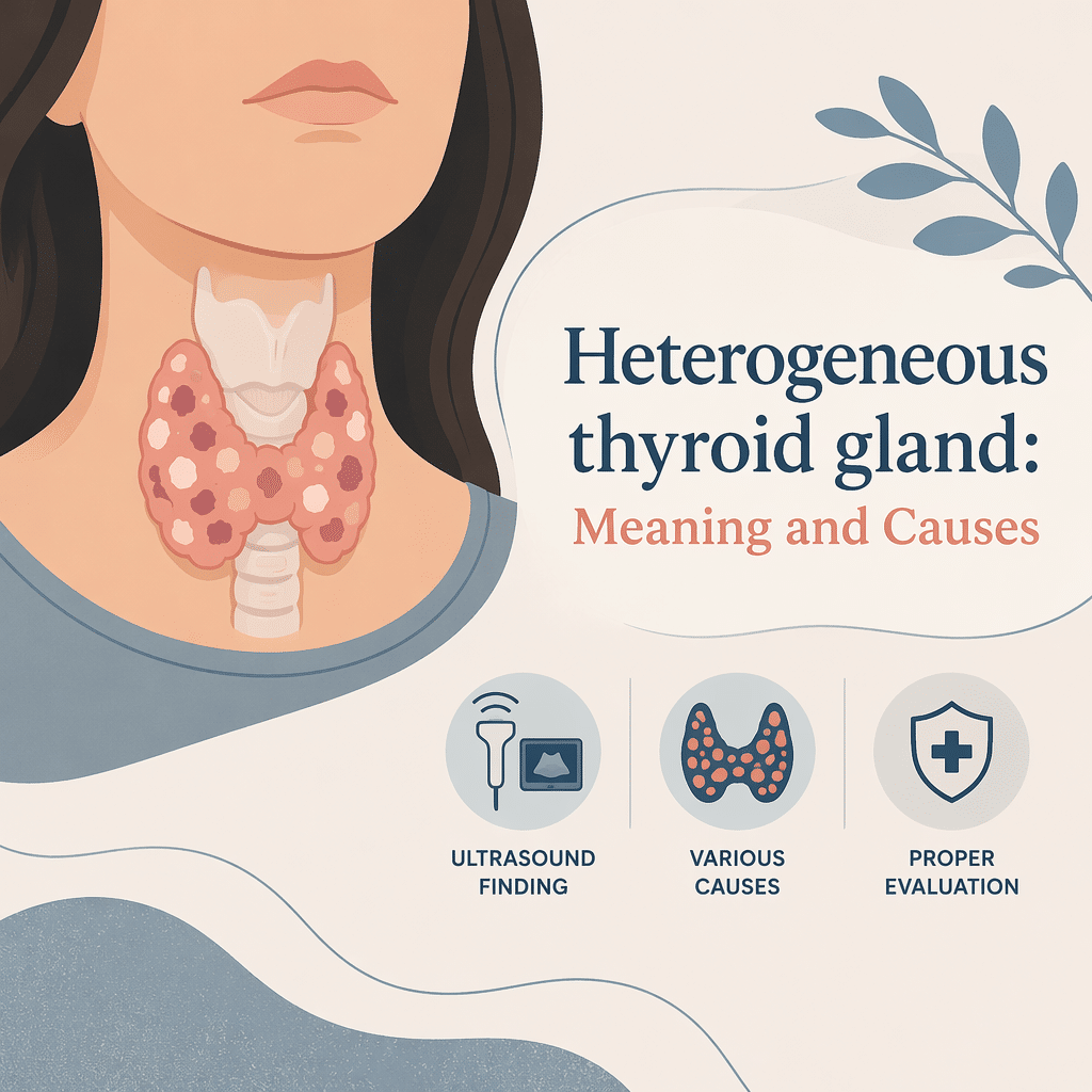
A heterogeneous thyroid gland describes an uneven or mixed texture of thyroid tissue seen on ultrasound. This finding often reflects inflammation, multiple nodules, or scarring rather than a single uniform change. In this article you will learn what a heterogeneous thyroid gland means, how clinicians evaluate it, common causes and symptoms, when further testing helps, and practical steps you can take while you wait for results.
What does heterogeneous thyroid gland mean?
A heterogeneous thyroid gland shows varied echotexture on ultrasound. Ultrasound images display areas that look different from the surrounding tissue. Radiologists use the term when the gland contains regions of mixed brightness, nodules, or fibrosis. This pattern does not itself diagnose a specific disease. Instead, clinicians combine imaging with blood tests and clinical history to reach a diagnosis.
How doctors detect a heterogeneous thyroid gland
Clinicians order a thyroid ultrasound when they find a lump, abnormal thyroid function tests, or a visibly enlarged gland. A sonographer performs the scan and a radiologist or endocrinologist reads the images. Ultrasound shows tissue texture, nodule size, blood flow, and calcifications. Doctors also run blood tests for TSH, free T4, and thyroid antibodies to assess function and cause. If ultrasound finds suspicious nodules, clinicians often recommend a fine-needle aspiration biopsy for cellular analysis.
Common causes and risk factors
Chronic autoimmune thyroiditis, often called Hashimoto’s thyroiditis, commonly produces a heterogeneous thyroid gland. Multiple benign nodules or a multinodular goiter can create a mixed texture. Subacute or silent thyroiditis and prior radiation therapy also alter gland appearance. Age and longstanding iodine deficiency increase the chance of nodularity. Smoking and certain medications can affect thyroid structure indirectly.
Symptoms linked to a heterogeneous thyroid gland
Many people with a heterogeneous thyroid gland feel no symptoms. However, symptoms reflect underlying function. Hypothyroidism may cause fatigue, weight gain, cold sensitivity, and dry skin. Hyperthyroidism may lead to weight loss, palpitations, heat intolerance, and anxiety. A large multinodular gland can cause a visible neck swelling or compressive symptoms, such as difficulty swallowing or breathing. If you notice rapid neck growth, hoarseness, or breathing trouble, seek prompt evaluation.
How doctors interpret a heterogeneous thyroid gland on ultrasound and lab tests
Doctors integrate ultrasound features with lab results and your clinical picture. If TSH remains normal and nodules look small and benign, clinicians often choose watchful waiting with periodic ultrasound. If antibodies appear, physicians consider autoimmune thyroiditis and monitor function over time. When nodules show suspicious features—irregular margins, microcalcifications, increased blood flow—clinicians usually recommend biopsy. Shared decision-making guides whether to treat, monitor, or remove tissue.
When a fine-needle biopsy helps
Fine-needle aspiration (FNA) provides cells for cytology. Doctors use FNA when ultrasound raises concern for cancer. The procedure uses a thin needle and typically causes only mild discomfort. Pathology results may report benign, indeterminate, suspicious, or malignant findings. If results remain indeterminate, clinicians may repeat the biopsy, use molecular testing, or recommend surgical removal.
Treatment options and follow-up
Treatment targets the underlying cause and symptoms. For autoimmune hypothyroidism, doctors prescribe levothyroxine to restore normal hormone levels. Clinicians treat hyperthyroidism with medications, radioactive iodine, or sometimes surgery. For benign nodules without symptoms, experts often monitor size and function with periodic exams and ultrasound. If a nodule causes compression or shows cancer risk, surgeons may remove part or all of the thyroid. Follow-up plans vary by diagnosis, but most patients receive periodic blood tests and imaging when necessary.
Lifestyle and self-care tips for thyroid health
Maintain a balanced diet that supports metabolic health. Avoid excessive iodine intake unless a clinician recommends it. Take prescribed thyroid medications at the same time daily and follow instructions about food and supplements. For example, calcium or iron supplements can interfere with levothyroxine absorption, so separate dosing times. Manage stress, sleep well, and get routine primary care. These steps support overall thyroid care while you pursue specific medical evaluation.
Frequently Asked Questions (FAQ)
Q: Does a heterogeneous thyroid gland mean cancer?
A: Not usually. Heterogeneity often reflects benign conditions. Doctors use ultrasound features and biopsy to assess cancer risk.
Q: Will I always need surgery for this finding?
A: No. Many people with a heterogeneous thyroid gland never need surgery. Clinicians use function tests and imaging to decide.
Q: How often will I need follow-up imaging?
A: Follow-up intervals vary. For small benign findings, clinicians often repeat ultrasound every 6 to 24 months based on risk.
Q: Can thyroid function tests be normal with this ultrasound finding?
A: Yes. You can have normal TSH and still show heterogeneous texture. Imaging and labs provide complementary information.
Q: When should I see a specialist?
A: See an endocrinologist if you have abnormal thyroid function, suspicious ultrasound features, compressive symptoms, or unclear biopsy results.
Q: Can lifestyle changes reverse the ultrasound findings?
A: Lifestyle changes support treatment and symptom control, but they rarely reverse structural changes caused by long-term disease.
Glossary of Key Terms
Heterogeneous echotexture: Mixed appearance of thyroid tissue on ultrasound.
Ultrasound: Imaging technique that uses sound waves to visualize internal structures.
Nodule: A lump or focal lesion within the thyroid.
Fine-needle aspiration (FNA): A biopsy method that samples cells from a thyroid nodule.
TSH: Thyroid-stimulating hormone, a blood test that guides thyroid evaluation.
Hashimoto’s thyroiditis: An autoimmune disease that often causes hypothyroidism.
Euthyroid: Normal thyroid function.
Calcification: Calcium deposits that appear on imaging and may affect risk assessment.
Understand Your Lab Test Results with AI DiagMe
Understanding thyroid imaging and lab results can feel overwhelming. AI DiagMe helps translate test numbers and imaging findings into clear, actionable explanations. Use AI DiagMe to compare your results with typical patterns and to prepare focused questions for your clinician.
➡️ Analyze Your Lab Results with AI DiagMe Now
