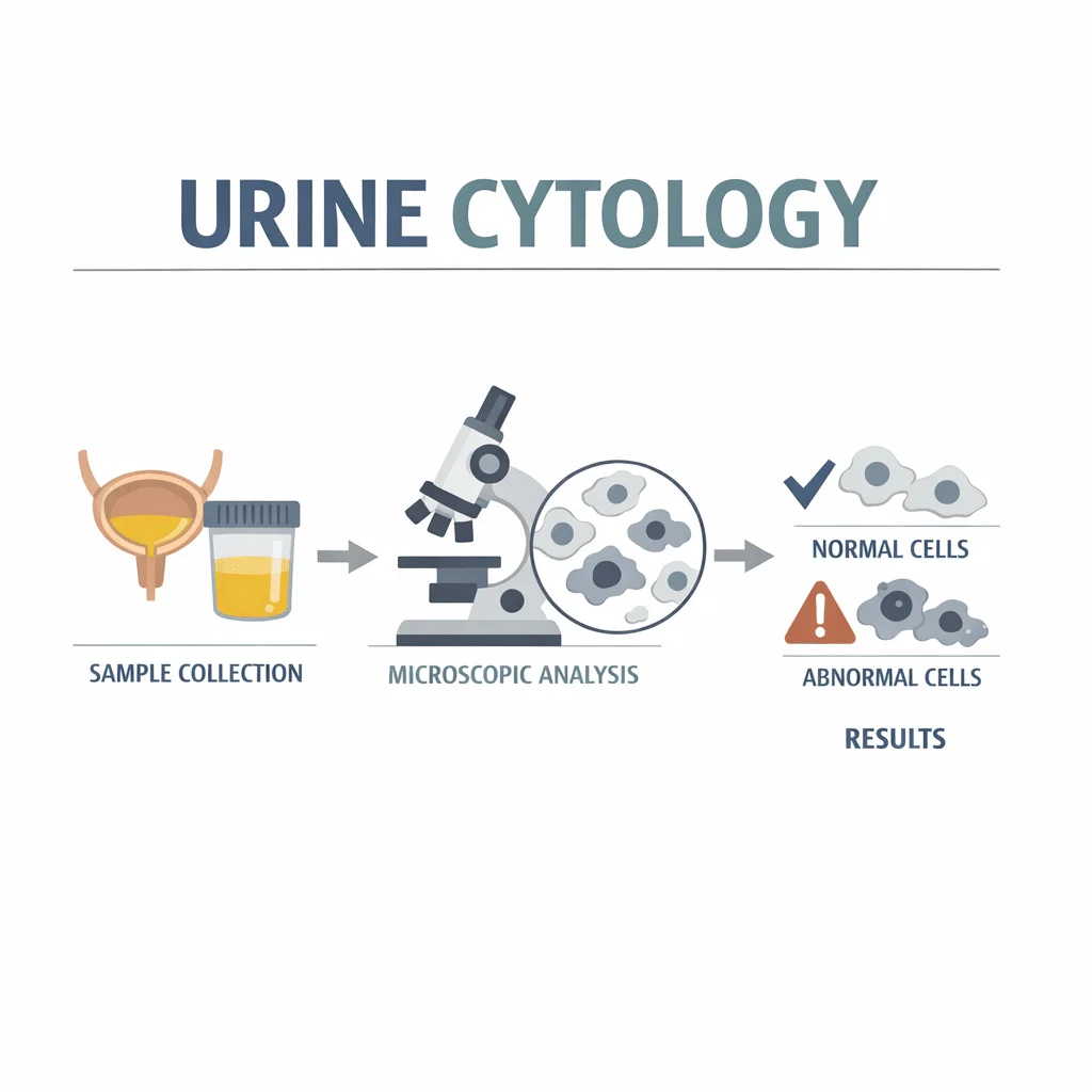
Sitologi urin adalah pemeriksaan laboratorium yang mengamati sel-sel yang dilepaskan oleh saluran kemih ke dalam urin, mencari sel-sel yang tampak seperti sel kanker di bawah mikroskop. Ini bukan pemeriksaan skrining rutin, dan hasilnya dilaporkan dalam bentuk kategori, bukan angka. Dokter paling sering memesannya ketika ditemukan darah dalam urin, serta untuk memantau kondisi setelah pengobatan kanker kandung kemih. Dalam artikel ini, Anda akan mempelajari apa yang dapat dan tidak dapat dilihat oleh pemeriksaan ini, mengapa laboratorium meminta Anda untuk tidak membawa sampel urin pertama di pagi hari, apa arti setiap kategori hasil dalam bahasa yang mudah dipahami, dan langkah selanjutnya yang biasanya mengikuti setiap hasil.
Apa itu sitologi urin, dan apa yang bukan
Kandung kemih, ureter, dan sistem drainase ginjal Anda dilapisi oleh lapisan khusus yang disebut urothelium. Seperti kulit, lapisan ini terus memperbarui dirinya sendiri, dan sel-sel yang sudah usang akan terlepas ke dalam urin yang mengalir di atasnya. Sitologi urin mengumpulkan sel-sel yang terlepas tersebut ke atas kaca objek, mewarnainya, lalu menyerahkannya kepada seorang sitopatologis — dokter yang terlatih untuk menilai sel berdasarkan penampilannya saja.
Pertanyaannya sangat spesifik: apakah ada sel-sel yang tampak seperti sel kanker urothelial yang agresif? Sampel tidak dapat menunjukkan dari bagian mana dalam saluran kemih sel-sel tersebut berasal.
Perbedaan sitologi urin dengan urinalisis
Urinalisis adalah pemeriksaan kimia dan mikroskopis yang menyeluruh. Pemeriksaan ini mengukur keasaman, protein, glukosa, dan darah, serta menghitung partikel dalam sedimen; kami menjelaskan apa saja yang terdapat dalam laporan urinalisis lengkap secara terpisah, dan menjelaskan cara membaca panel kimia dipstick dalam panduan tersendiri.
Sitologi tidak tumpang tindih dengan semua itu. Ini adalah pertanyaan tentang kanker yang ditujukan hanya pada sel. Urinalisis bisa sepenuhnya normal sementara sitologi menunjukkan kelainan, dan sebaliknya pun sama seringnya terjadi.
Sel-sel yang tidak menjadi perhatian laboratorium
Banyak sel lain yang dapat ditemukan dalam sampel urin, dan tidak satu pun yang menjadi target pemeriksaan ini. Sel skuamosa yang terlepas dari uretra atau vulva biasanya lebih mencerminkan cara pengambilan sampel daripada adanya penyakit, dan kami menjelaskan arti sel epitel dalam sampel urin dalam artikel khusus. Struktur silindris yang terbentuk di dalam tubulus ginjal termasuk dalam pembahasan yang berbeda, dan kami membahas apa yang ditunjukkan oleh silinder (cast) dalam urin tentang kondisi ginjal di tempat lain.
Mengapa dokter Anda meminta sitologi urin
Ada tiga alasan umum, dan alasan mana yang berlaku akan memengaruhi seberapa besar bobot hasil pemeriksaan tersebut.
Darah dalam urin
Darah yang terlihat secara kasat mata, atau darah yang hanya ditemukan di bawah mikroskop, adalah pemicu yang paling sering terjadi. Sebagian besar penyebabnya ternyata bersifat jinak, dan kami membahas berbagai penyebab darah dalam urin secara lengkap di tempat lain. Kanker kandung kemih tetap menjadi salah satu kemungkinan yang harus disingkirkan, dan sitologi urine membantu melakukan hal tersebut. Tes ini hampir tidak pernah digunakan sendiri dalam konteks ini.
Pemantauan setelah pengobatan kanker kandung kemih
Di sinilah tes ini benar-benar menunjukkan kegunaannya. Setelah kanker urotelial diangkat, lapisan yang menghasilkannya tetap berisiko, dan pasien menjalani program pemeriksaan kandung kemih secara berulang. Sitologi dilakukan bersamaan dengan pemeriksaan tersebut karena mengambil sampel dari seluruh lapisan, termasuk area datar dengan sel-sel abnormal yang tidak pernah membentuk benjolan yang terlihat dan mudah terlewat oleh kamera.
Paparan akibat pekerjaan dan faktor lainnya
Riwayat paparan jangka panjang terhadap bahan kimia industri tertentu, terutama amina aromatik yang pernah digunakan dalam pembuatan pewarna, karet, dan tekstil, meningkatkan risiko seumur hidup; merokok berat dalam jangka panjang adalah faktor utama lainnya. Sitologi dapat disertakan dalam pemantauan untuk kelompok-kelompok ini, meskipun National Cancer Institute mencatat bahwa tidak ada tes urine yang terbukti dapat mengurangi kematian bila digunakan untuk skrining populasi umum.
Cara pengambilan sampel sitologi urine, dan mengapa tidak menggunakan urine pertama pagi hari
Ini adalah langkah yang paling sering dilakukan keliru oleh pasien, biasanya karena mengikuti instruksi yang ditulis untuk tes yang berbeda.
Sampel urine yang dikeluarkan secara alami, dan jebakan urine pertama pagi hari
Sebagian besar sitologi dilakukan pada spesimen yang dikeluarkan secara alami, yaitu urine yang Anda keluarkan seperti biasa ke dalam wadah steril. Untuk banyak tes urine, sampel pertama pagi hari yang lebih pekat lebih disukai. Namun untuk sitologi, hal ini justru tidak dianjurkan. Sel-sel yang berada di kandung kemih semalaman mulai mengalami kerusakan, dan sel yang sudah rusak sulit untuk dinilai: ciri-ciri yang diandalkan oleh sitopatologis, seperti bentuk inti sel dan tekstur di dalamnya, menjadi kabur dan terdistorsi. Cleveland Clinic menyarankan untuk tidak menggunakan spesimen pertama pagi hari tepat karena alasan ini.
Laboratorium menginginkan sampel yang diambil pada pagi hari yang lebih siang, setelah Anda sudah beraktivitas dan buang air kecil beberapa kali, agar sel-selnya masih segar. Beberapa laboratorium meminta sampel pada tiga hari yang berbeda, karena pelepasan sel bersifat tidak menentu dan satu kali pengambilan bisa saja melewatkan sel-sel yang penting.
Pencucian dan penyikatan kandung kemih
Ketika hasil yang lebih banyak diperlukan, sel-sel dapat dikumpulkan secara langsung. Selama sistoskopi, cairan steril dimasukkan ke dalam kandung kemih dan ditarik keluar beberapa kali — teknik yang disebut barbotage — yang melepaskan sel-sel alih-alih menunggu sel tersebut luruh dengan sendirinya; American Cancer Society mencatat bahwa sitologi juga dilakukan pada bilasan kandung kemih ini. Jika kekhawatiran berada lebih ke atas, sel-sel dapat diambil secara selektif dari salah satu ureter selama ureteroskopi. Metode-metode ini menghasilkan lebih banyak material dan dalam kondisi yang lebih terjaga dibandingkan sampel urine yang dikeluarkan ke dalam wadah.
Hal-hal praktis pada hari pengambilan sampel
Hindari memberikan sampel saat menstruasi jika memungkinkan, karena darah membuat preparat lebih sulit dibaca. Beri tahu dokter yang meminta pemeriksaan jika Anda sedang mengalami infeksi saluran kemih aktif, baru saja menggunakan kateter atau menjalani prosedur kandung kemih, atau menerima pengobatan di dalam kandung kemih, karena semua hal ini dapat mengubah tampilan sel. Kami membahas cara infeksi saluran kemih didiagnosis dan ditangani secara terpisah.
Arti setiap kategori hasil sitologi urine, dan tindak lanjut yang biasanya dilakukan
Sebagian besar laboratorium di Amerika Serikat kini menggunakan kosakata bersama yang disebut Paris System for Reporting Urinary Cytology. Sistem ini dirancang untuk mencegah satu preparat yang sama dideskripsikan dengan lima cara berbeda di lima rumah sakit yang berbeda, dan mengorganisasikan segalanya di sekitar satu pertanyaan: apakah terdapat karsinoma urotelial derajat tinggi di sini? Tabel berikut menerjemahkan setiap kategori ke dalam bahasa yang mudah dipahami dan menunjukkan langkah yang biasanya dilakukan selanjutnya.
| Kategori laporan | Artinya dalam bahasa sederhana | Langkah yang biasanya dilakukan selanjutnya |
|---|---|---|
| Non-diagnostik (tidak memadai) | Sampel tidak mengandung cukup sel yang terawat baik untuk memberikan penilaian. Ini adalah komentar mengenai spesimen, bukan mengenai Anda. | Ulangi pengambilan sampel, biasanya dengan instruksi yang lebih jelas mengenai waktu dan volume. |
| Negatif untuk karsinoma urotelial derajat tinggi | Tidak ditemukan sel yang memiliki ciri-ciri kanker urotelial agresif. Ini adalah hasil yang paling umum sejauh ini. | Menenangkan, namun tidak menutup pemeriksaan. Jika perdarahan atau gejala berlanjut, pemeriksaan kandung kemih dan pencitraan yang direncanakan tetap dilanjutkan. |
| Sel urotelial atipikal | Beberapa sel tampak sedikit tidak biasa, tetapi tidak cukup tidak biasa untuk disebut sesuatu yang pasti. Ini adalah zona abu-abu, dan memiliki banyak penjelasan yang tidak berbahaya. | Dibaca bersama dengan pemeriksaan kandung kemih dan pencitraan; sering kali diperlukan sampel ulang setelah infeksi atau iritasi mereda. |
| Dicurigai karsinoma urotelial derajat tinggi | Sel-sel menunjukkan beberapa ciri yang mengkhawatirkan sekaligus, namun jumlahnya terlalu sedikit, atau terlalu rusak, untuk memberikan kesimpulan yang pasti. | Pemeriksaan kandung kemih segera dengan biopsi pada bagian yang tampak abnormal. |
| Karsinoma urotelial derajat tinggi | Sel-sel dengan tampilan kanker urotelial agresif terlihat jelas. | Pemeriksaan kandung kemih dan biopsi segera. Jika kandung kemih tampak normal, pencitraan ginjal dan ureter dilakukan selanjutnya, karena sel-sel tersebut mungkin berasal dari saluran yang lebih atas. |
| Neoplasma urotelial derajat rendah | Kategori yang sempit dan jarang digunakan, hanya dipakai ketika satu kelompok sel yang sangat spesifik ditemukan. Sebagian besar tumor yang tumbuh lambat tidak meninggalkan jejak sama sekali pada sitologi. | Pemeriksaan kandung kemih. Sitologi bukan alat yang menemukan pertumbuhan ini, sehingga hasil normal di sini tidak banyak berarti. |
Dua catatan penting. Kategori-kategori ini bukan skala keparahan yang berjenjang dari kanker ringan hingga berat; melainkan skala kepercayaan diagnostik. Dan rumusan kategori negatif itu penting. Bukan berarti negatif untuk kanker. Artinya negatif untuk karsinoma urotelial derajat tinggi, yang menjadi pokok bahasan bagian berikutnya.
Andal untuk tumor agresif, lemah untuk tumor yang tumbuh lambat
Hal terpenting yang perlu dipahami tentang sitologi urine adalah bahwa kinerjanya tidak seimbang, dan memang disengaja demikian. Kanker agresif derajat tinggi melepaskan sel-sel yang tampak jelas tidak normal: inti sel membesar dan memenuhi sebagian besar sel, tepinya menjadi tidak beraturan, dan materi di dalamnya menggumpal kasar. Seorang sitopatologis dapat mengenali hal ini dengan andal, dan laporan karsinoma derajat tinggi sangat jarang keliru.
Tumor yang tumbuh lambat dan berderajat rendah merupakan masalah yang berlawanan. Sel-selnya tampak hampir normal, mungkin sedikit lebih besar atau lebih banyak, tetapi tanpa perubahan yang memungkinkan diagnosis ditegakkan. Tumor berderajat rendah bisa terlihat jelas di kandung kemih sementara laporan sitologi menunjukkan hasil negatif, dan itu bukan kesalahan laboratorium: sel-sel tersebut memang benar-benar tidak tampak seperti kanker di bawah mikroskop.
Kesimpulan yang perlu diingat: hasil sitologi urine negatif tidak menyingkirkan kemungkinan tumor kandung kemih yang tumbuh lambat, dan memang tidak pernah dirancang untuk itu. Itulah mengapa tes ini merupakan pelengkap pemeriksaan kandung kemih, bukan penggantinya, dan mengapa hasil negatif tidak pernah membatalkan jadwal sistoskopi yang sudah direncanakan.
Sel urotelial atipikal: apa sebenarnya arti istilah tersebut
Jika laporan Anda menggunakan kata-kata ini, sitopathologis melihat sel-sel dengan beberapa ciri abnormal, tetapi terlalu sedikit cirinya, atau terlalu sedikit selnya, untuk dapat dikategorikan ke tingkat yang lebih tinggi. Ini adalah pernyataan ketidakpastian yang jujur, bukan cara halus untuk menyampaikan kabar buruk.
Banyak hal biasa yang membuat sel urotelial tampak tidak normal. Infeksi dan peradangan menyebabkan sel-sel tersebut membengkak; kami menjelaskan kapan bakteri dalam sampel urine perlu ditangani dan menjelaskan apa yang ditunjukkan oleh sel darah putih dalam urine dalam panduan terpisah. Organisme jamur mengiritasi dengan cara yang sama, dan kami menguraikan bagaimana ragi dalam sampel urine dinilai di tempat lain. Batu mengikis lapisan dinding saluran kemih. Kateter yang baru dipasang, instrumen yang baru digunakan, radioterapi panggul, kemoterapi, atau pengobatan yang dimasukkan langsung ke dalam kandung kemih semuanya membuat sel-sel tampak rusak selama berminggu-minggu.
Karena daftar tersebut, hasil atipikal hampir tidak pernah ditindaklanjuti sendiri. Hasil ini dibaca bersama gejala Anda, pemeriksaan kandung kemih, dan pencitraan Anda, dan biasanya mendorong pengambilan sampel ulang setelah iritasi yang dapat diobati mereda, atau kelanjutan pemeriksaan yang sudah direncanakan. Menunggu gambaran yang lebih lengkap adalah hal yang normal dan tepat, bukan suatu penundaan.
Apa yang terjadi setelah hasil sitologi urine yang abnormal
Sitologi tidak pernah menjadi kata terakhir. Tugasnya adalah meningkatkan atau menurunkan kecurigaan; konfirmasi dilakukan dengan cara lain.
Sistoskopi, standar acuan
Sistoskop adalah tabung fleksibel tipis dengan kamera yang dimasukkan melalui uretra untuk melihat langsung lapisan kandung kemih. Semua hal lain diukur berdasarkan pemeriksaan ini, karena sistoskop sekaligus melihat lapisan kandung kemih dan memungkinkan biopsi pada bagian yang mencurigakan. Prosedur ini dilakukan dengan gel anestesi lokal dan memakan waktu beberapa menit. Jika sitologi menunjukkan kelainan tetapi kandung kemih tampak normal, kombinasi tersebut sendiri sudah memberikan informasi: perhatian diarahkan ke bagian yang lebih atas.
Pencitraan saluran kemih bagian atas
Karena lapisan yang sama melapisi ureter dan sistem drainase ginjal, pemindaian seluruh saluran kemih biasanya dijadwalkan ketika hasil pemeriksaan mengkhawatirkan dan sistoskopi tidak menemukan kelainan. Pencitraan penampang lintang dengan kontras adalah pilihan yang umum digunakan.
Pengulangan sitologi dan tes biomarker urine
Mengulang pengambilan sampel sering kali berguna, terutama jika sampel pertama terlalu sedikit, sudah mengalami degenerasi, atau diambil saat terjadi infeksi. Selain itu, terdapat tes urine yang mencari sinyal molekuler, bukan penampilan sel: protein tumor, perubahan genetik, dan kelainan kromosom pada sel yang terlepas. Tes-tes ini melengkapi penilaian, bukan menggantikannya, dan kami menjelaskan bagaimana tes penanda tumor bekerja dan di mana keterbatasannya dalam artikel terpisah.
Pertanyaan yang layak diajukan
- Kategori pelaporan mana yang menjadi hasil sampel saya, dan apakah spesimennya memadai?
- Apakah ini sampel urine yang dikeluarkan secara alami atau hasil bilasan kandung kemih, dan apakah itu memengaruhi cara Anda membacanya?
- Apakah saya mengalami infeksi atau baru menjalani pengobatan saat sampel diambil?
- Berdasarkan hasil ini, apa yang akan mengubah rencana Anda, dan apa yang tidak?
Kemajuan ilmiah terkini
Penelitian selama tiga tahun terakhir berjalan ke dua arah: mengukur seberapa baik kosakata pelaporan bersama bekerja, dan menguji apakah perangkat lunak atau metode molekuler dapat menutupi apa yang terlewat oleh mikroskopi.
Kosakata bersama mengurangi laporan zona abu-abu, tetapi tidak menghilangkan titik buta
Sebuah tinjauan gabungan yang diterbitkan pada tahun 2024 menggabungkan 64 studi dan hampir 100.000 sampel urine untuk melihat perubahan yang terjadi setelah laboratorium mengadopsi Sistem Paris. Laboratorium yang menggunakannya memberi label lebih sedikit sampel sebagai atipikal dan memberikan sinyal yang lebih jelas ketika mereka menyebut sesuatu sebagai mencurigakan atau berderajat tinggi. Para penulis secara tegas menyatakan bahwa hasil negatif tetap tidak dapat secara andal menyingkirkan tumor yang agresif.
Apa artinya bagi Anda: laporan yang menunjukkan hasil negatif untuk karsinoma urotelial tingkat tinggi adalah kabar baik yang sesungguhnya, tetapi bukan berarti pemantauan selesai. Pemeriksaan kandung kemih yang dijadwalkan dokter Anda tetap merupakan bagian dari rencana yang menjawab pertanyaan tersebut.
Laporan zona abu-abu memiliki bobot nyata yang dapat diukur
Sebuah studi tahun 2026 mengikuti 170 sampel dan membandingkan setiap laporan sitologi urin dengan temuan akhir yang diperoleh. Dua ahli patologi yang membaca slide yang sama mencapai kesepakatan yang hampir sempurna, hal ini penting karena ketidakkonsistenan adalah masalah yang ingin diselesaikan oleh Sistem Paris. Laporan atipikal pada akhirnya diikuti oleh tumor agresif pada sekitar separuh kasus, sementara laporan negatif diikuti oleh tumor pada kurang dari satu dari seratus pasien. Ini adalah studi satu pusat, sehingga proporsi pastinya tidak dapat diterapkan di semua tempat.
Apa artinya bagi Anda: kategori atipikal adalah kategori yang benar-benar tidak pasti, itulah mengapa kategori ini memicu pemeriksaan lebih lanjut, bukan ketenangan. Laporan negatif membuat penyakit agresif menjadi tidak mungkin, dan itu adalah hal yang patut dipegang.
Perangkat lunak mulai membaca slide bersama ahli patologi
Sebuah tinjauan tahun 2026 mengumpulkan sebelas studi yang menerapkan perangkat lunak pengenalan gambar pada slide sitologi urin dan menemukan hasil yang secara konsisten menjanjikan dalam mendeteksi penyakit agresif, meskipun dicatat bahwa studi-studi tersebut berskala kecil dan menggunakan metode yang berbeda-beda. Sebuah studi terpisah tahun 2026 di sebuah rumah sakit besar di Amerika Serikat menguji salah satu sistem tersebut pada 200 slide. Pembacaan berbantuan perangkat lunak berhasil mendeteksi lebih banyak kanker dibandingkan mikroskopi saja dan memangkas waktu tinjauan per slide hingga sekitar dua pertiga, namun juga menandai lebih banyak sampel yang tidak berbahaya sebagai abnormal.
Apa artinya bagi Anda: di mana bantuan perangkat lunak digunakan, imbal baliknya adalah waktu pemrosesan yang lebih cepat dan kemungkinan kanker yang terlewat lebih kecil, dengan risiko sedikit lebih tinggi terjadinya alarm palsu yang kemudian diselesaikan melalui pemeriksaan lanjutan. Ini adalah hasil awal dan masih perlu dikonfirmasi.
Tes urine molekuler mendeteksi apa yang tidak terlihat oleh mikroskopi
Sebuah analisis besar tahun 2026 menggabungkan 37 studi yang mencakup lebih dari 7.000 pasien dan membandingkan tes urin yang mencari DNA tumor dengan sitologi konvensional. Pengujian DNA tumor mendeteksi kanker urotelial jauh lebih sering sambil menghasilkan jumlah hasil positif palsu yang sebanding — inilah tepatnya kelemahan mikroskop dalam mendeteksi tumor yang tumbuh lambat. Para penulis tidak sampai merekomendasikan penggunaan rutin. Sebuah panel panduan Eropa mencapai kesimpulan serupa untuk kanker saluran kemih bagian atas: beberapa tes urin molekuler tampak akurat, namun dasar buktinya masih tipis.
Apa artinya bagi Anda: tes-tes ini mungkin pada akhirnya dapat mengurangi seberapa sering orang dalam program pemantauan perlu menjalani prosedur kamera yang dimasukkan ke dalam kandung kemih. Itu bukan kondisi praktik saat ini, tetapi merupakan hal yang wajar untuk ditanyakan kepada urolog Anda.
Glosarium
| Ketentuan | Definisi |
|---|---|
| Uroteliun | Lapisan khusus kandung kemih, ureter, dan sistem drainase ginjal. Lapisan ini terus memperbarui dirinya sendiri, itulah mengapa sel-selnya muncul dalam urine. |
| Karsinoma urotelial | Kanker yang berasal dari lapisan tersebut. Ini adalah jenis kanker kandung kemih yang paling umum, dan juga dapat muncul di bagian atas ureter atau ginjal. |
| Sitopatologis | Dokter yang mengkhususkan diri dalam mendiagnosis penyakit berdasarkan tampilan sel-sel individual, bukan dari sepotong jaringan. |
| Sistem Paris | Kosakata pelaporan bersama yang digunakan oleh sebagian besar laboratorium di Amerika Serikat untuk sitologi urine, yang dirancang untuk mendeteksi kanker urotelial agresif. |
| Karsinoma urotelial derajat tinggi | Bentuk kanker yang agresif dengan sel-sel yang tampak sangat abnormal. Inilah penyakit yang menjadi target utama sitologi urin. |
| Neoplasma urotelial derajat rendah | Pertumbuhan lambat pada lapisan yang sama. Sel-selnya tampak mendekati normal, itulah mengapa sitologi biasanya tidak dapat mendeteksinya. |
| Spesimen urine spontan | Urine yang dikeluarkan secara normal ke dalam wadah, berbeda dengan urine yang dikumpulkan melalui alat. |
| Pencucian kandung kemih (barbotage) | Metode pengumpulan sampel di mana cairan steril dimasukkan ke dalam kandung kemih dan ditarik keluar berulang kali untuk melepaskan lebih banyak sel dibandingkan yang diperoleh hanya dari berkemih biasa. |
| Sistoskopi | Pemeriksaan langsung lapisan kandung kemih menggunakan kamera tipis yang dimasukkan melalui uretra. Ini adalah standar acuan untuk kanker kandung kemih. |
| Hematuria | Darah dalam urin, baik yang terlihat oleh mata maupun yang hanya terdeteksi melalui pemeriksaan. Ini adalah alasan paling umum untuk memesan sitologi urin. |
Pertanyaan yang sering diajukan
Apa sebenarnya yang dicari oleh tes sitologi urine?
Tes ini mencari sel-sel yang terlepas dari lapisan saluran kemih yang memiliki ciri khas visual kanker agresif: inti sel yang membesar, garis tepi inti yang tidak beraturan, dan tekstur internal yang kasar. Tes ini tidak mencari bakteri, protein, gula, darah, atau kristal — semua itu merupakan bagian dari urinalisis. Tes ini juga tidak mengukur apa pun, sehingga tidak ada rentang nilai normal dan tidak ada angka yang bisa dibandingkan. Hasilnya berupa kategori yang menggambarkan apa yang dilihat oleh sitopatolog, dan kategori tersebut dibangun berdasarkan satu pertanyaan, bukan survei umum terhadap urine Anda.
Seberapa akurat tes sitologi urine?
Akurasi sepenuhnya bergantung pada jenis penyakit yang sedang diperiksa. Untuk kanker urotelial tingkat tinggi yang agresif, sitologi cukup dapat diandalkan, dan hasil positif sangat jarang keliru. Untuk tumor tingkat rendah yang tumbuh lambat, kinerjanya kurang baik, karena sel-sel tersebut tampak hampir normal di bawah mikroskop. Cara mudah untuk memahaminya: hasil positif biasanya memang menandakan sesuatu, sementara hasil negatif jauh lebih dapat menyingkirkan penyakit agresif dibandingkan menyingkirkan tumor yang tumbuh lambat. Itulah mengapa sitologi digunakan bersama pemeriksaan kandung kemih, bukan sebagai penggantinya.
Mengapa laboratorium terkadang meminta tiga sampel terpisah?
Sel tidak terlepas dengan kecepatan yang tetap. Sebuah tumor mungkin melepaskan banyak sel pada satu hari dan hampir tidak ada pada hari berikutnya, sehingga satu spesimen bisa melewatkan sel-sel yang baru akan tertangkap pada sampel kedua atau ketiga. Pengambilan sampel pada tiga hari yang berbeda membantu menyebarkan risiko tersebut. Jika tiga sampel diminta, sebaiknya diambil pada hari yang berbeda — bukan tiga kali pengambilan dalam satu pagi — dan masing-masing harus mengikuti panduan yang sama: bukan urine pertama di pagi hari, dan dalam jumlah yang cukup, bukan sekadar sedikit.
Apa perbedaan sitologi urine dengan urinalisis atau kultur urine?
Ketiganya menggunakan urine dan menjawab pertanyaan yang berbeda. Urinalisis adalah pemeriksaan kimia dan mikroskopis menyeluruh yang digunakan untuk mendeteksi infeksi, masalah ginjal, dehidrasi, dan perubahan metabolik. Kultur urine menumbuhkan bakteri yang ada untuk mengidentifikasi infeksi dan menentukan pilihan antibiotik. Sitologi urine tidak memperhatikan semua itu, melainkan memeriksa tampilan sel untuk mencari tanda-tanda kanker. Seorang pasien dengan infeksi bisa saja memiliki hasil sitologi yang normal, dan pasien dengan hasil urinalisis yang sepenuhnya normal bisa memiliki hasil sitologi yang tidak normal.
Berapa lama hasil sitologi urine keluar?
Sitologi membutuhkan waktu lebih lama dibandingkan sebagian besar tes urine karena hasilnya dibuat oleh seseorang, bukan mesin. Sampel harus diproses, dipadatkan ke atas kaca objek, dan diwarnai sebelum diperiksa oleh sitopatolog, dan kasus yang sulit sering kali ditinjau oleh spesialis kedua sebelum laporan ditandatangani. Beberapa hari kerja hingga sekitar satu minggu adalah waktu yang umum, meskipun ini bervariasi tergantung laboratorium dan apakah pewarnaan tambahan diperlukan. Jika hasilnya mendesak, tim medis Anda biasanya dapat memberi tahu kapan hasilnya bisa diharapkan.
Apakah infeksi saluran kemih dapat memengaruhi hasil tes?
Ya, dan ini adalah salah satu penjelasan yang paling umum untuk laporan yang menunjukkan hasil atipikal. Peradangan menyebabkan sel-sel urotelial membengkak dan inti selnya membesar, yang menyerupai beberapa perubahan yang terlihat pada kanker. Batu saluran kemih, kateter yang baru dipasang, prosedur kandung kemih yang baru dilakukan, dan pengobatan yang diberikan langsung ke dalam kandung kemih memiliki efek yang sama. Jika Anda mengalami infeksi atau prosedur tertentu sekitar waktu pengambilan sampel, beri tahu dokter yang membaca laporan tersebut, karena hal itu mungkin menjadi alasan di balik kata-kata dalam laporan, dan sampel ulang setelah kondisi membaik sering kali memberikan hasil yang lebih jelas.
Sumber
- American Cancer Society — Tests for Bladder Cancer — American Cancer Society, 2024. American Cancer Society
- National Cancer Institute — Bladder and Other Urothelial Cancers Screening (PDQ) — National Cancer Institute, 2025. National Cancer Institute
- Cleveland Clinic — Urine Cytology: What Is It, Purpose, Procedure and Results — Cleveland Clinic, 2024. Klinik Cleveland
- Farahani SJ, Li J, Minder B, Vielh P, Glisic M, Muka T — Impact of implementing the first edition of the Paris system for reporting: a systematic review and meta-analysis — Cytopathology, 2024. PubMed
- Gupta A, Chandra S, Verma T, Gupta A, Singh P, Dhayal IR, Srivastava A — The Paris System for urine cytology: prospective assessment of reproducibility, diagnostic accuracy, and malignancy risk — Diagnostic Cytopathology, 2026. PubMed
- Nabiyouni F, Chiou PZ — Enhancing urine cytopathology with artificial intelligence: a systematic review — American Journal of Clinical Pathology, 2026. PubMed
- Lajara S, Cuda J, Geisler DL, Staniszewski C, Dhir R, Khader SN, et al. — Enhancing urothelial carcinoma diagnosis with artificial intelligence-integrated urine cytology: biopsy-validated accuracy and efficiency gain — Cancer Cytopathology, 2026. PubMed
- Yajima S, Imasato N, Hashimoto T, Kobayashi S, Ishii G, Masuda H — Diagnostic performance of urinary tumor DNA in urothelial carcinoma: a systematic review and network meta-analysis — Urologic Oncology, 2026. PubMed
- Rai BP, Rajan K, Rajan P, Pradere B, Capoun O, Soukup V, et al. — Diagnostic accuracy of liquid-based biomarkers for detecting and risk stratifying upper tract urothelial cancers — European Urology Oncology, 2026. PubMed
Bacaan lebih lanjut
- Apa yang bisa ditandai oleh perubahan warna urine
- Cara memahami hasil elektrolit urin
- Apa yang diukur oleh kreatinin urine dan mengapa ia muncul di begitu banyak laporan
- Apa yang diukur oleh tes kortisol urine
- Bagaimana hasil tes narkoba dalam urine dihasilkan dan dibaca
Pahami hasil lab Anda dengan AI DiagMe.
Hasil sitologi urine disampaikan dalam bentuk kata-kata, bukan angka, dan biasanya datang bersamaan dengan urinalisis, kultur urine, serta serangkaian tes darah yang memang melaporkan angka. Memahami bagaimana semua bagian ini saling berkaitan membuat kategori seperti 'atipikal' menjadi jauh tidak menakutkan, dan memperjelas pertanyaan apa yang perlu Anda bawa ke janji dokter Anda. AI DiagMe membaca laporan laboratorium Anda dan menjelaskan setiap baris dengan bahasa yang mudah dipahami. Layanan ini membantu Anda memahami hasil Anda; tidak mendiagnosis Anda, dan tidak menggantikan dokter Anda.
Dapatkan interpretasi hasil Anda dalam hitungan menit.