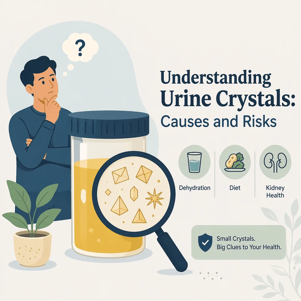
A vedea „cristale în urină" pe un buletin de analize este îngrijorător, și este totodată una dintre cele mai supra-interpretate informații dintr-un sumar de urină. Pentru majoritatea oamenilor, înseamnă foarte puțin. Cristalele sunt particule minerale minuscule pe care un tehnician de laborator sau un analizor automat le observă atunci când urina este examinată la microscop, iar mulți oameni complet sănătoși le au. Și mai surprinzător: multe dintre aceste cristale nu s-au aflat niciodată în vezica ta urinară. S-au format în eprubeta de recoltare, după ce proba a ieșit din corpul tău.
În acest articol vei afla ce sunt cu adevărat cristalele în urină, de ce proba în sine le produce atât de des, ce tipuri sunt practic întotdeauna inofensive, ce tipuri rare necesită cu adevărat atenție și cum se leagă cristalele de calculii renali. Vei vedea, de asemenea, ce anume urmărește medicul alături de acest rezultat și ce simptome ar schimba tabloul clinic.
Ce sunt de fapt cristalele în urină
Urina este o soluție concentrată de săruri, acizi și produse reziduale. Ca orice soluție concentrată, poate dizolva doar o anumită cantitate de substanțe înainte ca o parte din ele să iasă din soluție și să formeze un solid. Acel solid este un cristal.
Dacă apar sau nu cristale depinde de trei variabile fizice: cât de concentrată este urina, cât de acidă sau alcalină este și la ce temperatură se află. Niciunul dintre acești trei factori nu reprezintă o boală. Este pur și simplu chimie. O persoană bine hidratată, care a băut un pahar mare de apă cu o oră înainte de test, produce urină diluată care rareori cristalizează. Cineva care a dat prima urină de dimineață, după opt ore fără lichide, produce urină concentrată care cristalizează frecvent.
Cristalele sunt raportate împreună cu celelalte elemente observate în componenta microscopică a sumarului de urină: globule roșii și albe, celule epiteliale, bacterii și cilindri urinari. Majoritatea laboratoarelor le raportează ca o cantitate aproximativă — puține, moderate, multe — nu ca un număr precis, ceea ce este în sine un semnal că rezultatul nu trebuie citit ca o măsurătoare.
De ce proba în sine creează adesea cristalele
Acesta este cel mai important lucru de pe această pagină, și aproape niciodată nu este explicat pacienților. Cristalele găsite pe un buletin de analize sunt adesea un artefact al modului în care a fost manipulată proba, nu o reflectare a ceea ce se întâmpla în rinichii tăi.
Temperatura și întârzierea
Urina părăsește corpul la aproximativ temperatura corpului. Din momentul în care stă într-un recipient pe o suprafață sau este pusă la frigider pentru a fi transportată la laborator, se răcește. Solubilitatea scade odată cu temperatura, astfel că sărurile dizolvate care se aflau confortabil în soluție în vezica ta precipită în recipient. Cu cât trece mai mult timp între recoltarea probei și examinarea ei, cu atât se formează mai multe cristale.
Efectul nu este neglijabil. Studiile de laborator care răcesc deliberat probele de urină constată că probele care nu prezintă aproape niciun cristal la temperatura camerei dezvoltă cristale abundente după răcire. Cu alte cuvinte, o probă refrigerată sau analizată cu întârziere poate genera un buletin cu cristale care nu au existat în corpul tău în niciun moment.
pH-ul urinei
Fiecare tip de cristal are o chimie care favorizează fie condiții acide, fie alcaline. pH-ul urinei se modifică și el în timp ce proba stă: bacteriile care sunt contaminanți inofensivi de pe piele sau din zona genitală pot descompune ureea și pot crește pH-ul în câteva ore. O probă cu pH deplasat spre alcalin tinde să formeze cristale de fosfat care nu erau prezente când urina era proaspătă.
Metoda de recoltare și recipientul
Chiar și dispozitivul de recoltare contează. Comparațiile dintre sistemele cu tuburi vacuum și recipientele standard au evidențiat diferențe măsurabile în cristalele raportate de la aceiași pacienți. Acesta este un memento că un rezultat cu cristale reflectă un proces fizic dintr-un tub de plastic la fel de mult ca orice fenomen biologic.
Consecința practică este simplă. Medicii tratează cristalele izolate dintr-o probă analizată cu întârziere sau refrigerată ca informații cu valoare redusă, iar un test repetat pe o probă proaspătă, examinată prompt, nu arată adesea nimic.
Tipurile comune de cristale și ce sugerează de obicei fiecare
Majoritatea cristalelor aparțin unui număr mic de tipuri familiare. Denumirile lor sună clinic, dar marea majoritate sunt benigne.
Oxalatul de calciu este de departe cel mai frecvent. La microscop apare sub formă de mici plicuri sau gantere. Este asociat cu alimentele bogate în oxalați, precum spanacul, rubarbarul, nucile, sfecla roșie, ciocolata și ceaiul, dar este prezent și în urina unui număr mare de persoane sănătoase care nu vor forma niciodată un calcul.
Urații amorfi și fosfații amorfi sunt depozite granulare fără formă definită, nu cristale geometrice adevărate. Sunt aproape întotdeauna benigne și reprezintă unele dintre cele mai clare exemple de artefacte de precipitare, apărând cu ușurință în probele răcite sau lăsate să stea.
Fosfatul triplu, numit și struvit, formează cristale în formă de capac de coșciug în urina alcalină. Urina alcalină are multe cauze inofensive, printre care o dietă vegetariană sau simpla stagnare a probei. Cu toate acestea, struvitul poate fi asociat și cu infecții cauzate de bacterii care descompun ureea, astfel că un medic îl va interpreta împreună cu simptomele și markerii de infecție, cum ar fi nitrați și leucocitele în urină și nu izolat.
Cristalele de acid uric apar sub formă de diamante sau rozete în urina acidă. Pot fi observate în gută, în afecțiuni cu un ritm ridicat de distrugere celulară și la persoanele pur și simplu deshidratate. Prin ele însele nu reprezintă un diagnostic, deși medicul poate verifica nivelul de acidul uric din sânge dacă există un motiv clinic.
| Tipul de cristal | pH-ul tipic al urinei | Ce înseamnă de obicei | Este necesară o acțiune? |
|---|---|---|---|
| Oxalat de calciu | Orice pH | Cel mai frecvent tip; întâlnit la multe persoane sănătoase și după mese bogate în oxalați | De obicei, nu prin el însuși |
| Urați amorfi | Acid | Un artefact de precipitare al urinei răcite sau lăsate să stea | Nu |
| Fosfați amorfi | Alcalin | Un artefact de precipitare; fără semnificație clinică în sine | Nu |
| Acid uric | Acid | Urină concentrată, gută sau stări cu un ritm crescut de distrugere celulară | Doar dacă simptomele sau analizele de sânge indică un motiv |
| Fosfat triplu (struvit) | Alcalin | Adesea inofensiv; poate însoți infecțiile cu bacterii care descompun ureea | Interpretat împreună cu simptomele urinare și markerii de infecție |
| Fosfat de calciu | Alcalin | Frecvent și de obicei incidental | De obicei, nu prin el însuși |
| Cistină | Acid | Întotdeauna anormal; indică cistinurie, o tulburare ereditară cu formare de calculi | Da — necesită întotdeauna evaluare medicală |
| Cristale medicamentoase | Variază în funcție de medicament | Legat de anumite medicamente; poate indica o nefropatie cristalină | Da — informează medicul prescriptor |
Tipurile de cristale care contează întotdeauna
Două categorii ies din tiparul liniștitor. Sunt rare, tocmai de aceea merită menționate clar.
Cristale de cistină
Cristalele de cistină sunt plăci hexagonale plate și nu reprezintă niciodată o descoperire normală. Prezența lor indică cistinurie, o afecțiune ereditară în care tubii renali nu reușesc să reabsoarbă aminoacidul cistină. Cistina se dizolvă greu la aciditatea normală a urinei, astfel că precipită și formează calculi, adesea începând din copilărie sau la vârsta adultă tânără și recurând pe tot parcursul vieții. Cistinuria afectează aproximativ o persoană din zece mii.
Dacă un buletin de analiză menționează cristale de cistină, acest lucru justifică o discuție cu medicul, indiferent de cum te simți. Confirmarea diagnosticului și începerea urmăririi pe termen lung cât mai devreme este ceea ce protejează funcția renală de-a lungul deceniilor.
Cristale medicamentoase și nefropatie prin cristale
Unele medicamente se dizolvă greu în urină și pot cristaliza în tubii renali. Antibioticele sulfonamide, aciclovirul, indinavirul, atazanavirul, metotrexatul și vitamina C în doze mari sunt exemple recunoscute. Când se întâmplă acest lucru, cristalele pot obstrucționa tubii și pot leza rinichiul, un proces numit nefropatie cristalină.
Cristalele medicamentoase au, prin urmare, o importanță diferită față de oxalatul de calciu obișnuit. Dacă iei unul dintre aceste medicamente și sunt raportate cristale, informează medicul prescriptor. Acesta poate verifica creatinină și poate reevalua hidratarea sau dozajul. Tulburările ereditare mai rare, cum ar fi deficitul de adenin fosforiboziltransferază, produc și ele cristale distinctive, identificate în același mod.
Cristalele din urină și calculii renali: ce indică și ce nu indică această descoperire
Cristalele nu sunt calculi. Un calcul este o masă solidă care s-a format în decurs de săptămâni sau ani în interiorul tractului urinar; un cristal este o particulă microscopică ce poate exista doar câteva minute. Prezența cristalelor pe un buletin de analiză nu înseamnă că ai un calcul și nu înseamnă că vei dezvolta unul.
Relația există, dar este modestă și merge într-o direcție specifică. Persoanele care au format deja calculilor renali în mod repetat tind să prezinte cristalurie mai frecvent decât cele care nu au avut niciodată calculi. La persoanele cu calculi recurenți, tipul de cristal este cu adevărat util, deoarece sugerează din ce ar putea fi alcătuit calculul, orientând astfel investigațiile și planul de urmărire.
Această distincție contează pentru modul în care îți interpretezi propriul rezultat. Dacă nu ai avut niciodată un calcul și nu ai simptome, cristalele descoperite la o analiză de rutină reprezintă un semnal slab pe care medicul îl va nota cel mai probabil și va trece mai departe. Dacă ești un pacient cu calculi recurenți, același rezultat conține informații pe care urologul sau nefrologul tău le va folosi activ.
Ce face efectiv un medic cu cristalele din raportul tău
În majoritatea cazurilor, nu este necesară nicio acțiune bazată exclusiv pe prezența cristalelor. Medicii interpretează linia referitoare la cristale în context, iar contextul include mai mulți alți factori.
Verifică dacă ai simptome: durere în flanc, durere la urinare, urgență urinară, febră. Controlează pH-ul urinei, deoarece un tip de cristal care corespunde pH-ului este de obicei doar chimie care se comportă previzibil. Caută sânge în urină, care este mult mai relevantă decât cristalele. Verifică markerii de infecție și iau în considerare modul în care a fost recoltată proba și cât timp a așteptat. Pot arunca, de asemenea, o privire la culoarea urinei și concentrația ca indicator aproximativ al hidratării, sau la alte rezultate urinare, cum ar fi urină spumoasă dacă există proteine.
Urmează analizele de sânge, dacă există un motiv. Un panel de funcție renală care indică modificări ale BUN și creatinina, sau un calciu nivel, schimbă imaginea de la o constatare întâmplătoare la ceva ce merită investigat. Dacă se suspectează cu adevărat prezența calculilor, ecografia sau o tomografie computerizată cu doză redusă de radiații răspund direct la această întrebare, iar pentru persoanele cu calculi recurenți poate urma un bilanț metabolic care include colectarea urinei pe 24 de ore.
Ceea ce un medic nu va face de obicei este să prescrie o dietă pe baza cristalelor singure. Sfaturile dietetice pentru prevenirea calculilor sunt individualizate și vin după confirmarea diagnosticului de calculi și după o evaluare metabolică. Un lucru merită spus clar, deoarece credința contrară este frecventă și dăunătoare: restricționarea calciului din alimentație nu este un remediu general pentru cristalele de oxalat de calciu, iar reducerea aportului de calciu poate crește de fapt riscul de formare a calculilor. Orice modificare a dietei tale, a aportului de lichide sau a suplimentelor trebuie decisă împreună cu medicul tău, nu pe baza unui buletin de analize.
Când cristalele din urină sunt mai importante
Două situații cresc semnificația acestei constatări. Prima este simptomele. Cristalele însoțite de durere în flanc, sânge vizibil în urină, febră sau durere la urinare nu mai reprezintă un rezultat întâmplător; ele fac parte dintr-un tablou clinic care necesită evaluare, adesea pentru un calcul sau o infecție urinarăanormal. Al doilea este reducerea funcției renale. Dacă valorile tale renale sunt deja anormale, cristalele — în special cele medicamentoase — merită o atenție mai mare.
Solicită îngrijire medicală urgentă dacă ai oricare dintre următoarele:
- Durere severă în flanc sau spate care iradiază spre zona inghinală, care poate indica prezența unui calcul. Însoțită de febră, aceasta poate semnala un rinichi obstruat și infectat, ceea ce reprezintă o urgență medicală.
- Sânge vizibil în urină.
- Febră sau frisoane însoțite de simptome urinare.
- Imposibilitatea de a urina.
- O scădere clară a cantității de urină produse.
Cele mai recente progrese științifice în analiza cristalelor urinare
Cercetările recente de laborator s-au concentrat mai puțin pe semnificația cristalelor în organism și mai mult pe cât de fiabil le detectăm în primul rând. Patru teme se remarcă.
Cercetători japonezi au răcit deliberat probe de urină de la persoane care formaseră calculi renali și au comparat ce apărea înainte și după răcire. Foarte puține probe au arătat cristale la temperatura ambiantă, în timp ce marea majoritate au prezentat cristale după răcire — iar un cristal specific, brushita (un fosfat de calciu), a fost observat mai frecvent la persoanele ale căror calculi recidivaseră rapid. Ce înseamnă asta pentru tine: este o dovadă directă că răcirea unui specimen creează cristale care nu existau anterior și, totodată, un indiciu că răcirea controlată ar putea deveni într-o zi un test deliberat, nu un accident de transport.
Un raport de caz clinic de laborator a descris un pacient la care măsurarea acidului uric urinar a revenit fals scăzută, deoarece refrigerarea provocase cristalizarea uratului din soluție; ajustarea pH-ului pentru a redizolva cristalele a restabilit valoarea corectă. Ce înseamnă asta pentru tine: cristalizarea în eprubetă nu adaugă doar o linie în raportul de microscopie — poate denatura și alte valori din același eșantion. Testarea promptă contează.
Cercetători canadieni au testat sistematic cum rezistă în timp rezultatele dipstick-ului urinar și ale chimiei urinare, atât la temperatura camerei, cât și la frigider. Majoritatea parametrilor au fost stabili în intervale definite, dar pH-ul s-a numărat printre cei care au derivat. Ce înseamnă asta pentru tine: deoarece pH-ul este cel care determină ce cristale se pot forma, un pH în schimbare dintr-o probă întârziată ajută să explice de ce rapoartele privind cristalele și valorile pH-ului par uneori nepotrivite.
Echipe belgiene și olandeze au antrenat un model de învățare profundă pentru a clasifica particulele din sedimentul urinar pe baza imaginilor de la microscopul digital, testându-l ulterior prospectiv într-un laborator spitalicesc real. Acuratețea a fost ridicată pe setul de date original, dar a scăzut când sistemul a întâlnit probe reale, neselectate. Ce înseamnă asta pentru tine: analiza automată a sedimentului se îmbunătățește rapid și va face raportarea cristalelor mai consecventă, dar revizuirea umană rămâne importantă — acesta este unul dintre motivele pentru care un singur rezultat cu cristale nu este considerat definitiv.
În final, o recenzie clinică despre cistinurie la copii a prezentat modul în care această afecțiune este diagnosticată și gestionată, subliniind că tratamentul urmărește menținerea cistinei dizolvate în urină și că îngrijirea continuă pe tot parcursul vieții. Ce înseamnă asta pentru tine: cristalele de cistină sunt singurul rezultat frecvent raportat care schimbă întotdeauna conduita terapeutică, iar identificarea lor timpurie este cea care face posibilă protecția renală pe termen lung. Îndrumările privind diagnosticul și tratamentul vin de la medici; Articolul MedlinePlus Genetics despre cistinurie de la Biblioteca Națională de Medicină a SUA este un punct de plecare fiabil, în limbaj accesibil.
Întrebări frecvente
Cristalele în urină sunt grave?
De obicei, nu. La majoritatea oamenilor, acestea sunt o descoperire întâmplătoare, fără consecințe clinice, iar o parte semnificativă dintre ele s-au format în recipientul de recoltare, nu în organism. Tipurile comune — oxalat de calciu, urați amorfi și fosfați, fosfat de calciu — nu indică prin ele însele o boală. Excepțiile sunt cristalele de cistină, care sunt întotdeauna anormale, și cristalele asociate unor medicamente specifice. Cristalele devin mai relevante și atunci când apar împreună cu simptome precum durere în flanc, febră sau sânge vizibil în urină, sau când funcția renală este deja redusă. Medicul tău este persoana potrivită pentru a interpreta această descoperire în contextul tău.
Cristalele înseamnă că am calculi renali?
Nu. Cristalele sunt particule microscopice; calculii sunt mase solide care se formează în luni sau ani. Prezența cristalelor într-un rezultat nu înseamnă că există un calcul și nu prezice că se va forma unul. Legătura există în sens invers: persoanele care formează calculi în mod repetat prezintă cristale mai frecvent decât cele care nu au avut niciodată. Dacă nu ai avut niciodată un calcul și nu ai simptome, cristalele sunt o informație slabă. Dacă ești o persoană cu calculi recurenți, tipul de cristale ajută la orientarea investigațiilor. Imagistica, nu microscopia, este cea care confirmă sau exclude cu adevărat prezența unui calcul.
Ar trebui să îmi schimb dieta din cauza cristalelor din urină?
Nu pe cont propriu. Modificările alimentare pentru prevenirea calculilor se stabilesc după un diagnostic confirmat de calcul și o evaluare metabolică, și sunt adaptate fiecărei persoane în parte. Merită corectată o concepție greșită frecventă: mulți oameni presupun că prezența cristalelor de oxalat de calciu înseamnă că trebuie să reducă calciul din dietă. Medicii recomandă, în general, contrariul, deoarece reducerea calciului alimentar poate crește cantitatea de oxalat absorbită din alimente și poate ridica riscul de calculi. Medicii discută de obicei și despre aportul de lichide, sodiu și proteine, dar detaliile depind de rezultatele tale. Prezintă analizele medicului tău și decideți împreună.
De ce menționează rezultatul cristale dacă mă simt perfect bine?
Deoarece cristalele și simptomele sunt în mare parte fără legătură. Formarea cristalelor depinde de concentrație, aciditate și temperatură, iar niciunul dintre acești factori nu trebuie să provoace vreo senzație. O probă recoltată dimineața devreme de la o persoană sănătoasă este concentrată și acidă — condiții ideale pentru formarea cristalelor. Dacă proba a stat câteva ore sau a fost refrigerată înainte de analiză, în acest timp au apărut mai multe cristale. Faptul că te simți bine în timp ce un buletin de analize menționează cristale este combinația obișnuită, nu o contradicție.
Pot medicamentele să cauzeze cristale în urină?
Da. Mai multe medicamente sunt slab solubile în urină și pot cristaliza, printre care antibioticele sulfonamide, aciclovirul, indinavirul, atazanavirul și metotrexatul, precum și dozele foarte mari de vitamina C. Aceste cristale sunt mai îngrijorătoare decât cele obișnuite, deoarece se pot depune în tubii renali și pot provoca leziuni, o afecțiune numită nefropatie prin cristale. Dacă iei vreunul dintre aceste medicamente și cristalele apar în analize, spune-i medicului care ți le-a prescris. Nu opri niciodată un medicament prescris pe baza unui sumar de urină; decizia de a ajusta doza, de a modifica hidratarea sau de a schimba medicamentul îi aparține medicului tău.
Repetarea analizei va da un rezultat diferit?
Adesea, da — și asta este mai degrabă informativ decât îngrijorător. Dacă prima probă a fost refrigerată sau a așteptat câteva ore înainte de examinarea la microscop, o probă proaspătă analizată imediat poate arăta mult mai puține cristale sau deloc. Unele laboratoare vor sugera repetarea analizei tocmai din acest motiv. Dacă cristalele persistă pe o probă proaspătă, recoltată și procesată corect, sau dacă tipul de cristale este unul care contează întotdeauna — cum ar fi cistina — medicul tău va investiga mai departe, în loc să repete analiza la nesfârșit.
Glosar
| Termen | Definiție |
|---|---|
| Cristaluria | Termenul medical pentru cristalele prezente în urină. |
| Sediment urinar | Materialul solid care se depune dintr-o probă de urină și este examinat la microscop. |
| Faza pre-analitică | Tot ce se întâmplă cu o probă între recoltare și analiză, inclusiv transportul, întârzierea și temperatura. |
| pH-ul urinei | O măsură a gradului de aciditate sau alcalinitate a urinei, care determină ce tipuri de cristale se pot forma. |
| Cistinuria | O afecțiune ereditară în care cistina nu este reabsorbită de rinichi, provocând cristale de cistină și calculi recurenți. |
| Struvit | Un cristal de fosfat de magneziu și amoniu care se formează în urină alcalină, uneori asociat cu bacterii care descompun ureea. |
| Nefropatia prin cristale | Leziune renală cauzată de depunerea cristalelor în tubii renali, cel mai adesea legată de medicamente. |
| Suprasaturare | O stare în care urina conține mai multă substanță dizolvată decât poate menține stabil în soluție. |
| Depozit amorf | Material granular amorf în urină, fără o formă cristalină definită și, în general, fără semnificație clinică. |
| Bilanț metabolic | Un set de analize din sânge și urină pe 24 de ore folosit pentru a determina de ce cineva formează calculi renali. |
Surse
- MedlinePlus, Biblioteca Națională de Medicină a SUA. Crystals in Urine. https://medlineplus.gov/lab-tests/crystals-in-urine/
- National Institute of Diabetes and Digestive and Kidney Diseases (NIDDK), National Institutes of Health. Kidney Stones. https://www.niddk.nih.gov/health-information/urologic-diseases/kidney-stones
- Simhadri PK, Rout P, Leslie SW. Urinary Crystals Identification and Analysis. StatPearls, NCBI Bookshelf, National Library of Medicine. https://www.ncbi.nlm.nih.gov/books/NBK606103/
- MedlinePlus Genetics, Biblioteca Națională de Medicină a SUA. Cystinuria. https://medlineplus.gov/genetics/condition/cystinuria/
- Tanaka Y, Tsujino I, Yoshikawa HY, et al. Cooling-induced brushite crystallization in urine as a predictive risk marker for calcium kidney stone recurrence. Urolithiasis, 2025. Retrieved via PubMed. https://doi.org/10.1007/s00240-025-01820-2
- Han J, Jiang Y. Urine Uric Acid Pseudodeficiency from Urate Crystallization: a Pre-Analytical Laboratory Pitfall. Clinical Laboratory, 2025. Accesat prin PubMed. https://doi.org/10.7754/Clin.Lab.2025.250415
- Bohn MK, Fabros A, Di Meo A. Evaluation of pre-analytical factors impacting urine test strip and chemistry results. Clinical Chemistry and Laboratory Medicine, 2025. Accesat prin PubMed. https://doi.org/10.1515/cclm-2024-1233
- Mouslech SG, Wijnants S, van der Schagt AL, et al. Towards Clinical Integration of Deep Learning-Based Classification of Urinary Sediment Particles from Digital Microscopy Images: A Prospective Study. Clinical Chemistry, 2026. Retrieved via PubMed. https://doi.org/10.1093/clinchem/hvaf182
- Gökçe Mİ, Karaburun MC. Cystinuria in children: diagnosis and treatment. World Journal of Urology, 2025. Accesat prin PubMed. https://doi.org/10.1007/s00345-025-05604-6
- Korkmaz B, Kural A, Başman A, et al. Effect of vacuum and non-vacuum urine collection systems on urinalysis. Scandinavian Journal of Clinical and Laboratory Investigation, 2025. Accesat prin PubMed. https://doi.org/10.1080/00365513.2025.2524850
Lectură suplimentară
- Rezultatele sumarului de urină: ghid complet de interpretare
- Calculii renali: cauze, simptome și tratamente
- Valorile normale ale pH-ului urinar: cauze și semnificație
- Hematurie: sânge în urină, cauze și simptome
- Urina spumoasă explicată: cauze și riscuri
O analiză de urină rareori stă singură. Cristalele din urină au mult mai mult sens când le poți vedea alături de pH-ul urinei, creatinina și calciul tău din același raport. AI DiagMe îți citește rezultatele de laborator și explică fiecare linie în limbaj simplu, astfel încât să ajungi la consultație cu întrebări mai bune. Te ajută să îți înțelegi rezultatele; nu pune diagnostice și nu înlocuiește medicul tău.
Înțelege-ți rezultatele analizelor cu AI DiagMe
Obține interpretarea rezultatelor în câteva minute