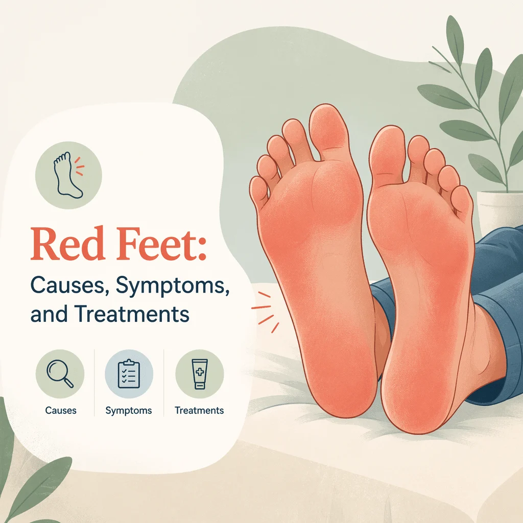
Picioarele roșii — roșeața care afectează unul sau ambele picioare — poate să nu însemne nimic îngrijorător sau poate fi un semn serios. Căldura, o baie fierbinte, o tură lungă în picioare sau o alergare pot înroși pielea fără niciun pericol, iar culoarea dispare de la sine. Dar același aspect poate fi primul semn al unei infecții cutanate, al unei probleme de circulație sau al unui picior aflat în pericol la o persoană cu diabet.
În acest articol vei afla care situații obișnuite pot înroși picioarele, ce afecțiuni ale pielii și ale circulației produc același efect, cum deosebesc medicii o infecție reală de afecțiunile care îi seamănă, de ce piciorul diabetic este o categorie aparte și ce examinează și testează un medic.
Începe cu lista de semne de alarmă de mai jos: dacă vreunul dintre ele se potrivește cu ce observi, nu mai citi și mergi să fii consultat.
Semne de alarmă: când picioarele roșii necesită evaluare urgentă
Solicită ajutor medical — în aceeași zi sau urgență, acolo unde este indicat
⚠️ Dacă ai diabet și un picior roșu, cald sau umflat, ai nevoie de o evaluare în aceeași zi — chiar dacă nu doare. Leziunile nervoase cauzate de diabet reduc senzația de durere, astfel că o infecție care poate pune în pericol membrul sau un picior Charcot se pot dezvolta cu un disconfort foarte mic. Institutul Național pentru Diabet și Boli Digestive și Renale (NIDDK) recomandă să îți contactezi imediat medicul dacă pielea de pe picior devine roșie, caldă sau dureroasă și precizează că un picior Charcot poate debuta exact cu aceste semne. “Nu mă doare” nu este un motiv de liniște în cazul piciorului diabetic.
Solicită îngrijire urgentă sau de urgență și dacă ai oricare dintre următoarele:
- Roșeață care se extinde, însoțită de febră, frisoane sau stare generală alterată.
- O margine roșie care avansează vizibil în câteva ore.
- Pielea care devine neagră sau vineție, sau apariția de bășici pe zona roșie.
- Durere mult mai intensă decât sugerează aspectul pielii — durerea disproporționată față de aspect poate semnala o infecție necrozantă (care distruge țesuturile), o urgență medicală.
- Un picior rece și palid, fără puls perceptibil de tine sau de un medic.
- Umflarea gambei însoțită de durere sau sensibilitate la atingere, care poate indica o tromboză venoasă profundă (un cheag într-o venă profundă a piciorului).
- Roșeață în jurul unei răni deschise, al unei bășici sau al unui ulcer la picior.
- Orice articulație roșie, fierbinte și umflată — posibilă artrită septică (infecție în interiorul unei articulații).
- Roșeață la orice persoană cu circulație deficitară, sistem imunitar slăbit sau cu antecedente de ulcer la picior ori amputație.
Roșeața însoțită de căldură locală, umflătură, durere, febră, rană deschisă sau apărută la o persoană cu diabet ori boală arterială este o problemă medicală. Nu poate fi evaluată de pe o pagină web.
Cauze obișnuite ale picioarelor roșii
Majoritatea cazurilor de picioare roșii au o explicație banală. Pielea se înroșește atunci când vasele mici de la suprafață se dilată, iar căldura este cel mai frecvent factor declanșator: o baie fierbinte, asfaltul încins, un calorifer, o cameră caldă. Culoarea apare de obicei la ambele picioare, este uniformă, nu în pete, și dispare în mai puțin de o oră după ce te răcorești.
Gravitația este al doilea factor. Dacă stai în picioare ore întregi sau stai jos cu picioarele atârnate, sângele se acumulează în venele picioarelor și pielea capătă o nuanță roșiatică închisă care dispare când ridici picioarele. Această acumulare obișnuită diferă de roșeața de declivitate ischemică descrisă mai jos, iar modul în care răspunde la ridicarea picioarelor este cel care le deosebește.
Efortul fizic înroșește picioarele din același motiv: mușchii au nevoie de sânge, vasele se dilată, pielea se înfierbântă. Frecarea cauzată de pantofii noi sau de șosetele care freacă lasă pete roșii pe zonele de presiune, iar alcoolul și unele medicamente dilată și ele vasele de sânge. Arsura solară este ușor de trecut cu vederea pe dosul picioarelor: piele roșie, sensibilă și caldă, strict limitată la zona expusă, adesea cu o linie clară acolo unde a stat curelușa sandalei.
Aceste cauze obișnuite au un tipar comun: ambele picioare, fără febră, fără umflătură persistentă, fără durere reală și ameliorare treptată odată ce factorul declanșator este eliminat. Orice altceva merită o consultație la un medic.
Afecțiuni ale pielii care înroșesc picioarele
Dermatita de contact este o inflamație cauzată de ceva care atinge pielea — cauciuc și adezivi din pantofi, coloranți din șosete, nichel din catarame, ingrediente din cremele pentru picioare. Roșeața urmărește forma obiectului care a provocat-o, provoacă mâncărime și rămâne localizată acolo unde a avut loc contactul, în loc să se extindă spre exterior ca o infecție.
Piciorul atletului (tinea pedis) este o infecție fungică care debutează de obicei între degete cu piele albă și macerată, roșeață, fisuri și mâncărime. Importanța sa depășește simpla mâncărime: fisurile dintre degete reprezintă o poartă de intrare frecventă pentru bacteriile care provoacă celulita. Dacă mâncărimea este simptomul dominant, consultă ghidul nostru despre picioare care mănâncă noaptea.
Degerăturile superficiale (pernioza) apar după expunerea la frig și umezeală — nu îngheț, ci doar frig. Pete roșii sau violacee, pruriginoase și cu senzație de arsură se dezvoltă pe degete la câteva ore după reîncălzire și dispar în una-două săptămâni.
Eritromelalgia este o afecțiune distinctivă și frecvent nerecunoscută. Provoacă episoade de durere intensă cu senzație de arsură, cu picioare roșii și fierbinți, declanșate de căldură, efort fizic sau atârnarea picioarelor în jos, și ameliorate prin răcire — mulți oameni descriu că își cufundă picioarele în apă rece. Între atacuri, picioarele arată normal. Deoarece acest tipar este atât de caracteristic, descrierea lui clară unui medic scurtează adesea drumul spre diagnostic.
Psoriazisul, eczema și reacțiile la medicamente pot înroși și ele picioarele, de obicei cu semne vizibile și în alte zone ale corpului.
Circulația și picioarele roșii: vene, artere și roșeața de declivitate
Probleme venoase: dermatita de stază și insuficiența venoasă cronică
Când valvele venelor din picioare nu mai funcționează corect, sângele are dificultăți să urce înapoi și presiunea crește în vasele mici de la gleznă. În timp, aceasta duce la insuficiență venoasă cronică: umflătură mai accentuată seara, senzație de greutate și durere surdă, piele maronie în jurul gleznelor, descuamare uscată și pruriginoasă și o decolorare roșiatică difuză.
Această ultimă manifestare — dermatita de stază — este cel mai frecvent lucru confundat cu o infecție la nivelul gambei. De obicei apare la ambele picioare, se instalează în săptămâni, nu în zile, provoacă mai mult mâncărime decât durere și nu este însoțită de febră.
Un aspect important de siguranță: măsurile care ajută în problemele venoase pot fi dăunătoare atunci când arterele sunt și ele îngustate, astfel că nimeni nu ar trebui să înceapă compresia din proprie inițiativă. Dacă este sau nu indicată depinde de evaluarea arterelor.
Boala arterială și roșeața de declivitate: o roșeață care nu este liniștitoare
Boala arterială periferică îngustează arterele care alimentează picioarele și tălpile. Conform Bibliotecii Naționale de Medicină, semnele sale includ pulsuri slabe sau absente la nivelul piciorului, răni care se vindecă greu sau deloc, piele palidă sau albăstruie, un picior mai rece decât celălalt și creștere deficitară a unghiilor și a părului.
Când îngustarea devine severă, se întâmplă ceva contraintuitiv. Vasele mici din picior rămân permanent dilatate, încercând să atragă orice cantitate de sânge posibilă. Lasă piciorul în jos și gravitația le umple, producând o nuanță roșie-închisă, vineție pe antepicior și degete. Ridică piciorul deasupra nivelului inimii și culoarea dispare, lăsând pielea izbitoare de palidă.
Acest semn — roșu când piciorul e jos, alb când e ridicat — se numește rubor de declivitate și nu este un semn de circulație bună. Indică ischemie cronică amenințătoare de membru, un stadiu al bolii arteriale în care piciorul este în pericol fără restabilirea fluxului sanguin. Apare adesea însoțit de dureri la nivelul antepiciorului noaptea, care se ameliorează când piciorul atârnă peste marginea patului, și de răni pe degete sau călcâi care nu se vindecă. Este un motiv să mergi urgent la medic, nu să te relaxezi.
Tromboza venoasă profundă se încadrează și ea aici. Un cheag într-o venă profundă a piciorului provoacă cel mai adesea umflarea și durerea unui gambă; roșeața poate fi sau nu prezentă, motiv pentru care nu poate confirma sau exclude un cheag. Umflarea gambei însoțită de durere necesită o evaluare urgentă, iar Analiză de sânge D-dimer cu ecografie face adesea parte din aceasta.
Celulita — și de ce două picioare roșii nu sunt, de obicei, celulită
Celulita este o infecție bacteriană a pielii și a țesutului de sub aceasta. MedlinePlus descrie o piele dureroasă, roșie și sensibilă la atingere, care poate face bășici și cruste, adesea însoțită de febră, frisoane și ganglioni limfatici umflați. Bacteriile pătrund printr-o leziune a pielii.
Caracteristica importantă de reținut este asimetria. Celulita este aproape întotdeauna limitată la un singur membru, deoarece bacteriile nu infectează de obicei ambele picioare în aceeași zi. Când ambele picioare sau ambele gambe sunt roșii în același timp, probabilitatea se îndreaptă puternic spre o cauză sistemică sau mecanică: dermatită de stază, retenție de lichide din cauza problemelor cardiace, renale sau hepatice, ori o reacție de contact sau la medicamente.
Medicii grupează aceste afecțiuni asemănătoare sub denumirea de pseudocelulită. Distincția contează în ambele sensuri: tratarea unui imitator ca infecție înseamnă antibiotice inutile, în timp ce ignorarea unei infecții reale îi permite să se răspândească.
Nimic din cele de mai sus nu înseamnă că roșeața bilaterală este inofensivă. Afecțiuni grave pot afecta ambele picioare — insuficiența cardiacă severă, bolile inflamatorii generalizate și, rar, infecția bilaterală. Simetria este un indiciu pe care trebuie să îl raportezi medicului tău, nu o concluzie la care să ajungi singur acasă.
Piciorul diabetic: o categorie aparte
Tot ce s-a spus mai sus se schimbă atunci când persoana are diabet. Trei factori se cumulează: leziunile nervoase elimină durerea care ar trage un semnal de alarmă, fluxul sanguin redus încetinește vindecarea, iar glicemia crescută face infecția mai probabilă și mai agresivă.
NIDDK spune clar: poți avea probleme la picioare fără să simți durere. De aceea, scurtătura obișnuită — “dacă ar fi grav, ar durea” — eșuează complet în acest caz, și de aceea regula de la începutul acestei pagini este evaluarea în aceeași zi, indiferent de durere.
Două situații explică o mare parte din pericol. Prima este infecția piciorului legată de diabet, care poate evolua de la o mică leziune a pielii până la afectarea țesuturilor profunde și a osului în câteva zile.
A doua este neuro-osteoartropatia Charcot, sau piciorul Charcot. Într-un picior care și-a pierdut sensibilitatea protectoare, oasele și articulațiile se pot inflama, înmuia și prăbuși. Stadiul incipient arată ca un picior roșu, fierbinte și umflat — adesea vizibil mai cald decât celălalt — cu durere surprinzător de redusă, și este confundat frecvent cu o infecție sau o entorsă. Depistat devreme, deformarea permanentă poate fi adesea evitată; nedepistat, poate duce la un picior prăbușit, predispus la ulcerații.
Dacă trăiești cu diabet, verifică-ți picioarele zilnic, inclusiv între degete și tălpi, și tratează orice roșeață nouă, căldură, umflătură sau rană ca pe un motiv să îți contactezi echipa medicală în aceeași zi. Schimbările unghiilor contează și ele — vezi unghii de la picioare la diabetici. Controlul este urmărit cu HbA1c și glicemia à jeun.
Articulații roșii, fierbinți și umflate: guta și artrita septică
Uneori roșeața este concentrată pe o articulație, nu răspândită pe toată pielea. Guta este exemplul clasic. Conform National Institute of Arthritis and Musculoskeletal and Skin Diseases, mulți oameni au primul puseu la nivelul halucelui, atacurile încep adesea brusc noaptea și pot fi suficient de intense pentru a-i trezi din somn, iar articulația este umflată, roșie și caldă.
Guta apare atunci când cristalele de urat se formează în și în jurul unei articulații. Un test de acid uric din sânge este adesea verificat, deși un rezultat normal în timpul unui atac nu exclude guta, iar unul crescut nu o confirmă. Articolul nostru despre managementul gutei merge mai departe.
Guta este importantă aici deoarece o articulație fierbinte, roșie și umflată nu poate fi considerată automat gută. Artrita septică — infecția din interiorul articulației — arată aproape identic și distruge cartilajul rapid. Este o urgență, iar diferențierea celor două necesită de obicei extragerea de lichid din articulație. Nu aștepta niciodată ca o articulație roșie, fierbinte și umflată să treacă de la sine, presupunând că este o criză de gută.
Cum diferențiază un medic cauzele picioarelor roșii
Examinarea face cea mai mare parte din muncă. Un medic va întreba cât de repede a apărut roșeața, dacă afectează un picior sau ambele, dacă există durere, mâncărime sau amorțeală, dacă ai avut febră și care este istoricul tău medical. Apoi privește și palpează: marginile roșeții, căldura comparativ cu celălalt picior, umflătura și dacă există o rană sau o crăpătură între degete.
Două manevre simple au o importanță deosebită. Compararea picioarelor unul lângă altul evidențiază asimetria, iar ridicarea piciorului pentru a urmări culoarea diferențiază simpla acumulare de sânge de ruborul de declivitate. Palparea pulsurilor piciorului și măsurarea indicelui gleznă-braț (tensiunea arterială la gleznă comparată cu cea de la braț) evaluează circulația arterială.
Analizele răspund la întrebări specifice, nu sunt solicitate ca un set standard. Markerii inflamatori, precum proteina C reactivă (PCR) și viteza de sedimentare a hematiilor (VSH) arată că există inflamație, dar nu și cauza acesteia. Un hemoleucogramă completă poate arăta un număr crescut de globule albe, iar procalcitonina poate ajuta la evaluarea infecției bacteriene. Glicemia și HbA1c verifică prezența diabetului. Investigațiile imagistice — ecografia pentru cheaguri, radiografia sau RMN-ul pentru modificări osoase și Charcot — se adaugă atunci când istoricul medical indică această direcție.
Tabelul de mai jos rezumă tiparele pe care le evaluează un medic. Este un ghid despre ce ar trebui să includă descrierea ta, nu un substitut pentru examinarea clinică.
| Ce observi | La ce indică de obicei | Ce să faci |
|---|---|---|
| Ambele picioare roșii după stat în picioare, căldură sau baie fierbinte; dispare la răcire sau ridicarea picioarelor; fără durere sau febră | Dilatare vasculară normală și acumulare gravitațională a sângelui | Răcește, ridică picioarele, observă; mergi la medic dacă roșeața nu dispare |
| Un picior roșu, fierbinte, umflat și dureros, care se extinde în câteva ore, cu sau fără febră | Posibilă celulită sau altă infecție acută | Consultație medicală în aceeași zi; urgență dacă există febră sau răspândire rapidă |
| Ambele gambe și picioare roșii-închis de săptămâni, cu mâncărime, umflarea gleznelor și pete maronii | Dermatită de stază cauzată de insuficiență venoasă, frecvent confundată cu celulita | Programează o consultație medicală; nu începe compresia fără o evaluare arterială prealabilă |
| Roșeață care se intensifică când piciorul atârnă în jos și se estompează când piciorul este ridicat | Rubor de declivitate — semn al unei boli arteriale severe, nu al unei circulații bune | Consultație medicală promptă, cu trimitere pentru evaluare vasculară |
| Picior roșu, cald sau umflat la o persoană cu diabet, cu durere redusă sau absentă | Posibilă infecție a piciorului legată de diabet sau un picior Charcot activ | Consultație medicală în aceeași zi, indiferent cât de ușoare par simptomele |
| Roșeață în jurul unei răni deschise, a unei bășici sau a unui ulcer la picior | Posibilă infecție a plăgii, care poate afecta țesuturile profunde sau osul | Consultație medicală în aceeași zi; nu trata acasă |
| Durere bruscă și intensă într-o articulație, adesea la degetul mare de la picior, roșie, fierbinte și umflată | Criză de gută, dar artrita septică arată similar | Consultație medicală urgentă pentru a diferenția cele două afecțiuni |
| Picioare roșii și fierbinți cu senzație de arsură în episoade, ameliorate prin răcire, normale între episoade | Sugestiv pentru eritromelalgie | Programează o consultație medicală și descrie explicit ameliorarea prin răcire |
Cele mai recente progrese științifice în evaluarea picioarelor roșii
Cercetările recente s-au concentrat mai puțin pe tratament și mai mult pe problema mai dificilă: diferențierea afecțiunilor care arată la fel.
Un studiu de validare prospectivă publicat în JAMA Dermatology în 2024 de Pulia și colaboratori a studiat adulți care s-au prezentat la camera de urgență cu un gambă acut roșie. Ce s-a descoperit: temperatura suprafeței pielii măsurată prin imagistică termică (o cameră care detectează căldura, nu lumina) a fost constant mai ridicată în celulita adevărată față de pseudocelulită, iar combinarea acesteia cu scorul ALT-70 — o listă de verificare bazată pe asimetrie, numărul de globule albe, frecvența cardiacă și vârstă — a identificat mai bine afecțiunile similare decât oricare dintre ele separat. Autorii notează că celulita este diagnosticată greșit în până la 30% din cazuri. Ce înseamnă asta pentru tine: dacă un medic pune la îndoială dacă piciorul tău roșu este cu adevărat infectat, aceasta reflectă dovezile actuale, nu îndoiala față de simptomele tale.
O recenzie sistematică și meta-analiză publicată în Archives of Dermatological Research în 2024 de Lin și colaboratori a analizat paisprezece studii despre celulita suspectată. Ce s-a constatat: atunci când un dermatolog a revizuit diagnosticul, planul inițial de tratament cu antibiotice s-a schimbat într-o proporție mare a cazurilor, îmbunătățind acuratețea și reducând utilizarea inutilă a antibioticelor, fără rate mai mari de reinternare. Ce înseamnă asta pentru tine: o a doua opinie de specialitate pentru un picior roșu este considerată o îngrijire medicală corectă, nu o întârziere — mai ales când roșeața apare pe ambele picioare sau durează de săptămâni.
Ghidurile IWGDF/IDSA din 2023 privind infecțiile piciorului diabetic, coordonate de Senneville și colaboratori, au actualizat standardul internațional. Ce s-a constatat: infecția la piciorul diabetic trebuie diagnosticată clinic — pe baza roșeții, căldurii, umflăturii, sensibilității și secrețiilor — și clasificată după severitate, iar această clasificare determină urgența investigațiilor. Ce înseamnă asta pentru tine: semnele vizibile de pe piciorul tău reprezintă punctul de plecare al unei evaluări formale a severității, ceea ce este un motiv în plus să nu aștepți și să vezi cum evoluează.
IWGDF a emis, de asemenea, primul său ghid dedicat neuro-osteoartropatiei Charcot active în 2023, coordonat de Wukich și colaboratori. Ce s-a constatat: acesta stabilește cum trebuie identificat și gestionat piciorul Charcot activ, tratând recunoașterea timpurie ca prioritate, deoarece faza acută se prezintă ca un picior roșu, cald și umflat, ușor de confundat cu o infecție. Ce înseamnă asta pentru tine: dacă ai diabet și neuropatie și un picior devine brusc roșu, fierbinte și umflat, menționează explicit piciorul Charcot — semnalarea lui din timp schimbă cursul evenimentelor.
Întrebări frecvente
Când sunt picioarele roșii o urgență?
Tratează picioarele roșii ca o urgență dacă roșeața se extinde vizibil în timp ce privești, dacă ai febră sau te simți rău în același timp, dacă pielea face bășici sau devine neagră sau vineție, sau dacă durerea este mult mai intensă decât ar sugera aspectul pielii — această combinație poate indica o infecție necrozantă. Un picior rece, palid și fără puls este, de asemenea, o urgență, la fel ca un gambă care se umflă brusc și devine dureroasă. Oricine are diabet și un picior roșu, cald sau umflat are nevoie de o evaluare în aceeași zi, chiar și fără niciunul dintre aceste semne, deoarece durerea poate lipsi.
De ce îmi sunt ambele picioare roșii?
Roșeața care afectează ambele picioare în același timp nu este, de obicei, o infecție. Celulita este aproape întotdeauna limitată la un singur membru, astfel că simetria indică mai degrabă cauze care acționează asupra întregului organism sau a ambelor picioare în mod egal: căldura, statul prelungit în picioare sau așezat cu picioarele în jos, dermatita de stază cauzată de insuficiența venoasă, retenția de lichide din cauza problemelor cardiace, renale sau hepatice, sau o reacție la ceva cu care au intrat în contact ambele picioare. Acestea fiind spuse, roșeața bilaterală nu este automat inofensivă — necesită în continuare un consult medical, mai ales dacă este recentă, dureroasă sau însoțită de umflături sau dificultăți de respirație.
Am diabet și piciorul meu este roșu, dar nu mă doare. Pot să aștept?
Nu. Neuropatia diabetică diminuează sau elimină senzația de durere, astfel că absența durerii nu îți spune nimic despre gravitatea problemei. Un picior roșu, cald sau umflat la o persoană cu diabet poate fi o infecție deja profundă sau un picior Charcot activ, în care oasele se înmoaie. Ambele situații sunt urgențe și pot apărea cu disconfort minim. Contactează în aceeași zi echipa de diabet, podiatrul sau medicul tău și nu te baza pe cum simți piciorul pentru a decide cât de urgent este.
De ce mi se înroșesc tălpile când mă ridic în picioare?
Gravitația atrage sângele în vasele piciorului atunci când acesta se află sub nivelul inimii, iar la majoritatea oamenilor acest lucru produce o roșeață ușoară și uniformă care dispare în mai puțin de un minut după ridicarea picioarelor. Dacă roșeața este intensă, închisă la culoare sau violacee, apare mai ales pe partea din față a piciorului și pe degete și se transformă într-o paloare accentuată atunci când ridici piciorul, acest tipar se numește rubor de declivitate și poate semnala o îngustare severă a arterelor piciorului. Întrebarea esențială nu este dacă apare culoarea, ci cât de dramatică este schimbarea atunci când piciorul este ridicat.
Poate roșeața picioarelor să fie un semn de cheag de sânge?
Tromboza venoasă profundă provoacă mai frecvent umflarea și durerea unui singur gambă decât roșeața piciorului în sine, iar atunci când apare decolorarea, aceasta poate fi roșie, albăstruie sau chiar absentă. Deoarece roșeața este un indicator nesigur în acest caz, semnele la care trebuie să reacționezi sunt: umflarea unui singur gambă, sensibilitate la nivelul gambei și un picior care se simte greu sau doare la mers, mai ales după o intervenție chirurgicală, imobilizare sau o călătorie lungă. Dacă acestea sunt prezente, solicită o evaluare urgentă în loc să încerci să judeci situația după culoare.
Pielea roșie de pe picioare înseamnă întotdeauna infecție?
Nu, și eroarea inversă este la fel de frecventă. Multe picioare roșii sunt cauzate de căldură, gravitație, frecare, dermatită sau boli venoase, nu de bacterii, iar studiile arată că afecțiunile non-infecțioase care imită celulita sunt adesea tratate ca atare. În același timp, o infecție reală poate debuta discret, mai ales la un picior cu sensibilitate redusă. Concluzia practică este că roșeața singură nu confirmă și nici nu exclude o infecție: elementele din jur — un picior sau ambele, febra, viteza de apariție, durerea, o rană, diabetul — sunt cele care determină răspunsul, iar acestea necesită evaluarea unui medic.
Glosar
| Termen | Definiție |
|---|---|
| Celulită | O infecție bacteriană a pielii și a țesutului de sub ea, care provoacă piele roșie, dureroasă și sensibilă la atingere, de obicei la un singur membru |
| Pseudocelulita | Un termen general pentru afecțiunile non-infecțioase care seamănă cu celulita, cum ar fi dermatita de stază sau o reacție la medicamente |
| Dermatită de stază | Piele roșie, pruriginoasă și scuamoasă în jurul gleznelor, cauzată de presiunea crescută de lungă durată în venele picioarelor |
| Rubor decliv | Roșeață intensă a piciorului când acesta atârnă în jos, care dispare când piciorul este ridicat — semn al unei îngustări severe a arterelor |
| Eritromelalgie | Episoade de durere arsură cu picioare roșii și fierbinți, declanșate de căldură și ameliorate de obicei prin răcire |
| Degerături (pernioză) | Pete roșii sau violacee, pruriginoase, pe degete, care apar la câteva ore după expunerea la frig și umezeală |
| Neuropatie diabetică | Leziuni ale nervilor cauzate de diabet, care reduc sau elimină sensibilitatea la nivelul picioarelor, inclusiv capacitatea de a simți durerea |
| Piciorul Charcot | Inflamație, înmuiere și prăbușire a oaselor și articulațiilor unui picior care și-a pierdut sensibilitatea protectoare, debutând adesea ca un picior roșu, cald și umflat |
| Boală arterială periferică | Îngustarea arterelor care irigă picioarele, reducând fluxul sanguin și încetinind vindecarea rănilor |
| Tromboză venoasă profundă | Un cheag de sânge format într-o venă profundă, de obicei la nivelul gambei, coapsei sau pelvisului |
Surse
- National Institute of Diabetes and Digestive and Kidney Diseases (NIDDK). Diabetes & Foot Problems
- MedlinePlus, National Library of Medicine. Cellulitis
- MedlinePlus, National Library of Medicine. Peripheral Arterial Disease
- National Institute of Arthritis and Musculoskeletal and Skin Diseases (NIAMS). Gout
- National Heart, Lung, and Blood Institute (NHLBI). Venous Thromboembolism
- Senneville É, Albalawi Z, van Asten SA, et al. IWGDF/IDSA Guidelines on the Diagnosis and Treatment of Diabetes-related Foot Infections (IWGDF/IDSA 2023). Clinical Infectious Diseases. 2023 (via PubMed)
- Wukich DK, Schaper NC, Gooday C, et al. Guidelines on the diagnosis and treatment of active Charcot neuro-osteoarthropathy in persons with diabetes mellitus (IWGDF 2023). Diabetes/Metabolism Research and Reviews. 2023;40(3):e3646 (via PubMed)
- Pulia MS, Schwei RJ, Alexandridis R, et al. Validation of Thermal Imaging and the ALT-70 Prediction Model to Differentiate Cellulitis From Pseudocellulitis. JAMA Dermatology. 2024;160(5):511-517 (via PubMed)
- Lin V, Callado GY, Pardo I, et al. Diagnostic stewardship and dermatology consultation in cellulitis management: a systematic literature review and meta-analysis. Archives of Dermatological Research. 2024;317(1):61 (via PubMed)
Lectură suplimentară
- Diabetul: cauze, simptome și tratamente
- Unghiile de la picioare în diabet: simptome, cauze și tratamente
- Gută: ameliorarea durerii, tratamente și management
- Picioare care mănâncă noaptea: cauze și tratamente
- Niveluri de glucoză: cauze, simptome și tratamente
Înțelege-ți rezultatele analizelor cu AI DiagMe
Dacă un medic investighează picioarele roșii, investigațiile pot include markeri inflamatori precum CRP sau VSH, hemoleucogramă completă, glicemie sau HbA1c, acid uric sau D-dimer. Aceste rezultate vin sub formă de cifre cu valori de referință și cu puține explicații.
AI DiagMe te ajută să înțelegi ce înseamnă acele cifre odată ce le ai, astfel încât să poți purta o conversație mai bine informată cu medicul tău. Nu pune diagnostice, nu poate vedea sau examina piciorul tău și nu este destinat urgențelor — dacă vreunul dintre semnalele de alarmă menționate ți se aplică, solicită îngrijire medicală imediat.
Obține interpretarea rezultatelor în câteva minute