



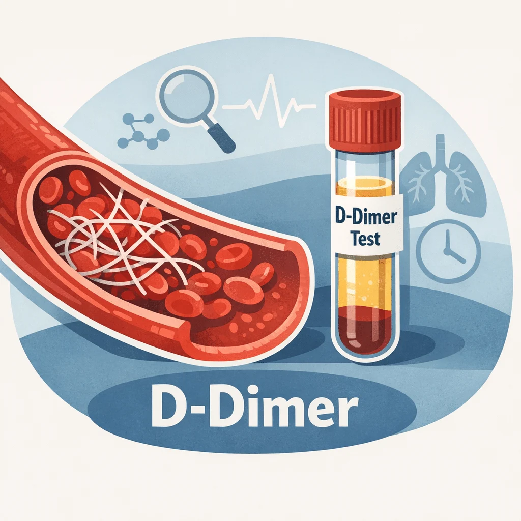
La prueba de dímero D mide un pequeño fragmento de proteína que aparece en tu sangre cuando un coágulo se desintegra. Los médicos la solicitan con mayor frecuencia para ayudar a descartar un coágulo peligroso, como una trombosis venosa profunda (TVP) o una embolia pulmonar (EP), en combinación con una evaluación clínica del riesgo. Esta guía explica qué mide la prueba, cómo interpretar tu resultado en relación con el rango de referencia, por qué importan los valores de corte ajustados por edad y qué otras condiciones pueden elevar este marcador incluso sin la presencia de un coágulo. También encontrarás un resumen en lenguaje sencillo de las investigaciones más recientes, un glosario y orientación clara sobre cuándo un resultado requiere atención médica oportuna.
¿Qué es una prueba de dímero D?
El dímero D es un fragmento que queda después de que tu cuerpo disuelve un coágulo de sangre. No existe en cantidades significativas en la sangre de una persona sana, a menos que la formación y disolución de coágulos estén ocurriendo activamente en algún lugar del cuerpo. Cuando se forma un coágulo, la proteína fibrina crea una red que lo mantiene unido. Una vez que la lesión sana, una enzima llamada plasmina disuelve esa red, y los residuos incluyen fragmentos de dímero D que circulan en la sangre hasta que los riñones y el hígado los eliminan.
Una prueba de dímero D detecta esos fragmentos en una muestra de sangre de rutina. Como el fragmento solo aparece cuando se está formando y disolviendo un coágulo, un nivel elevado le indica al médico que hay actividad de coagulación en algún lugar del cuerpo. No dice dónde ni por qué — esas respuestas requieren más estudios.
Por qué esta prueba es importante en la práctica diaria
La principal utilidad de este marcador está en lo que un resultado normal descarta, no en lo que un resultado alto confirma. Cuando el médico ya considera poco probable un coágulo con base en tus síntomas e historial, un dímero D normal ayuda a descartar una trombosis venosa profunda (TVP) o una embolia pulmonar (EP) sin necesidad de estudios de imagen. Esta combinación — criterio clínico más un análisis de sangre — es lo que hace tan útil al dímero D en urgencias y consulta externa, evitando que muchas personas se sometan a una tomografía o ultrasonido innecesarios.
Cómo leer los resultados de tu prueba de dímero D
Tu reporte muestra un número, una unidad y un rango de referencia. Un resultado típico se ve así: Dímero D 320 ng/mL FEU (rango de referencia: menor de 500 ng/mL FEU). FEU significa unidades equivalentes de fibrinógeno, una de las dos formas más comunes en que los laboratorios reportan esta prueba; algunos laboratorios usan en cambio unidades de dímero D (DDU), que tienen un punto de corte numérico diferente. Si comparas un resultado con información que encuentres en otro lugar, verifica siempre qué unidad usó tu propio laboratorio.
La mayoría de los laboratorios para adultos establecen un punto de corte estándar alrededor de 500 ng/mL FEU. Un resultado por debajo de ese umbral generalmente se considera negativo, lo que significa que no se detectó actividad de coagulación en un nivel significativo. Un resultado igual o por encima del umbral se considera positivo, lo que lleva al médico a investigar más, en lugar de diagnosticar un coágulo de inmediato.
Por qué la edad cambia el panorama
El dímero D aumenta de forma natural con la edad, incluso en personas que se sienten completamente bien. Usar el mismo valor de corte para una persona de 30 años y otra de 80 significa que los adultos mayores son marcados como "positivos" con mucha más frecuencia sin que realmente tengan un coágulo. Para corregir esto, muchos laboratorios y servicios de urgencias utilizan ahora un valor de corte ajustado por edad para pacientes mayores de 50 años: multiplica tu edad en años por 10 para obtener tu valor de corte personal en ng/mL FEU. Una persona de 70 años, por ejemplo, tendría un valor de corte de alrededor de 700 ng/mL en lugar del estándar de 500.
Este ajuste ha sido estudiado en grandes grupos de pacientes y actualmente cuenta con el respaldo de varias sociedades médicas, aunque la FDA no lo ha aprobado formalmente como un cambio en el etiquetado. Su propósito es específico y concreto: mejora la capacidad de un resultado normal para descartar un coágulo en adultos mayores, sin dejar de detectar coágulos reales de manera significativa. No es un cuadro general de "rango normal según tu edad" — solo aplica para interpretar un resultado que se usa para investigar una posible TVP o EP.
Un flujo de decisión sencillo para leer tu resultado
Usa esta secuencia como punto de partida, no como sustituto del consejo médico.
| Paso | Qué revisar |
|---|---|
| 1. Identifica tu unidad | Confirma si tu laboratorio reporta en ng/mL FEU o DDU, indicado junto a tu resultado |
| 2. Compara con el rango impreso | Usa el rango de referencia de tu propio reporte, no un número de otra fuente |
| 3. Revisa tu edad | Si tienes más de 50 años, pregunta si aplica un valor de corte ajustado por edad (edad x 10 ng/mL) |
| 4. Considera el contexto reciente | Una cirugía reciente, el embarazo, una infección o una enfermedad prolongada pueden elevar el resultado por sí solos |
| 5. Habla con tu médico | Solo un médico puede combinar este resultado con tus síntomas y factores de riesgo |
Por qué el dímero D es sensible pero no específico
Un estudio es sensible cuando detecta de manera confiable una afección cuando está presente, y específico cuando se mantiene normal de manera confiable cuando la afección está ausente. El dímero D es un ejemplo clásico de marcador altamente sensible pero poco específico: casi siempre se eleva cuando se está formando o disolviendo un coágulo significativo, pero muchas otras situaciones también lo elevan, por lo que un resultado positivo por sí solo demuestra poco.
Las condiciones y situaciones que pueden elevar el dímero D sin que haya un coágulo peligroso incluyen el embarazo, una cirugía o traumatismo reciente, una infección activa o sepsis, cáncer, enfermedad hepática y simplemente la edad avanzada. El ejercicio físico intenso, como correr un maratón, también puede provocar un aumento temporal que se normaliza en uno o dos días. Por eso los médicos combinan el análisis con una evaluación estructurada del riesgo —como la escala de Wells, que considera factores como la inmovilidad reciente, coágulos previos y síntomas específicos— en lugar de actuar solo con base en el número.
Condiciones relacionadas con un dímero D elevado
Un dímero D alto es una señal para investigar más, no un diagnóstico. Las secciones siguientes describen las situaciones que los médicos consideran con mayor frecuencia.
Trombosis venosa profunda (TVP)
La TVP es un coágulo que se forma en una vena profunda, generalmente en la pantorrilla o el muslo. Mientras el cuerpo intenta disolver el coágulo, los fragmentos de dímero D pasan al torrente sanguíneo y elevan el resultado del análisis. Los síntomas típicos incluyen hinchazón, dolor, calor o enrojecimiento en una pierna. El ultrasonido de la pierna sigue siendo el método estándar para confirmar o descartar una TVP una vez que se sospecha.
Embolia pulmonar (EP)
Una embolia pulmonar ocurre cuando un coágulo —generalmente uno que comenzó como una TVP— se desprende y se aloja en una arteria del pulmón. La falta de aire repentina, el dolor agudo en el pecho que empeora al respirar y la frecuencia cardíaca acelerada son señales de alerta comunes. Debido a que una embolia pulmonar puede ser potencialmente mortal, los médicos suelen pasar rápidamente de un dímero D positivo a estudios de imagen con una angiotomografía pulmonar. Nuestra guía detallada sobre síntomas y tratamiento de la embolia pulmonar cubre todo el proceso de diagnóstico y tratamiento.
Coagulación intravascular diseminada (CID)
La CID es una condición grave y poco frecuente en la que el sistema de coagulación se activa en todo el cuerpo al mismo tiempo, formando muchos coágulos pequeños que consumen los factores de coagulación más rápido de lo que el cuerpo puede reponerlos. Esto, paradójicamente, aumenta el riesgo tanto de coagulación como de sangrado al mismo tiempo. Los niveles de dímero D en la CID suelen ser muy altos, y el análisis se usa junto con el conteo de plaquetas y otros valores de coagulación para confirmar el diagnóstico y monitorear la respuesta al tratamiento.
Infección, inflamación y cáncer
Cualquier proceso que active la inflamación tiende a incrementar la actividad de coagulación, por eso las infecciones graves, las enfermedades inflamatorias crónicas y algunos tipos de cáncer pueden producir un dímero D elevado sin que se haya formado un coágulo en algún lugar peligroso. En estas situaciones, el médico generalmente revisa el dímero D junto con otros marcadores de inflamación, verificando tus niveles de proteína C reactiva para ayudar a distinguir una inflamación común de un problema real de coagulación.
Qué significa un D-dímero normal
Un D-dímero normal es un resultado genuinamente tranquilizador cuando el médico ya considera que tu riesgo de tener un coágulo es bajo o intermedio. En ese contexto, la prueba tiene un valor predictivo negativo muy alto, lo que significa que es excelente para confirmar que una TVP o una TEP son poco probables, y muchas veces evita que te hagan una tomografía o un ultrasonido. Este es el uso más valioso de la prueba en la medicina del día a día.
Vale la pena conocer algunas advertencias. En ocasiones, la prueba puede dar un resultado normal muy al inicio de un coágulo, antes de que se haya producido suficiente degradación para elevar el nivel. Tampoco descarta todos los tipos de problemas vasculares, y si tu médico tiene una fuerte sospecha clínica a pesar de un resultado normal, es posible que de todas formas indique más estudios. Los medicamentos anticoagulantes tomados antes de la prueba también pueden producir un resultado falsamente normal, por lo que es importante informarle a tu equipo de salud sobre cualquier uso de anticoagulantes.
El panel de coagulación: dónde encaja el D-dímero
El D-dímero rara vez aparece solo en un reporte de laboratorio. Por lo general forma parte de un panel de coagulación que también incluye TP, TTP e INR, cada uno de los cuales mide una parte diferente del proceso de coagulación. La tabla a continuación muestra cómo se compara el D-dímero con las otras pruebas de coagulación más comunes.
| Prueba | Qué mide | Uso frecuente |
|---|---|---|
| Dímero D | Fragmento que queda cuando un coágulo se disuelve | Ayuda a descartar TVP o TEP cuando el riesgo es bajo |
| TP / INR | Velocidad de la vía extrínseca de coagulación | Monitorea la warfarina y evalúa la función hepática |
| TTP / TTPa | Velocidad de la vía intrínseca de coagulación | Monitorea la heparina e investiga trastornos hemorrágicos |
| Fibrinógeno | La proteína que forma la malla de fibrina del coágulo | Se evalúa junto con el D-dímero en la CID |
Dos de los anticoagulantes naturales que mantienen la coagulación bajo control se miden por separado cuando se sospecha un trastorno hereditario de la coagulación: Proteína C y Proteína S. Los médicos también revisan los resultados del TTPa con más detalle cuando se necesita analizar más de cerca una tendencia al sangrado o a la coagulación, y los coágulos sin explicación a veces llevan a realizar estudios para detectar deficiencia de antitrombina III.
Situaciones especiales que elevan el D-dímero
Embarazo
El embarazo aumenta de forma natural la actividad de coagulación para preparar al cuerpo para el parto, por lo que los niveles de dímero D suben de manera constante a lo largo de cada trimestre y generalmente están muy por encima del valor de referencia estándar para adultos en el tercer trimestre. Por esta razón, los médicos no utilizan el rango de referencia general para evaluar el resultado de una paciente embarazada; cuando se sospecha un coágulo durante el embarazo, habitualmente se investiga con estudios de imagen y no solo con el dímero D. Si quieres tener un panorama más amplio de los análisis de laboratorio prenatales, nuestra guía cubre el conjunto completo de los análisis de sangre durante el embarazo, incluyendo qué marcadores cambian y por qué.
Cirugía reciente u hospitalización
Cualquier operación activa la reparación de tejidos y la actividad de coagulación, por lo que es común y esperado que el dímero D suba durante una a varias semanas después de la cirugía. Los equipos quirúrgicos ya toman en cuenta este patrón esperado al interpretar tus estudios de laboratorio prequirúrgicos, por lo que un resultado elevado poco después de una operación rara vez genera alarma por sí solo.
Edad avanzada
Como se mencionó antes, el envejecimiento por sí solo eleva el dímero D basal, que es exactamente la razón por la que existe el punto de corte ajustado por edad para pacientes mayores de 50 años.
Cuándo consultar a tu médico
La mayoría de los resultados anormales de dímero D se siguen con calma con tu médico habitual, y no como una emergencia. Sin embargo, ciertas combinaciones de síntomas requieren atención inmediata independientemente del valor en tus estudios.
Busca atención urgente o de emergencia si tienes:
- Dificultad repentina para respirar, dolor en el pecho que empeora al respirar, o tos con sangre.
- Hinchazón, dolor, calor o enrojecimiento en una pierna o brazo que aparece de repente.
- Frecuencia cardíaca acelerada con mareo o desmayo junto con cualquiera de los síntomas anteriores.
Comunícate con tu médico para hablar sobre tu resultado, sin necesidad de atención de emergencia, si tienes:
- Un dímero D levemente elevado sin síntomas y con una causa reciente conocida, como cirugía o embarazo.
- Un resultado moderadamente elevado que tu médico quiere monitorear con un estudio de seguimiento en algunas semanas.
- Preguntas sobre cómo un medicamento o suplemento podría estar afectando tu resultado.
Avances científicos recientes
La investigación sobre la interpretación del dímero D ha avanzado rápidamente en los últimos años, principalmente refinando cómo los médicos usan puntos de corte ajustados, en lugar de cambiar lo que el estudio en sí mide. Una revisión sistemática y metaanálisis de 2023 que agrupó 68 estudios y más de 140,000 pacientes comparó varias formas de ajustar el punto de corte del dímero D: por edad, por probabilidad clínica previa a la prueba y mediante un algoritmo combinado llamado YEARS. Todas las estrategias de ajuste mantuvieron prácticamente sin cambios la capacidad del estudio para detectar coágulos reales, al mismo tiempo que mejoraron de manera significativa la capacidad de un resultado normal para descartar un coágulo, aunque las estrategias variaron en la consistencia de su rendimiento entre diferentes grupos de pacientes. Lo que esto significa para ti: los puntos de corte ajustados por edad y por probabilidad parecen seguros y no son solo una idea teórica — están respaldados por datos combinados de un número muy grande de pacientes (Gerber et al., Journal of Internal Medicine, 2023; DOI).
Una revisión narrativa de 2024 se enfocó específicamente en comparar el valor de corte ajustado por edad con el valor de corte adaptado a la probabilidad clínica, los dos refinamientos más utilizados. La revisión encontró que el enfoque ajustado por edad tiende a ser el más cauteloso de los dos: deja pasar muy pocos coágulos reales, pero descarta a un número algo menor de personas como libres de coágulos, mientras que el enfoque basado en la probabilidad clínica descarta a más personas, aunque con una probabilidad ligeramente mayor de pasar por alto algún caso. Lo que esto significa para ti: ninguno de los dos métodos es universalmente “mejor” — la elección del valor de corte en tu servicio de urgencias refleja un balance cuidadoso entre seguridad y evitar estudios innecesarios, no un atajo arbitrario (Righini et al., Journal of Clinical Medicine, 2024).
Un análisis secundario de 2025 del estudio de referencia ADJUST-PE evaluó si el valor de corte ajustado por edad funciona de manera consistente con diferentes pruebas de laboratorio para el dímero D, ya que no todos los hospitales utilizan el mismo equipo. Al repetir la prueba original en muestras almacenadas con varios métodos alternativos, se encontró que dos de los cuatro métodos alternativos tuvieron un desempeño igualmente seguro al del método original, mientras que los otros dos clasificaron a más pacientes como de bajo riesgo de una manera que habría dejado pasar un pequeño número de coágulos reales. Lo que esto significa para ti: el método específico que usa tu laboratorio es importante, y este es un hallazgo preliminar, aún en proceso de confirmación, que explica por qué los hospitales validan su propio equipo antes de adoptar un valor de corte ajustado por edad, en lugar de asumir que todas las pruebas de dímero D se comportan de la misma manera (Robert-Ebadi et al., Journal of Thrombosis and Haemostasis, 2025).
Más allá del ajuste del valor de corte, una revisión de 2024 publicada en la revista Haematologica analizó cómo los hematólogos deben abordar el caso de pacientes ambulatorios aparentemente sanos en quienes se detecta un dímero D elevado sin causa aparente y sin signos de coágulo, una situación cada vez más frecuente dado que el estudio se solicita con mayor regularidad. La revisión destacó que un dímero D elevado de forma aislada en una persona sin síntomas rara vez requiere una investigación agresiva, y que lo más adecuado es realizar un seguimiento estructurado pero mesurado —en lugar de alarmarse—, tomando en cuenta la edad, los medicamentos y cualquier factor de riesgo sutil de cáncer o de una tendencia subyacente a la coagulación. Lo que esto significa para ti: si te dicen que tu dímero D está "un poco elevado" sin otros hallazgos, las guías actuales de expertos se inclinan por un seguimiento tranquilo y escalonado en lugar de pruebas invasivas inmediatas (Franchini et al., Haematologica, 2024).
Glosario
| Término | Definición |
|---|---|
| Valor de corte ajustado por edad | Un umbral personalizado de dímero D para pacientes mayores de 50 años, calculado multiplicando la edad por 10 ng/mL |
| Dímero D | Un fragmento de proteína que se libera en la sangre cuando se descompone un coágulo |
| Trombosis venosa profunda (TVP) | Un coágulo sanguíneo que se forma en una vena profunda, con mayor frecuencia en la pierna |
| Coagulación intravascular diseminada (CID) | Una afección grave en la que la coagulación y el sangrado ocurren en todo el cuerpo al mismo tiempo |
| Fibrina | Una proteína que forma la estructura en malla de un coágulo sanguíneo |
| UEF (unidades equivalentes de fibrinógeno) | Una de las dos unidades de medida más comunes para reportar los resultados del dímero D |
| Valor predictivo negativo | Qué tan confiablemente un resultado normal del estudio descarta la afección que se está investigando |
| Embolia pulmonar (EP) | Un coágulo sanguíneo que bloquea una arteria en el pulmón, generalmente después de desplazarse desde una vena de la pierna |
| Sensibilidad y especificidad | Qué tan confiablemente un estudio detecta una afección cuando está presente (sensibilidad) y da resultado normal cuando está ausente (especificidad) |
| Puntuación de Wells | Una herramienta clínica de puntuación que estima la probabilidad de una TVP o TEP antes de realizar los estudios |
Preguntas frecuentes sobre el estudio de dímero D
¿El estrés o el ejercicio intenso pueden elevar los niveles de dímero D?
El estrés psicológico ordinario generalmente no eleva el dímero D de manera significativa. El ejercicio físico muy intenso, como correr un maratón u otra prueba de resistencia, puede provocar un aumento temporal debido al incremento en la actividad de coagulación y descomposición en los músculos. Este efecto suele regresar a los valores normales en un plazo de 24 a 48 horas, por lo que vale la pena mencionarle a tu médico si hiciste un entrenamiento fuerte poco antes de que te realizaran el estudio.
¿Los anticonceptivos hormonales afectan los niveles de dímero D?
Sí. Los anticonceptivos hormonales combinados que contienen estrógeno y progestina pueden producir un aumento leve en el dímero D, ya que el estrógeno influye en el sistema de coagulación. El efecto suele ser moderado, no dramático. Los métodos solo con progestina generalmente tienen poco o ningún impacto medible. Si tomas anticonceptivos hormonales, coméntaselo a quien te solicite o interprete el estudio.
¿Qué medicamentos pueden afectar los resultados del dímero D?
Los anticoagulantes, conocidos comúnmente como anticoagulantes o "adelgazadores de la sangre", se recetan con frecuencia precisamente por un resultado elevado de dímero D, pero también pueden provocar un resultado falsamente normal si el estudio se repite mientras ya estás en tratamiento. Los fármacos fibrinolíticos, que disuelven coágulos activamente en situaciones de emergencia, generan un aumento temporal brusco mientras actúan. Siempre dale a tu equipo médico una lista completa de tus medicamentos y suplementos antes de este estudio.
¿Por qué un médico pediría un estudio de dímero D por dolor en el pecho?
Los síntomas de una embolia pulmonar, como la falta de aire repentina y el dolor en el pecho, pueden parecerse mucho a un problema cardíaco. Pedir una prueba de dímero D junto con marcadores cardíacos ayuda al médico a distinguir rápidamente entre las dos posibilidades. Un resultado normal, combinado con una baja probabilidad clínica, hace menos probable una embolia pulmonar y permite enfocar el estudio en otras causas de molestia en el pecho de manera más eficiente.
¿Puedes tener una embolia pulmonar con el dímero D normal?
Es poco común, pero posible, sobre todo con un coágulo muy pequeño o de larga data (crónico) que no se está disolviendo activamente al momento del análisis. También puede ocurrir si ya estás tomando un anticoagulante que suprime el resultado. Por eso el criterio clínico del médico siempre acompaña al número: si la sospecha sigue siendo alta a pesar de un resultado normal, generalmente se ordenarán estudios de imagen de todas formas.
¿Los resultados del dímero D pueden variar entre distintos laboratorios?
Sí, y de manera significativa. Diferentes laboratorios usan distintos métodos de análisis y pueden reportar en unidades diferentes, lo que significa que tanto el número como el rango de referencia que aparece en tu reporte pueden variar de un lugar a otro. Siempre interpreta tu resultado comparándolo con el rango que indica el laboratorio específico que realizó tu análisis, y menciona el tipo de prueba o las unidades si te están comparando entre visitas en distintos lugares.
Fuentes
- Prueba de dímero D — MedlinePlus, Biblioteca Nacional de Medicina de EE. UU.
- Prueba de dímero D: qué es, para qué sirve, riesgos y resultados — Cleveland Clinic
- Trombosis venosa profunda (TVP): diagnóstico y tratamiento, incluyendo el análisis de sangre de dímero D — Mayo Clinic
- Gerber et al., Utilidad y limitaciones de los niveles de corte de dímero D ajustados por paciente para el diagnóstico de tromboembolismo venoso — una revisión sistemática y metaanálisis, Journal of Internal Medicine, 2023 (PubMed PMID 37143392) — DOI
- Righini, Robert-Ebadi y Le Gal, Puntos de corte de dímero D ajustados por edad y probabilidad clínica para descartar embolia pulmonar: una revisión narrativa de ensayos clínicos, Journal of Clinical Medicine, 2024 (PubMed PMID 38929970) — DOI
- Robert-Ebadi et al., Diferentes pruebas de dímero D con puntos de corte ajustados por edad para descartar embolia pulmonar: análisis secundario del estudio ADJUST-PE, Journal of Thrombosis and Haemostasis, 2025 (PubMed PMID 40252844) — DOI
- Franchini, Focosi, Pezzo y Mannucci, Cómo manejamos un dímero D elevado, Haematologica, 2024 (PubMed PMID 37881856) — DOI
Lecturas recomendadas
- Panel de coagulación: TP, TTP, INR y dímero D explicados
- Embolia pulmonar: síntomas, diagnóstico y tratamiento
- Prueba de TTPa en sangre: cómo entender tus niveles
- Cómo leer tus resultados de análisis de sangre
- Marcadores de laboratorio: glosario completo
Los resultados de coagulación rara vez cuentan la historia completa por sí solos, y el dímero D es más fácil de entender junto con el resto de tu reporte. AI DiagMe puede ayudarte a comprender valores relacionados como tu conteo de plaquetas, fibrinógeno y TP/INR en un lenguaje claro, mostrando cómo se relacionan con tu resultado de dímero D. Está diseñado para ayudarte a entender tus estudios de laboratorio, no para diagnosticarte ni reemplazar a tu médico.
Entiende tus resultados de laboratorio con AI DiagMe
Obtén la lectura de tus resultados en minutos