


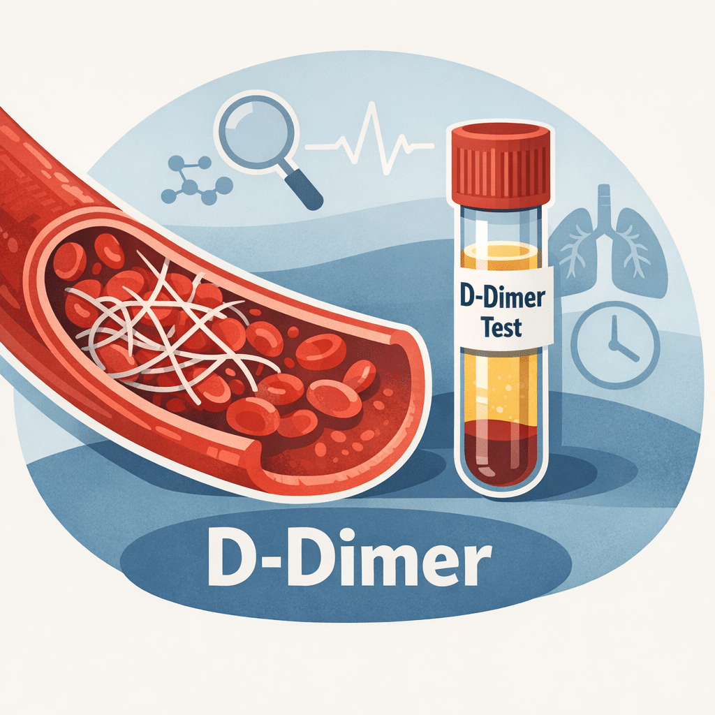
A D-dimer test measures a small protein fragment that appears in your blood when a clot breaks down. Doctors order it most often to help rule out a dangerous clot, such as a deep vein thrombosis (DVT) or a pulmonary embolism (PE), when combined with a clinical risk assessment. This guide explains what the test measures, how to read your number against the reference range, why age-adjusted cutoffs matter, and which other conditions can raise this marker even without a clot. You will also find a plain-language summary of the latest research, a glossary, and clear guidance on when a result deserves prompt medical attention.
What is a D-dimer test?
D-dimer is a fragment left over after your body breaks down a blood clot. It does not exist in meaningful amounts in healthy blood unless clotting and clot breakdown are actively happening somewhere in your body. When a clot forms, the protein fibrin builds a mesh that holds it together. Once the injury heals, an enzyme called plasmin dissolves that mesh, and the debris includes D-dimer fragments that circulate until your kidneys and liver clear them.
A D-dimer test picks up those fragments in a standard blood sample. Because the fragment only appears when a clot is being made and broken down, a raised level tells a clinician that clotting activity is happening somewhere. It does not say where, and it does not say why — those answers require more testing.
Why the test matters in everyday practice
The main strength of this marker is what a normal result rules out, not what a high result confirms. When a doctor already judges a clot unlikely based on your symptoms and history, a normal D-dimer helps exclude a DVT or PE without further imaging. This combination — clinical judgment plus a blood test — is what makes the D-dimer so useful in emergency and outpatient settings, sparing many people an unnecessary CT scan or ultrasound.
D-dimeerin testitulosten lukeminen
Laboratorioraporttisi sisältää luvun, yksikön ja viitevälin. Tyypillinen tulos näyttää tältä: D-dimeeri 320 ng/mL FEU (viiteväli: alle 500 ng/mL FEU). FEU tarkoittaa fibrinogeenivastaavia yksiköitä (fibrinogen equivalent units) – yksi kahdesta yleisestä tavasta, joilla laboratoriot ilmoittavat tämän testin tuloksen. Jotkut laboratoriot käyttävät sen sijaan D-dimeeriyksiköitä (DDU), joilla on eri numeerinen raja-arvo. Jos vertaat tulostasi muualta löytämääsi tietoon, tarkista aina, mitä yksikköä oma laboratoriosi on käyttänyt.
Most adult laboratories set a standard cutoff around 500 ng/mL FEU. A result below that threshold is usually called negative, meaning clot activity was not detected at a meaningful level. A result at or above the threshold is called positive, which prompts your doctor to look further rather than to diagnose a clot outright.
Why age changes the picture
D-dimer naturally climbs with age, even in people who feel completely well. Using the same flat cutoff for a 30-year-old and an 80-year-old means older adults are flagged “positive” far more often without actually having a clot. To correct for this, many labs and emergency departments now use an age-adjusted cutoff for patients over 50: multiply your age in years by 10 to get your personal cutoff in ng/mL FEU. A 70-year-old, for example, would have a cutoff around 700 ng/mL instead of the standard 500.
This adjustment has been studied in large groups of patients and is now supported by several professional societies, though it has not been formally approved by the FDA as a labeling change. Its purpose is narrow and specific: it improves how well a normal result rules out a clot in older adults, without meaningfully missing real clots. It is not a general “normal range for your age” chart — it only applies to interpreting a result that is being used to investigate a possible DVT or PE.
A simple decision flow for reading your number
Use this sequence as a starting point, not a substitute for medical advice.
| Step | What to check |
|---|---|
| 1. Find your unit | Confirm whether your lab reports in ng/mL FEU or DDU, printed next to your result |
| 2. Compare to the printed range | Use the reference range on your own report, not a number from another source |
| 3. Check your age | If you are over 50, ask whether an age-adjusted cutoff (age x 10 ng/mL) applies |
| 4. Note recent context | Recent surgery, pregnancy, infection, or a long illness can raise the result on their own |
| 5. Discuss with your doctor | Only a clinician can combine this result with your symptoms and risk factors |
Why D-dimer is sensitive but not specific
A test is sensitive when it reliably picks up a condition when present, and specific when it reliably stays normal when the condition is absent. D-dimer is a textbook example of a highly sensitive but poorly specific marker: it almost always rises when a significant clot is forming or dissolving, but plenty of other situations raise it too, so a positive result alone proves little.
Conditions and situations known to raise D-dimer without any dangerous clot include pregnancy, recent surgery or trauma, active infection or sepsis, cancer, liver disease, and simply being older. Intense physical exertion, such as running a marathon, can also cause a temporary rise that settles within a day or two. This is precisely why doctors combine the test with a structured risk assessment — such as the Wells score, which weighs factors like recent immobility, prior clots, and specific symptoms — rather than acting on the number in isolation.
Conditions linked to a high D-dimer
A raised D-dimer is a signal to investigate further, not a diagnosis. The sections below cover the situations doctors consider most often.
Syvä laskimotukos (DVT)
DVT is a clot that forms in a deep vein, most often in the calf or thigh. As your body attempts to break the clot down, D-dimer fragments enter your bloodstream and raise your test result. Typical symptoms include swelling, pain, warmth, or redness in one leg. A leg ultrasound remains the standard way to confirm or rule out DVT once suspicion is raised.
Keuhkoembolia (PE)
A pulmonary embolism happens when a clot — usually one that started as a DVT — breaks loose and lodges in an artery in the lung. Sudden shortness of breath, sharp chest pain that worsens with breathing, and a fast heart rate are common warning signs. Because a PE can be life-threatening, doctors often move quickly from a positive D-dimer to imaging with a CT pulmonary angiogram. Our detailed guide on pulmonary embolism symptoms and treatment covers the full diagnostic and treatment pathway.
Disseminoitunut intravaskulaarinen koagulaatio (DIC)
DIC is a rare, serious condition in which the clotting system activates throughout the body at once, forming many small clots that consume clotting factors faster than the body can replace them. This paradoxically raises the risk of both clotting and bleeding at the same time. D-dimer levels in DIC are usually very high, and the test is used alongside a platelet count and other clotting values to confirm the diagnosis and track treatment response.
Infection, inflammation, and cancer
Any process that activates inflammation tends to nudge clotting activity upward, which is why severe infections, chronic inflammatory conditions, and some cancers can all produce an elevated D-dimer without a clot ever forming somewhere dangerous. In these situations, a doctor typically reviews the D-dimer together with other inflammation markers, checking your C-reactive protein levels erottamaan tavallinen tulehdus todellisesta hyytymisongelmasta.
Mitä normaali D-dimeeri tarkoittaa
Normaali D-dimeeri on aidosti rauhoittava löydös, kun lääkäri arvioi veritulpan riskin jo valmiiksi matalaksi tai kohtalaiseksi. Tässä tilanteessa testillä on erittäin korkea negatiivinen ennustearvo – se on erinomainen vahvistamaan, että syvä laskimotukos tai keuhkoembolia on epätodennäköinen, ja säästää usein sinut TT-kuvaukselta tai ultraäänitutkimukselta. Tämä on testin ylivoimaisesti tärkein käyttötarkoitus jokapäiväisessä lääketieteessä.
Muutama asia on hyvä tietää. Testi voi toisinaan antaa normaalin tuloksen hyvin varhain tukoksen alussa, ennen kuin hajoamistuotteita on kertynyt riittävästi tason nostamiseksi. Se ei myöskään sulje pois kaikkia verisuoniongelmia, ja jos lääkärillä on vahva kliininen epäily normaalista tuloksesta huolimatta, lisätutkimuksia voidaan silti tehdä. Ennen testiä otetut verenohennuslääkkeet voivat myös antaa virheellisesti normaalin tuloksen, joten on tärkeää kertoa hoitotiimillesi kaikesta antikoagulanttien käytöstä.
Hyytymistutkimuspaneeli: D-dimeerin rooli
D-dimeeri esiintyy harvoin yksinään laboratoriovastauksessa. Se on yleensä osa laajempaa hyytymistutkimuspaneelia, johon kuuluvat myös PT, PTT ja INR, joista kukin mittaa eri osaa hyytymisprosessista. Alla oleva taulukko näyttää, miten D-dimeeri vertautuu muihin yleisiin hyytymistesteihin.
| Testata | Mitä se mittaa | Tavallinen käyttötarkoitus |
|---|---|---|
| D-dimeeri | Hyytymän hajoamisesta jäävä fragmentti | Auttaa sulkemaan pois syvän laskimotukoksen tai keuhkoembolian, kun riski on matala |
| PT / INR | Ulkoisen hyytymisreitin nopeus | Varfariinihoidon seuranta, maksan toiminnan arviointi |
| PTT / aPTT | Sisäisen hyytymisreitin nopeus | Hepariinihoidon seuranta, verenvuotohäiriöiden tutkiminen |
| Fibrinogeeni | Proteiini, joka muodostaa hyytymän fibriiniverkon | Arvioidaan yhdessä D-dimeerin kanssa DIC:ssä |
Kaksi luonnollista antikoagulanttia, jotka pitävät hyytymisen kurissa, mitataan erikseen, kun epäillään perinnöllistä hyytymishäiriötä: Proteiini C ja Proteiini S. Lääkärit tarkastelevat myös aPTT-tuloksia tarkemmin kun verenvuoto- tai hyytymistendenssi vaatii lähempää tarkastelua, ja selittämättömät tukokset johtavat joskus antitrombiini III:n puutoksen testaamiseen.
Erityistilanteet, jotka nostavat D-dimeeriä
Raskaus
Raskaus lisää luonnostaan hyytymisaktiviteettia valmistaen kehoa synnytykseen, joten D-dimeerin taso nousee tasaisesti joka raskauskolmanneksella ja on tyypillisesti selvästi yli tavallisen aikuisviiterajan kolmannella kolmanneksella. Tämän vuoksi lääkärit eivät käytä yleistä viitearvoa arvioidessaan raskaana olevan potilaan tulosta; epäiltyä tukosta raskauden aikana tutkitaan yleensä kuvantamisella eikä pelkällä D-dimeeriarvolla. Jos haluat laajemman kuvan raskaudenaikaisista laboratoriotutkimuksista, oppaamme kattaa koko joukon verikokeet raskauden aikana, including which markers shift and why.
Recent surgery or hospitalization
Any operation triggers tissue repair and clotting activity, so it is common and expected for D-dimer to rise for one to several weeks after surgery. Surgical teams already factor this expected pattern into how they read your pre-surgical blood work, so an elevated result soon after an operation rarely triggers alarm on its own.
Older age
As covered above, aging alone raises baseline D-dimer, which is exactly why the age-adjusted cutoff exists for patients over 50.
Milloin mennä lääkäriin
Most abnormal D-dimer results are followed up calmly with your regular doctor rather than as an emergency. However, certain combinations of symptoms need prompt attention regardless of your lab number.
Seek urgent or emergency care if you have:
- Sudden shortness of breath, chest pain that worsens with breathing, or coughing up blood.
- Swelling, pain, warmth, or redness in one leg or arm that appears suddenly.
- A fast heart rate with dizziness or fainting alongside any of the symptoms above.
Contact your doctor to discuss your result, without needing emergency care, if you have:
- A mildly elevated D-dimer with no symptoms and a known recent cause, such as surgery or pregnancy.
- A moderately elevated result that your doctor wants to monitor with a repeat test in a few weeks.
- Questions about how a medication or supplement might be affecting your result.
Uusimmat tieteelliset edistysaskeleet
Research on D-dimer interpretation has moved quickly in the past few years, largely refining how clinicians use adjusted cutoffs rather than changing what the test itself measures. A 2023 systematic review and meta-analysis pooling 68 studies and more than 140,000 patients compared several ways of adjusting the D-dimer cutoff — by age, by pretest clinical probability, and using a combined algorithm called YEARS. All of the adjustment strategies kept the test’s ability to catch real clots essentially unchanged while meaningfully improving how well a normal result ruled out a clot, though the strategies varied in how consistent their performance was across different patient groups. What this means for you: age-adjusted and probability-adjusted cutoffs appear safe and are not just a theoretical idea — they are backed by very large combined patient data (Gerber et al., Journal of Internal Medicine, 2023; DOI-koodi).
A 2024 narrative review focused specifically on comparing the age-adjusted cutoff against the clinical-probability-adapted cutoff, the two most widely used refinements. The review found that the age-adjusted approach tends to be the more cautious of the two, missing very few real clots but ruling out somewhat fewer people as clot-free, while the clinical-probability approach rules out more people but carries a slightly higher chance of missing a case. What this means for you: neither method is universally “better” — your emergency department’s choice of cutoff reflects a considered trade-off between safety and avoiding unnecessary scans, not an arbitrary shortcut (Righini et al., Journal of Clinical Medicine, 2024).
A 2025 secondary analysis of the landmark ADJUST-PE study tested whether the age-adjusted cutoff works consistently across different D-dimer laboratory assays, since hospitals do not all use the same testing equipment. Repeating the original test on stored samples with several alternative assays found that two of the four alternative assays performed similarly safely to the original method, while two others classified more patients as low-risk in a way that would have missed a small number of real clots. What this means for you: the specific assay your lab uses matters, and this is an early finding, still being confirmed, that explains why hospitals validate their own equipment before adopting an age-adjusted cutoff rather than assuming all D-dimer tests behave identically (Robert-Ebadi et al., Journal of Thrombosis and Haemostasis, 2025).
Beyond cutoff refinement, a 2024 review in the journal Haematologica examined how hematologists should approach otherwise healthy outpatients found to have an unexplained high D-dimer with no signs of a clot — an increasingly common scenario as the test is ordered more widely. The review emphasized that an isolated high D-dimer in someone without symptoms rarely needs aggressive investigation and should prompt a structured but measured work-up rather than alarm, taking into account age, medications, and any subtle risk factors for cancer or an underlying clotting tendency. What this means for you: if you are told your D-dimer is “a bit high” with no other findings, current expert guidance leans toward calm, stepwise follow-up rather than immediate invasive testing (Franchini et al., Haematologica, 2024).
Sanasto
| Termi | Määritelmä |
|---|---|
| Age-adjusted cutoff | A personalized D-dimer threshold for patients over 50, calculated as age multiplied by 10 ng/mL |
| D-dimeeri | A protein fragment released into the blood when a clot is broken down |
| Syvä laskimotukos (DVT) | A blood clot that forms in a deep vein, most often in the leg |
| Disseminoitunut intravaskulaarinen koagulaatio (DIC) | A serious condition where clotting and bleeding happen throughout the body at once |
| Fibriini | A protein that forms the mesh structure of a blood clot |
| FEU (fibrinogen equivalent units) | One of two common reporting units for D-dimer results |
| Negative predictive value | How reliably a normal test result rules out the condition being investigated |
| Keuhkoembolia (PE) | A blood clot that blocks an artery in the lung, usually after traveling from a leg vein |
| Sensitivity and specificity | How reliably a test detects a condition when present (sensitivity) and stays normal when absent (specificity) |
| Wellsin pisteytysjärjestelmä | A clinical scoring tool that estimates the probability of a DVT or PE before testing |
Usein kysyttyjä kysymyksiä D-dimeeritestistä
Can stress or intense exercise raise D-dimer levels?
Ordinary psychological stress does not typically raise D-dimer to a meaningful degree. Very intense physical exertion, such as running a marathon or another endurance event, can cause a temporary rise due to increased clotting and breakdown activity in the muscles. This effect usually settles back to baseline within 24 to 48 hours, so a recent hard workout is worth mentioning to your doctor if your test was scheduled soon afterward.
Vaikuttaako hormonaalinen ehkäisy D-dimeeritasoihin?
Kyllä. Sekä estrogeenia että progestiinia sisältävät yhdistelmähormonaaliset ehkäisymenetelmät voivat nostaa D-dimeeriarvoa hieman, koska estrogeeni vaikuttaa veren hyytymiseen. Vaikutus on yleensä lievä eikä kovin merkittävä. Pelkkää progestiinia sisältävillä menetelmillä on yleensä vain vähäinen tai olematon vaikutus mitattavaan arvoon. Jos käytät hormonaalista ehkäisyä, mainitse siitä lääkärille tai muulle terveydenhuollon ammattilaiselle, joka määrää tai tulkitsee testiäsi.
Mitkä lääkkeet voivat vaikuttaa D-dimeerin testituloksiin?
Anticoagulant medications, commonly called blood thinners, are often prescribed precisely because of a high D-dimer finding, but they can also cause a falsely normal result if the test is repeated while you are already treated. Fibrinolytic drugs, which actively dissolve clots in emergency settings, cause a sharp temporary surge as they work. Always give your care team a complete list of medications and supplements before this test.
Why would a doctor order a D-dimer test for chest pain?
Symptoms of a pulmonary embolism, including sudden breathlessness and chest pain, can closely resemble a heart problem. Ordering a D-dimer test alongside cardiac markers helps a doctor quickly distinguish between the two possibilities. A normal result, combined with a low clinical probability, makes a PE less likely and allows the workup to focus on other causes of chest discomfort more efficiently.
Voiko keuhkoemboliaa saada, jos D-dimeeri on normaali?
Tämä on harvinaista mutta mahdollista, erityisesti hyvin pienen tai pitkäaikaisen (kroonisen) hyytymän kohdalla, joka ei aktiivisesti hajoa testaushetkellä. Näin voi käydä myös, jos käytät jo antikoagulanttia, joka vaimentaa tulosta. Juuri siksi lääkärin kliininen arvio kulkee aina luvun rinnalla – jos epäily pysyy korkeana normaalista testituloksesta huolimatta, kuvantamistutkimus järjestetään tyypillisesti silti.
Voivatko D-dimeerin tulokset vaihdella eri laboratorioiden välillä?
Kyllä, merkittävästikin. Eri laboratoriot käyttävät erilaisia testausmenetelmiä ja voivat ilmoittaa tulokset eri yksiköissä, minkä vuoksi sekä luku että raportissasi näkyvä viiteväli voivat vaihdella laboratoriosta toiseen. Tulkitse tuloksesi aina sen laboratorion ilmoittaman viitevälin perusteella, joka teki testin. Jos tuloksiasi verrataan eri käyntikerroilta eri paikoissa, mainitse käytetty menetelmä tai yksikkö.
Lähteet
- D-dimeeri-testi — MedlinePlus, U.S. National Library of Medicine
- D-dimeeri-testi: mitä se on, mihin sitä käytetään, riskit ja tulokset — Cleveland Clinic
- Syvä laskimotukos (DVT): diagnoosi ja hoito, mukaan lukien D-dimeerin verikoe — Mayo Clinic
- Gerber ym., Utility and limitations of patient-adjusted D-dimer cut-off levels for diagnosis of venous thromboembolism — a systematic review and meta-analysis, Journal of Internal Medicine, 2023 (PubMed PMID 37143392) — DOI-koodi
- Righini, Robert-Ebadi ja Le Gal, Age-Adjusted and Clinical Probability Adapted D-Dimer Cutoffs to Rule Out Pulmonary Embolism: A Narrative Review of Clinical Trials, Journal of Clinical Medicine, 2024 (PubMed PMID 38929970) — DOI-koodi
- Robert-Ebadi ym., Different D-dimer assays with age-adjusted cutoffs to exclude pulmonary embolism: secondary analysis of ADJUST-PE study, Journal of Thrombosis and Haemostasis, 2025 (PubMed PMID 40252844) — DOI-koodi
- Franchini, Focosi, Pezzo ja Mannucci, How we manage a high D-dimer, Haematologica, 2024 (PubMed PMID 37881856) — DOI-koodi
Lisälukemista
- Coagulation panel: PT, PTT, INR and D-dimer explained
- Keuhkoembolia: oireet, diagnoosi ja hoito
- aPTT-verikoe: tasojesi tulkinta
- Verikokeen tulosten lukeminen
- Verimerkkiaineet: kattava sanasto
Hyytymistulokset kertovat harvoin koko tarinan yksinään, ja D-dimeerin on helpoin ymmärtää muun laboratorioraporttisi yhteydessä. AI DiagMe auttaa sinua hahmottamaan liittyvät arvot – kuten verihiutalemäärän, fibrinogeenin ja PT/INR:n – selkokielellä ja näyttää, miten ne liittyvät D-dimeerin tulokseesi. Se on suunniteltu auttamaan sinua ymmärtämään laboratoriotuloksesi – ei diagnosoimaan sinua tai korvaamaan lääkäriäsi.
Ymmärrä laboratoriotuloksiasi tekoälyn DiagMen avulla
Tulkitse tuloksesi muutamassa minuutissa