

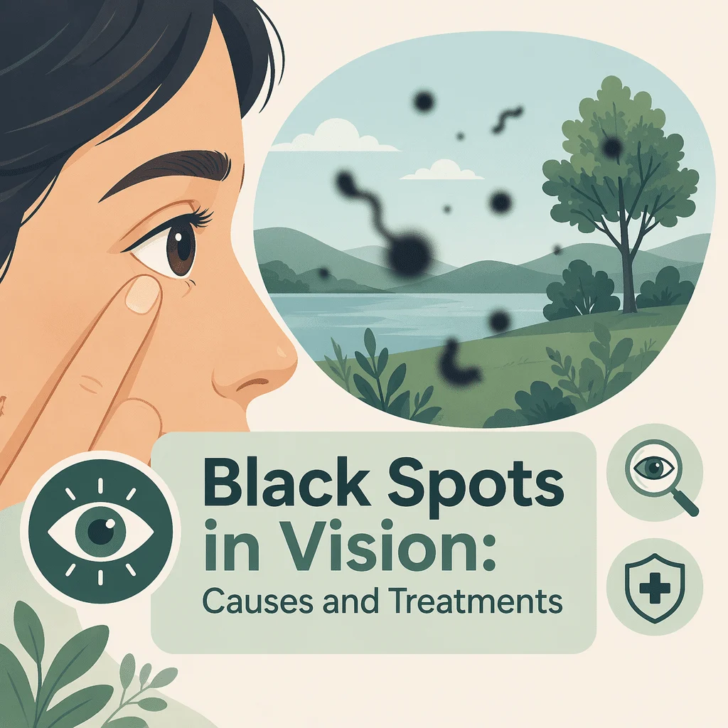
Black spots in vision are one of the most common reasons people ask for an urgent eye appointment, and in most cases they turn out to be harmless. These dark specks, threads and cobweb shapes drift across your sight, dart away when you try to look straight at them, and stand out most against a bright wall or a clear sky. They are usually shadows cast by tiny clumps floating inside the jelly that fills the eye.
A small but important minority, however, signal a tear in the retina that needs treating within hours rather than weeks. In this article you will learn what causes these spots, which patterns count as an emergency, how eye doctors examine you, which treatments genuinely work, and how conditions such as diabetes and high blood pressure change the picture.
When black spots in vision are an emergency
Read this section first. Most people who notice black spots in vision need reassurance, not surgery. But three patterns mean you should be examined the same day, ideally within a few hours, because they can indicate a retinal tear or a detaching retina.
- A sudden shower of many new spots, arriving over minutes or hours rather than months.
- Flashes of light, often like a camera flash or a lightning streak at the edge of your sight, especially in a dark room.
- A curtain, shadow or dark veil moving across part of your field of view, or a patch of vision that has simply gone missing.
None of these is usually painful, which is exactly why they get ignored: a detaching retina rarely hurts. If you have any of them, contact an ophthalmologist, an eye emergency department or your eye doctor straight away and say you have new flashes and floaters. That phrase is understood everywhere and will get you triaged quickly. The National Eye Institute gives the same advice.
Harmless pattern or urgent pattern
The table below separates the everyday, age-related version from the patterns that need urgent attention. It is a guide to how quickly to act, not a way to diagnose yourself.
| What you see | How fast it came on | What it usually means | What to do and how quickly |
|---|---|---|---|
| One or two grey specks or threads, stable for months or years | Gradual, over months | Ordinary age-related clumping in the vitreous jelly | Mention it at your next routine eye exam |
| A single large ring or circle that appeared over a few days, with mild flashes | Days | Often a posterior vitreous detachment, the jelly peeling away from the retina | Dilated eye exam within about a week |
| A sudden burst of dozens or hundreds of new spots, like soot or pepper | Minutes to hours | Possible retinal tear or bleeding inside the eye | Same-day emergency examination |
| Repeated flashes plus a curtain or shadow creeping in from one side | Hours | Possible retinal detachment | Emergency, same day, do not wait overnight |
| A shimmering zigzag arc that grows and fades within an hour, both eyes | Builds over 20 to 30 minutes, then clears | More typical of migraine aura than of a retinal problem | Routine appointment, urgent if it is your first episode |
What black spots in vision actually are
The eye is filled with a clear gel called the vitreous, which gives the eyeball its shape. In youth this gel is firm and evenly transparent. With age it slowly liquefies, and the fine collagen fibres inside it stick together into strands and knots. Light entering the eye passes around these clumps and casts a shadow on the retina, the light-sensitive film at the back of the eye. Your brain interprets that shadow as a dark shape floating in front of you.
Why they drift when you move your eyes
Because the clumps float freely in a gel, they lag behind your eye movements and glide onward when your eye stops. That is why they seem to swim away whenever you try to look straight at them.
Common causes of black spots in vision
Age-related vitreous changes and posterior vitreous detachment
This is by far the most frequent explanation. As the gel shrinks it eventually peels away from the retina, an event called a posterior vitreous detachment. It typically happens between the ages of 50 and 70, earlier in short-sighted eyes, and often produces one prominent ring-shaped spot along with a burst of brief flashes.
Retinal tear and retinal detachment
As the gel separates, it can pull hard enough on a weak point in the retina to tear it. Fluid then seeps through and lifts the retina off the back wall of the eye, which is a detachment. Untreated, this permanently destroys sight in that area. A tear caught early is usually sealed in a short outpatient laser session, which is why the same-day rule matters so much.
Bleeding inside the eye
If a tearing retina rips a small blood vessel, red blood cells spill into the vitreous and are seen as a sudden cloud of tiny dark dots, often described as soot, smoke or a swarm of insects. Bleeding can also come from fragile abnormal vessels caused by advanced diabetic eye disease.
Inflammation inside the eye
Uveitis, inflammation of the middle layer of the eye, releases white blood cells and debris into the vitreous. It tends to cause spots alongside light sensitivity, redness, aching or blurred vision, and needs treatment rather than watchful waiting.
Migraine aura and other look-alikes
Not every visual disturbance is a floater. Migraine aura produces a shimmering, expanding zigzag or a blind spot with jagged bright edges, usually affecting both eyes, building over twenty to thirty minutes and then clearing. It moves with your gaze rather than drifting independently. Our library covers migraine symptoms, causes and treatments. A brief greying-out of vision in one eye that resolves within minutes is different again and should be assessed promptly, because it can reflect a circulation problem.
Health conditions that change the risk
Diabetes and blood sugar control
Long-term raised blood sugar damages the small vessels feeding the retina, a condition called diabetic retinopathy. In its advanced form the eye grows fragile new vessels that bleed easily, and that bleeding is felt as a sudden storm of black spots in vision. Good glucose control lowers the risk substantially, which is why regular monitoring matters. We cover the HbA1c blood test and its target levels, and you can also read our complete guide to the fasting blood glucose test. Our team explains the full range of diabetes symptoms and tests as well.
High blood pressure
Sustained hypertension narrows and stiffens retinal arteries and can cause small haemorrhages at the back of the eye. It rarely produces floaters alone, but it compounds the damage in someone who also has diabetes, and a hypertensive crisis can cause abrupt visual change. We also describe the warning signs of high blood pressure.
Short-sightedness, eye surgery and injury
A short-sighted eye is longer than average, so the retina is stretched thinner and the gel separates earlier, raising the lifetime risk of tears. Cataract surgery slightly increases that risk in the year that follows. A blow to the eye or head can also shake the gel loose and trigger sudden spots, and warrants an examination even if vision seems normal.
Anaemia and general circulation
Low haemoglobin does not create floaters, but it can cause the greyness, visual dimming and spots-before-the-eyes sensation that many people describe in the same words. Distinguishing the two matters, because the treatments are entirely different. We explain the symptoms and blood tests used for anaemia, and our guide details what a hemoglobin result means.
How eye doctors evaluate black spots in vision
The dilated eye exam
The essential test is a dilated examination. Drops widen the pupil for a few hours, then the doctor inspects the whole retina, including the far edges where tears usually hide. This involves gently pressing on the outside of the eyelid to bring the extreme periphery into view. It feels odd but is not painful, and it finds tears that a photograph or a quick slit-lamp look can miss. Expect blurred near vision and light sensitivity for several hours, so arrange not to drive home.
Imaging and ultrasound
Optical coherence tomography, a scan that builds a cross-section of the retina using light, shows whether the gel is still attached. If a dense haemorrhage blocks the view, an ultrasound scan through the closed eyelid can reveal a detachment hiding behind the blood.
Blood tests that support the picture
Blood work does not diagnose a floater, but it explains the background risk and is often ordered alongside an eye assessment. A glucose and HbA1c pair shows whether diabetes is present or poorly controlled, a lipid profile maps cardiovascular risk, and a blood count picks up anaemia or infection. We break down the lipid panel and its cholesterol numbers, we describe the complete blood count and how to read it, and you can also read our guide to diabetes blood tests.
Treatment options and what to expect
Watchful waiting, the usual answer
For ordinary age-related spots, doing nothing is the recommended approach, and it works better than most people expect. The clumps sink slowly out of the central line of sight, and the brain progressively suppresses the shadow, a process called neural adaptation. Within three to six months most people report that the spots no longer intrude.
Laser sealing of a retinal tear
If a tear is found, laser photocoagulation places a ring of tiny burns around it. These scar down over a couple of weeks and weld the retina to the wall behind it, stopping fluid getting underneath. It is done in clinic under anaesthetic drops, takes minutes, and is highly effective at preventing a detachment. This treats the tear, not the spots themselves.
Laser vitreolysis
A different laser technique aims at the clumps themselves, breaking large ones into fragments too small to cast a noticeable shadow. It suits a single well-defined clump sitting clear of the retina and lens, and is unsuitable for diffuse cobweb-like floaters. Reported results are mixed, and it is not offered everywhere.
Vitrectomy
Vitrectomy removes the vitreous gel altogether and replaces it with a clear fluid. It is the most reliable way to eliminate severe floaters, and satisfaction among carefully selected patients is high. It is also real surgery inside the eye, carrying a risk of cataract, retinal detachment and, rarely, infection, so it is reserved for people whose spots seriously interfere with reading, driving or work despite months of waiting.
Living with floaters and protecting your vision
If your examination was clear, the most useful habit is simple self-monitoring. Once a week, cover one eye and look at a plain light-coloured wall, then swap eyes. You are checking for a change in the number of spots, any new flashing, or a missing patch. Because both eyes normally compensate for each other, a problem in one eye can go unnoticed for weeks unless you test them separately.
Beyond that, keep routine dilated eye exams, particularly if you are short-sighted, diabetic or over fifty, and manage blood pressure and blood sugar, since both drive the retinal disease behind the dangerous kind of bleeding. Wear eye protection for sport and DIY. Our team explains high cholesterol causes and treatment for the wider vascular picture.
Latest scientific advances
Research published over the past three years has sharpened the advice around floaters. Here is what it found and, more importantly, what it changes for you.
How often new floaters really mean a tear
A prospective study of just over a thousand people who came to clinic with new flashes and floaters found that roughly one in ten already had a retinal tear or detachment at the first examination, and a further small group developed one over the next two months. Prospective means the researchers followed people forward in time rather than reviewing old records, which makes the figure more trustworthy. For you this cuts two ways: the odds are strongly in your favour, so this is a reason to be seen rather than to panic, but one examination is not always the end of the story. If your symptoms change after a normal check, go back.
Not every tear announces itself the same way
The same study found that a sizeable minority of tears were visible only with the indented peripheral technique, and that the ring-shaped floater doctors look for was absent in about a third of eyes that had a tear. This argues for insisting on a proper dilated examination of the retinal edges rather than a quick look alone.
Imaging helps, but it does not replace the exam
Researchers comparing slit-lamp examination, ultrasound and retinal scanning against what surgeons actually found during operations reported that each method missed a fair number of cases on its own, while rarely raising false alarms. In plain terms, a normal scan does not guarantee the gel is still attached. Good clinics therefore combine methods and review you again if doubt remains.
Where the treatments stand
A recent review of floater management concluded that observation remains the default, that removing the gel surgically is the most definitive option but carries genuine risks including cataract and retinal detachment, and that laser vitreolysis is a less invasive alternative with variable success. A separate overview noted a persistent difficulty: there is still no agreed way to measure objectively how bad someone’s floaters are, which makes treatments hard to compare.
A small 2025 study of laser vitreolysis in thirty eyes reported reduced floaters with no detected harm to retinal function after one month. Thirty eyes and one month is a very small, very short study, so treat it as encouraging rather than settled. For you, this means it is fair to ask a clinic offering laser treatment how many clumps you have, whether they sit safely clear of the lens and retina, and what happens if it does not work.
Surgery on the eye slightly raises the risk
An analysis of a large American cataract-surgery registry found a detachment in roughly one in five hundred eyes within a year, with the risk concentrated in younger patients, in men, and above all in eyes with lattice degeneration, a thinning at the retinal edge visible on examination. The practical point is that new flashes or a shower of black spots in vision after cataract surgery deserve the same urgent response as at any other time.
Automated screening for diabetic eye disease
A meta-analysis, which combines many separate studies for a more reliable overall answer, pooled twenty-one prospective trials and found that software reading retinal photographs identified diabetic retinopathy correctly in the large majority of cases. The consequence for you is that automated screening is spreading fast, making an annual retinal photograph easier to obtain if you have diabetes. It remains a filter, and you still need an ophthalmologist when new symptoms appear.
Glossary
| Term | Definition |
|---|---|
| Vitreous | The clear gel that fills the main cavity of the eye and holds its shape. It liquefies gradually with age. |
| Floater | A speck, thread or ring you see drifting across your sight. It is the shadow of a clump inside the vitreous, not a mark on the eye surface. |
| Retina | The light-sensitive layer lining the back of the eye. It converts light into the signals sent to the brain. |
| Posterior vitreous detachment (PVD) | The normal age-related separation of the vitreous gel from the retina. It commonly causes a sudden new floater and brief flashes. |
| Retinal tear | A break in the retina, usually caused by the gel tugging on it. Sealed early with laser, it rarely progresses further. |
| Retinal detachment | Fluid passing through a tear and lifting the retina off the back of the eye. It is a surgical emergency and can cause permanent sight loss. |
| Vitrectomy | An operation that removes the vitreous gel and replaces it with a clear fluid. It is the most definitive treatment for severe floaters. |
| Laser vitreolysis | A clinic laser procedure that breaks a large floater into fragments too small to notice. It suits only selected, well-defined clumps. |
| Optical coherence tomography (OCT) | A quick, painless scan that uses light to build a cross-section image of the retina and the gel in front of it. |
| Diabetic retinopathy | Damage to the small retinal blood vessels caused by long-term high blood sugar. Advanced stages can bleed into the vitreous. |
Frequently asked questions
Why am I suddenly seeing black spots in my vision?
A sudden change almost always reflects something happening in the vitreous gel. The most common event is a posterior vitreous detachment, where the gel peels away from the retina and its edge casts a new shadow. That usually produces one prominent floater, sometimes with brief flashes. Less often, the separating gel tears the retina or catches a blood vessel, releasing a cloud of tiny dots. Because you cannot tell these apart yourself, any sudden onset should be examined. If the spots arrived as a shower, or came with flashes or a shadow at the edge of your sight, treat it as a same-day emergency.
Do black spots in vision ever go away on their own?
Often, yes, in the sense that matters. The clumps themselves rarely disappear, but they sink out of the direct line of sight over weeks, and the brain steadily learns to ignore the shadow. Most people find that spots which felt intolerable at first have faded into the background within three to six months. If a floater is still disrupting reading or driving after that period, it is reasonable to ask an ophthalmologist about the options. Spots that are increasing in number rather than settling should be re-examined rather than waited out.
Can high blood sugar cause black spots in vision?
Indirectly, yes. Years of raised blood sugar damage the tiny vessels supplying the retina. In advanced diabetic retinopathy the eye grows fragile replacement vessels that leak and bleed, and blood in the gel is seen as a sudden swarm of dark dots. Short-term blood sugar swings can also blur vision by changing the shape of the lens, though that causes haziness rather than distinct spots. If you have diabetes and notice new spots, get examined promptly and have your control reviewed, since treatment of the retinopathy is far more effective when started early.
Are floaters in one eye more serious than in both?
New spots in a single eye deserve more attention, not less. Retinal tears and detachments affect one eye at a time, so a one-sided change fits that pattern. Symptoms that appear in both eyes simultaneously and resolve within an hour point more towards migraine aura, which originates in the brain rather than the eye. That said, age-related gel changes also start in one eye and often follow in the other a year or two later, so one-sided floaters are common and usually benign. The deciding factors remain how fast they appeared and whether flashes or a shadow came with them.
Why do black spots in vision come and go?
True floaters never entirely vanish. They drift out of your central sight and hide against busy backgrounds, then reappear when you look at a bright wall or a screen, which feels like coming and going. A dark patch that blocks part of your vision for several minutes and then clears completely is a different symptom: it can follow a migraine, or reflect a brief interruption of blood flow to the eye, and it deserves prompt assessment rather than watching. Spots that arrive with dizziness on standing point more towards blood pressure or circulation than the vitreous.
Will glasses, eye drops or supplements get rid of floaters?
No. Floaters sit inside the eye, so nothing applied to the surface reaches them, and no drop, vitamin or supplement has been shown to dissolve them. Glasses cannot correct them either, because the problem is a shadow rather than a focusing error. Eye exercises do not remove them, although moving your eyes up and down can temporarily shift a clump out of the central line of sight. The only interventions with real evidence are surgical removal of the gel and, in selected cases, laser treatment.
Sources
- National Eye Institute — Floaters — National Institutes of Health, 2025 — nei.nih.gov
- American Academy of Ophthalmology — What Are Floaters and Flashes? — 2025 — aao.org
- MedlinePlus Medical Encyclopedia — Retinal detachment — U.S. National Library of Medicine, 2025 — medlineplus.gov
- Mayo Clinic — Eye floaters: symptoms and causes — 2025 — mayoclinic.org
- Nixon TRW, Davie RL, Snead MP — Posterior vitreous detachment and retinal tear: a prospective study of community referrals — Eye (London), 2023 — doi.org/10.1038/s41433-023-02779-3
- Aleman AI, Kiryakoza L, Sridhar J, Sengillo J — Management of vitreous floaters: a review — Current Opinion in Ophthalmology, 2024 — doi.org/10.1097/ICU.0000000000001075
- Gill N, Singh A — Management of visually significant vitreous opacities — Missouri Medicine, 2024 — pmc.ncbi.nlm.nih.gov/articles/PMC11482840
- Liu J, Liu H, Zhang Y, et al. — Efficacy and safety of Nd:YAG laser vitreolysis in patients with physiological vitreous floaters — BMC Ophthalmology, 2025 — doi.org/10.1186/s12886-025-04311-5
- Zvorničanin J, Zvorničanin E, Popović M — Accuracy of biomicroscopy, ultrasonography and spectral-domain OCT in detection of complete posterior vitreous detachment — BMC Ophthalmology, 2023 — doi.org/10.1186/s12886-023-03233-4
- Morano MJ, Khan MA, Zhang Q, et al. — Incidence and risk factors for retinal detachment and retinal tear after cataract surgery: IRIS Registry analysis — Ophthalmology Science, 2023 — doi.org/10.1016/j.xops.2023.100314
- Wang Z, Li Z, Li K, et al. — Performance of artificial intelligence in diabetic retinopathy screening: a systematic review and meta-analysis of prospective studies — Frontiers in Endocrinology, 2023 — doi.org/10.3389/fendo.2023.1197783
Further reading
- Hypertension and headache: causes and warning signs
- The kidney function panel and how to read it
- The iron studies panel explained
- Low vitamin B12: symptoms, causes and treatments
- Normal thyroid levels and their ranges
Understand your lab results with AI DiagMe
Changes in your sight rarely stand alone. The conditions that put the retina at risk, above all diabetes and high blood pressure, show up in ordinary blood work long before they reach the eye, which is why an HbA1c and fasting glucose pair, a lipid panel, a complete blood count and a kidney function check are so often requested alongside an eye assessment. AI DiagMe reads those results back to you in plain language, so you arrive at your appointment knowing which numbers are outside their range and which questions to ask. It helps you understand your results, it does not diagnose you, and it does not replace your eye doctor or your physician.
Get your results interpreted in minutes