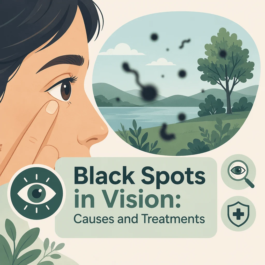
Svarta fläckar i synfältet är ett av de vanligaste skälen till att folk söker akut ögonvård, och i de flesta fall visar det sig vara ofarligt. Dessa mörka prickar, trådar och spindelvävsliknande former driver runt i ditt synfält, glider undan när du försöker titta direkt på dem och syns tydligast mot en ljus vägg eller en klar himmel. De är vanligtvis skuggor från små klumpar som flödar runt i den gelé som fyller ögat.
En liten men viktig andel av fallen signalerar dock en näthinnespricka som behöver behandlas inom timmar snarare än veckor. I den här artikeln får du lära dig vad som orsakar dessa fläckar, vilka mönster som räknas som ett akut tillstånd, hur ögonläkare undersöker dig, vilka behandlingar som verkligen fungerar, och hur sjukdomar som diabetes och högt blodtryck förändrar bilden.
När svarta fläckar i synen är ett nödläge
Läs det här avsnittet först. De flesta som märker svarta fläckar i synen behöver lugnas, inte opereras. Men tre mönster innebär att du bör undersökas samma dag, helst inom några timmar, eftersom de kan tyda på en näthinnespricka eller en lossnad näthinna.
- En plötslig skur av många nya fläckar som dyker upp på minuter eller timmar snarare än månader.
- Ljusblixtar, ofta som ett kameraflash eller en blixt i utkanten av synen, särskilt i ett mörkt rum.
- En ridå, skugga eller mörk slöja som rör sig över en del av ditt synfält, eller ett område i synen som helt enkelt har försvunnit.
Inget av dessa är vanligtvis smärtsamt, vilket är precis varför de ignoreras: en lossnad näthinna gör sällan ont. Om du har något av dem, kontakta en ögonläkare, en ögonakutmottagning eller din ögonläkare omedelbart och säg att du har nya ljusblixtar och flygande flugor. Den frasen förstås överallt och gör att du snabbt får rätt prioritet. National Eye Institute ger samma råd.
Ofarligt mönster eller akut mönster
Tabellen nedan skiljer den vardagliga, åldersrelaterade varianten från de mönster som kräver akut uppmärksamhet. Den är en vägledning för hur snabbt du bör agera, inte ett sätt att diagnostisera dig själv.
| Vad du ser | Hur snabbt det kom | Vad det vanligtvis betyder | Vad du ska göra och hur snabbt |
|---|---|---|---|
| En eller två grå fläckar eller trådar, stabila i månader eller år | Gradvis, under månader | Vanlig åldersrelaterad klumpning i glaskroppen | Ta upp det vid din nästa rutinmässiga ögonkontroll |
| En enstaka stor ring eller cirkel som uppstod under några dagar, med milda ljusblixtar | Dagar | Ofta en bakre glaskroppsavlossning, där geléet lossnar från näthinnan | Utvidgad ögonundersökning inom ungefär en vecka |
| En plötslig skur av dussintals eller hundratals nya fläckar, som sot eller peppar | Minuter till timmar | Möjlig näthinnespricka eller blödning inuti ögat | Akut undersökning samma dag |
| Upprepade ljusblixtar plus en ridå eller skugga som kryper in från ena sidan | Timmar | Möjlig näthinneavlossning | Akut, samma dag, vänta inte till nästa dag |
| En skimrande sicksackbåge som växer och försvinner inom en timme, i båda ögonen | Byggs upp under 20 till 30 minuter och klingar sedan av | Mer typiskt för migrän med aura än för ett näthinnaproblem | Vanlig tid, akut om det är ditt första anfall |
Vad svarta fläckar i synen egentligen är
Ögat är fyllt med ett klart gelé som kallas glaskroppen, vilket ger ögongloben dess form. I unga år är detta gelé fast och jämnt genomskinligt. Med åldern flytande det långsamt, och de fina kollagenfibrerna inuti det klumpar ihop sig till trådar och knutar. Ljus som träffar ögat passerar runt dessa klumpar och kastar en skugga på näthinnan, det ljuskänsliga skiktet längst bak i ögat. Din hjärna tolkar den skuggan som en mörk form som flyter framför dig.
Varför de rör sig när du rör på ögonen
Eftersom klumparna flödar fritt i en gel halkar de efter dina ögonrörelser och glider vidare när ögat stannar. Det är därför de verkar simma undan varje gång du försöker titta direkt på dem.
Vanliga orsaker till svarta fläckar i synfältet
Åldersrelaterade förändringar i glaskroppen och bakre glaskroppslösning
Detta är den överlägset vanligaste förklaringen. När glaskroppen krymper lossnar den till slut från näthinnan – ett tillstånd som kallas bakre glaskroppsavlossning. Det inträffar vanligtvis mellan 50 och 70 års ålder, tidigare hos personer med närsynthet, och ger ofta upphov till en tydlig ringformad fläck tillsammans med en serie korta ljusblixtar.
Näthinnebristning och näthinneavlossning
När gelen lossnar kan den dra tillräckligt hårt i en svag punkt på näthinnan för att orsaka en spricka. Vätska tränger då igenom och lyfter näthinnan från ögats bakvägg, vilket kallas näthinneavlossning. Utan behandling förstörs synen i det området permanent. En spricka som upptäcks tidigt kan vanligtvis tätas i en kort laserbehandling på öppenvård, vilket är anledningen till att regeln om samma dag är så viktig.
Blödning inuti ögat
Om en bristande näthinnan river av ett litet blodkärl spills röda blodkroppar ut i glaskroppen och upplevs som ett plötsligt moln av små mörka prickar – ofta beskrivna som sot, rök eller ett svärm av insekter. Blödning kan också uppstå från sköra, onormala blodkärl till följd av avancerad diabetisk ögonsjukdom.
Inflammation inuti ögat
Uveit, en inflammation i ögats mellersta lager, frigör vita blodkroppar och cellavfall i glaskroppen. Det brukar ge prickar i synfältet tillsammans med ljuskänslighet, rodnad, värk eller dimsyn, och kräver behandling snarare än avvaktande hållning.
Migrän med aura och liknande tillstånd
Inte alla synstörningar är glaskroppsflak. Migrän med aura ger ett flimrande, utvidgande sicksackmönster eller en blind fläck med taggiga, ljusa kanter – vanligtvis i båda ögonen – som byggs upp under tjugo till trettio minuter och sedan försvinner. Det rör sig med din blick i stället för att driva fritt. Vårt bibliotek tar upp migrän – symtom, orsaker och behandling. En kortvarig gråning av synen på ett öga som försvinner inom några minuter är något annat och bör bedömas snabbt, eftersom det kan tyda på ett cirkulationsproblem.
Hälsotillstånd som påverkar risken
Diabetes och blodsockerkontroll
Långvarigt förhöjt blodsocker skadar de små blodkärl som försörjer näthinnan – ett tillstånd som kallas diabetisk retinopati. I sitt avancerade stadium bildar ögat sköra nya blodkärl som lätt börjar blöda, och den blödningen upplevs som en plötslig storm av svarta prickar i synfältet. God blodsockerkontroll minskar risken avsevärt, vilket är anledningen till att regelbunden uppföljning är viktig. Vi tar upp HbA1c-blodprovet och dess målvärden, och du kan också läsa vår kompletta guide till fasteblodsocker-testet. Vårt team förklarar hela spektrumet av diabetessymtom och tester också.
Högt blodtryck
Långvarigt högt blodtryck trångar ihop och stelnar näthinneartärerna och kan orsaka små blödningar i ögonbotten. Det ger sällan glaskroppsflak ensamt, men förvärrar skadan hos den som även har diabetes, och en hypertensiv kris kan ge plötsliga synförändringar. Vi beskriver också varningstecknen på högt blodtryck.
Närsynthet, ögonkirurgi och skada
Ett närsyntöga är längre än genomsnittet, vilket gör att näthinnan sträcks ut tunnare och glaskroppen lossnar tidigare – det ökar den livslånga risken för näthinnehål. Kataraktoperation ökar den risken något under det efterföljande året. Ett slag mot ögat eller huvudet kan också skaka loss glaskroppen och utlösa plötsliga fläckar, och bör undersökas även om synen verkar normal.
Anemi och allmän cirkulation
Lågt hemoglobin skapar inte glaskroppsflak, men det kan ge en känsla av grå dimma, försämrad syn och prickar framför ögonen som många beskriver med samma ord. Att skilja de två åt är viktigt, eftersom behandlingarna är helt olika. Vi förklarar symtom och blodprover vid anemi, och vår guide beskriver i detalj vad ett hemoglobinvärde betyder.
Hur ögonläkare bedömer svarta fläckar i synen
Undersökning med vidgade pupiller
Det viktigaste undersökningsverktyget är en ögonbottenundersökning med vidgade pupiller. Ögondroppar vidgar pupillen under några timmar, varefter läkaren undersöker hela näthinnan – inklusive de yttersta kanterna där hål och bristningar brukar gömma sig. Det innebär att läkaren försiktigt trycker mot ögonlockets utsida för att få den yttersta periferin i blickfånget. Det känns lite konstigt men gör inte ont, och metoden hittar bristningar som ett fotografi eller en snabb undersökning i spaltlampa kan missa. Räkna med suddig närsyn och ljuskänslighet under flera timmar efteråt, så se till att du inte behöver köra hem.
Bilddiagnostik och ultraljud
Optisk koherenstomografi (OCT) är en skanning som bygger upp ett tvärsnitt av näthinnan med hjälp av ljus och visar om glaskroppen fortfarande sitter fast. Om en kraftig blödning skymmer sikten kan ett ultraljud genom det slutna ögonlocket avslöja en näthinneavlossning som döljer sig bakom blodet.
Blodprover som ger en helhetsbild
Blodprover diagnostiserar inte glaskroppsgrumlingar, men de förklarar den bakomliggande risken och beställs ofta tillsammans med en ögonundersökning. Ett glukos- och HbA1c-par visar om diabetes föreligger eller är dåligt kontrollerad, en lipidprofil kartlägger hjärt-kärlrisken och ett blodstatus fångar upp anemi eller infektion. Vi går igenom lipidpanelen och dess kolesterolvärden, vi beskriver fullständig blodstatus och hur du läser den, och du kan också läsa vår guide till blodprover vid diabetes.
Behandlingsalternativ och vad man kan förvänta sig
Avvaktande hållning – det vanligaste rådet
Vid vanliga åldersrelaterade fläckar är det rekommenderade tillvägagångssättet att inte göra något – och det fungerar bättre än de flesta tror. Klumparna sjunker långsamt ut ur det centrala synfältet, och hjärnan undertrycker gradvis skuggan i en process som kallas neural adaptation. Inom tre till sex månader brukar de flesta uppleva att fläckarna inte längre stör.
Laserbehandling av ett näthinnahål
Om ett hål hittas placerar laserkoagulation en ring av små brännskador runt det. Dessa läker ihop under ett par veckor och svetsar fast näthinnan mot väggen bakom, vilket hindrar vätska från att tränga in under den. Behandlingen utförs på mottagningen med bedövningsdroppar, tar bara några minuter och är mycket effektiv för att förhindra näthinneavlossning. Den behandlar hålet – inte fläckarna i sig.
Laservitreolys
En annan laserteknik riktar sig mot klumparna själva och bryter upp stora sådana i fragment som är för små för att kasta en märkbar skugga. Den lämpar sig för en enstaka väldefinierad klump som sitter fritt från näthinnan och linsen, och är olämplig för diffusa spindelvävsliknande grumlingar. Rapporterade resultat är varierande, och metoden erbjuds inte överallt.
Vitrektomi
Vid vitrektomi tar man bort hela glaskroppen och ersätter den med en klar vätska. Det är det mest tillförlitliga sättet att bli av med besvärliga glaskroppsgrumlingar, och nöjdheten bland noggrant utvalda patienter är hög. Det är dock ett riktigt kirurgiskt ingrepp inne i ögat, med risk för grå starr, näthinneavlossning och, i sällsynta fall, infektion – varför det reserveras för personer vars fläckar allvarligt stör läsning, bilkörning eller arbete trots månaders avvaktande.
Att leva med glaskroppsgrumlingar och skydda din syn
Om din undersökning var normal är den mest användbara vanan enkel självövervakning. En gång i veckan täcker du ett öga och tittar på en ljus, enfärgad vägg – sedan byter du öga. Du letar efter förändringar i antalet fläckar, nya ljusblixtar eller ett mörkt område i synfältet. Eftersom båda ögonen normalt kompenserar för varandra kan ett problem i det ena ögat gå obemärkt förbi i veckor om du inte testar dem var för sig.
Utöver det bör du hålla regelbundna ögonundersökningar med vidgade pupiller, särskilt om du är närsynt, diabetiker eller över femtio, och hålla blodtrycket och blodsockret under kontroll, eftersom båda driver den näthinnesjukdom som ligger bakom den farliga typen av blödning. Använd ögonskydd vid sport och gör-det-själv-arbete. Vårt team förklarar högt kolesterol orsaker och behandling för en bredare bild av kärlhälsan.
Senaste vetenskapliga framstegen
Forskning publicerad under de senaste tre åren har skärpt råden kring glaskroppsgrumlingar. Här är vad den kom fram till och, viktigare, vad det innebär för dig.
Hur ofta nya grumlingar verkligen innebär en näthinnespricka
En prospektiv studie av drygt tusen personer som sökte vård med nya ljusblixtar och grumlingar visade att ungefär en av tio redan hade ett näthinnehål eller en näthinneavlossning vid den första undersökningen, och ytterligare en liten grupp utvecklade det under de följande två månaderna. Prospektiv innebär att forskarna följde personerna framåt i tid i stället för att gå igenom gamla journaler, vilket gör siffran mer tillförlitlig. För dig innebär detta två saker: oddsen talar starkt till din fördel, så detta är en anledning att söka vård snarare än att få panik – men en undersökning är inte alltid slutet på historien. Om dina symtom förändras efter ett normalt besök, sök vård igen.
Inte varje spricka ger samma varningssignaler
Samma studie fann att en betydande andel av sprickorna bara syntes med den indragna perifera undersökningstekniken, och att den ringformade grumling som läkare letar efter saknades i ungefär en tredjedel av ögonen med en spricka. Det talar för att man bör kräva en ordentlig vidgad undersökning av näthinnekanterna snarare än en snabb titt.
Bilddiagnostik hjälper, men ersätter inte undersökningen
Forskare som jämförde spaltlampundersökning, ultraljud och näthinneskanningar med vad kirurger faktiskt hittade under operationer rapporterade att varje metod missade ett ganska stort antal fall på egen hand, medan falskt larm var sällsynt. I klartext: ett normalt fynd på en skanning garanterar inte att glaskroppen fortfarande sitter fast. Bra kliniker kombinerar därför flera metoder och följer upp om osäkerhet kvarstår.
Var behandlingarna står i dag
En nylig genomgång av behandling av glaskroppsgrumlingar drog slutsatsen att observation fortfarande är förstahandsvalet, att kirurgiskt avlägsnande av glaskroppen är det mest definitiva alternativet men medför verkliga risker inklusive katarakt och näthinneavlossning, samt att laservitreolys är ett mindre invasivt alternativ med varierande framgång. En separat översikt noterade en bestående svårighet: det finns fortfarande inget vedertaget sätt att objektivt mäta hur besvärliga någons grumlingar är, vilket gör det svårt att jämföra behandlingar.
En liten studie från 2025 om laservitreolys på trettio ögon rapporterade färre glaskroppsgrumlingar utan påvisbar skada på näthinnan efter en månad. Trettio ögon och en månad är en mycket liten och mycket kort studie, så betrakta resultaten som lovande snarare än fastslagna. För dig innebär det att det är rimligt att fråga en klinik som erbjuder laserbehandling hur många grumlingar du har, om de befinner sig på säkert avstånd från linsen och näthinnan, och vad som händer om behandlingen inte fungerar.
Ögonkirurgi ökar risken något
En analys av ett stort amerikanskt register för kataraktoperationer fann en näthinneavlossning i ungefär ett av femhundra ögon inom ett år, med störst risk hos yngre patienter, hos män och framför allt i ögon med gitterdegeneration – en uttunning vid näthinnekanterna som syns vid undersökning. Den praktiska slutsatsen är att nya ljusblixtar eller en skur av svarta prickar i synen efter en kataraktoperation kräver samma snabba reaktion som vid alla andra tillfällen.
Automatiserad screening för diabetisk ögonsjukdom
En metaanalys, som sammanväger många separata studier för ett mer tillförlitligt samlat svar, samlade tjugoett prospektiva studier och fann att programvara som läser näthinnefoton identifierade diabetisk retinopati korrekt i de allra flesta fall. Konsekvensen för dig är att automatiserad screening sprids snabbt, vilket gör det lättare att få ett årligt näthinnesfoto om du har diabetes. Det är fortfarande ett filter, och du behöver ändå en ögonläkare när nya symtom uppstår.
Ordlista
| Kalla | Definition |
|---|---|
| Glaskropp | Den klara gel som fyller ögats huvudhålighet och håller dess form. Den blir gradvis mer flytande med åldern. |
| Glaskroppsgrunling | En fläck, tråd eller ring som du ser driva genom ditt synfält. Det är skuggan av en grumling inuti glaskroppen – inte en fläck på ögats yta. |
| Näthinnan | Det ljuskänsliga lagret som bekläder ögats bakre del. Det omvandlar ljus till signaler som skickas till hjärnan. |
| Bakre glaskroppslossning (PVD) | Den normala åldersrelaterade lossningen av glaskroppsgelén från näthinnan. Den orsakar ofta en plötslig ny grumling och kortvariga ljusblixtar. |
| Näthinnespricka | En spricka i näthinnan, vanligtvis orsakad av att glaskroppen drar i den. Om den tätas tidigt med laser leder den sällan till ytterligare komplikationer. |
| Näthinneavlossning | Vätska som tränger igenom en spricka och lyfter näthinnan från ögats bakvägg. Det är ett kirurgiskt akuttillstånd och kan orsaka permanent synförlust. |
| Vitrektomi | En operation där glaskroppen avlägsnas och ersätts med en klar vätska. Det är den mest definitiva behandlingen vid svåra glaskroppsgrumlingar. |
| Laservitreolys | Ett laserbaserat klinikprocedur som bryter upp en stor grumling i fragment som är för små för att märkas. Det passar bara utvalda, väldefinierade grumlingar. |
| Optisk koherenstomografi (OCT) | En snabb och smärtfri undersökning som använder ljus för att skapa en tvärsnittsbild av näthinnan och glaskroppen framför den. |
| Diabetisk retinopati | Skada på näthinnas små blodkärl orsakad av långvarigt högt blodsocker. I avancerade stadier kan blödning ske in i glaskroppen. |
Vanliga frågor
Varför ser jag plötsligt svarta fläckar i synfältet?
En plötslig förändring speglar nästan alltid något som händer i glaskroppen. Den vanligaste orsaken är en bakre glaskroppslösning, där gelen lossnar från näthinnan och dess kant kastar en ny skugga. Det ger vanligtvis en tydlig flytande fläck, ibland med kortvariga ljusblixtar. Mer sällan river den lossnade gelen upp näthinnan eller drar med sig ett blodkärl, vilket frigör ett moln av små prickar. Eftersom du inte kan avgöra skillnaden själv bör ett plötsligt debut alltid undersökas. Om prickarna kom som ett regn, eller åtföljdes av ljusblixtar eller en skugga i synfältets kant, ska du behandla det som ett akut läge samma dag.
Försvinner svarta fläckar i synen av sig själva?
Ofta, ja, i den mening som spelar roll. Klumparna försvinner sällan helt, men de sjunker undan från den direkta synlinjen under loppet av några veckor, och hjärnan lär sig gradvis att ignorera skuggan. De flesta upplever att fläckar som kändes outhärdliga till en början har tonat ut i bakgrunden inom tre till sex månader. Om en flytare fortfarande stör läsning eller bilkörning efter den perioden är det rimligt att fråga en ögonläkare om vilka alternativ som finns. Fläckar som ökar i antal i stället för att lägga sig bör undersökas på nytt snarare än avvaktas.
Kan högt blodsocker orsaka svarta fläckar i synen?
Indirekt, ja. Många år av förhöjt blodsocker skadar de små blodkärlen som försörjer näthinnan. Vid avancerad diabetisk retinopati bildar ögat skört ersättningskärl som läcker och blöder, och blod i glaskroppen upplevs som en plötslig svärm av mörka prickar. Kortvariga blodsockersvängningar kan också göra synen suddig genom att förändra linsens form, men det ger en dimsyn snarare än tydliga fläckar. Om du har diabetes och märker nya fläckar bör du undersökas snarast och se över din blodsockerkontroll, eftersom behandling av retinopatin är betydligt effektivare när den sätts in tidigt.
Är flytande fläckar i ett öga allvarligare än i båda?
Nya fläckar på ett enda öga förtjänar mer uppmärksamhet, inte mindre. Näthinnebristningar och näthinneavlossningar drabbar ett öga i taget, så en ensidig förändring passar det mönstret. Symtom som uppträder i båda ögonen samtidigt och försvinner inom en timme talar mer för migränaura, som uppstår i hjärnan snarare än i ögat. Med det sagt börjar åldersrelaterade förändringar i glaskroppen också i ett öga och följs ofta av det andra ett eller två år senare, så ensidiga flytare är vanliga och oftast ofarliga. Det avgörande är fortfarande hur snabbt de uppträdde och om ljusblixtar eller en skugga följde med dem.
Varför kommer och går svarta fläckar i synen?
Äkta flytare försvinner aldrig helt. De driver ut ur ditt centrala synfält och gömmer sig mot röriga bakgrunder, för att sedan dyka upp igen när du tittar på en ljus vägg eller en skärm – vilket känns som att de kommer och går. En mörk fläck som skymmer en del av synen i flera minuter och sedan försvinner helt är ett annat symtom: det kan följa på en migrän eller spegla ett kortvarigt avbrott i blodflödet till ögat, och det bör bedömas snabbt snarare än avvaktas. Fläckar som uppträder tillsammans med yrsel vid uppresning talar mer för blodtrycks- eller cirkulationsproblem än för glaskroppen.
Kan glasögon, ögondroppar eller kosttillskott bli av med flytare?
Nej. Flygkroppar sitter inuti ögat, så ingenting som appliceras på ytan når dem, och inga droppar, vitaminer eller kosttillskott har visats kunna lösa upp dem. Glasögon kan inte heller korrigera dem, eftersom problemet är en skugga snarare än ett fokusfel. Ögonövningar tar inte bort dem, även om det att röra ögonen upp och ned tillfälligt kan flytta en klump ur den centrala synlinjen. De enda åtgärder med verkligt stöd i forskningen är kirurgiskt avlägsnande av glaskroppen och, i utvalda fall, laserbehandling.
Källor
- National Eye Institute — Floaters — National Institutes of Health, 2025 — nei.nih.gov
- American Academy of Ophthalmology — What Are Floaters and Flashes? — 2025 — aao.org
- MedlinePlus Medical Encyclopedia — Retinal detachment — U.S. National Library of Medicine, 2025 — medlineplus.gov
- Mayo Clinic — Eye floaters: symptoms and causes — 2025 — mayoclinic.org
- Nixon TRW, Davie RL, Snead MP — Posterior vitreous detachment and retinal tear: a prospective study of community referrals — Eye (London), 2023 — doi.org/10.1038/s41433-023-02779-3
- Aleman AI, Kiryakoza L, Sridhar J, Sengillo J — Management of vitreous floaters: a review — Current Opinion in Ophthalmology, 2024 — doi.org/10.1097/ICU.0000000000001075
- Gill N, Singh A — Management of visually significant vitreous opacities — Missouri Medicine, 2024 — pmc.ncbi.nlm.nih.gov/articles/PMC11482840
- Liu J, Liu H, Zhang Y, et al. — Efficacy and safety of Nd:YAG laser vitreolysis in patients with physiological vitreous floaters — BMC Ophthalmology, 2025 — doi.org/10.1186/s12886-025-04311-5
- Zvorničanin J, Zvorničanin E, Popović M — Accuracy of biomicroscopy, ultrasonography and spectral-domain OCT in detection of complete posterior vitreous detachment — BMC Ophthalmology, 2023 — doi.org/10.1186/s12886-023-03233-4
- Morano MJ, Khan MA, Zhang Q, et al. — Incidence and risk factors for retinal detachment and retinal tear after cataract surgery: IRIS Registry analysis — Ophthalmology Science, 2023 — doi.org/10.1016/j.xops.2023.100314
- Wang Z, Li Z, Li K, et al. — Performance of artificial intelligence in diabetic retinopathy screening: a systematic review and meta-analysis of prospective studies — Frontiers in Endocrinology, 2023 — doi.org/10.3389/fendo.2023.1197783
Vidare läsning
- Högt blodtryck och huvudvärk: orsaker och varningstecken
- Njurfunktionspanelen och hur du läser den
- Järnstatuspanelen förklarad
- Lågt vitamin B12: symtom, orsaker och behandling
- Normala sköldkörtelvärden och deras referensintervall
Förstå dina labresultat med AI DiagMe
Förändringar i synen uppstår sällan isolerat. De tillstånd som utgör en risk för näthinnan – framför allt diabetes och högt blodtryck – syns i vanliga blodprover långt innan de når ögat. Därför beställs HbA1c och fasteglukos, ett lipidpanel, ett stort blodstatus och en njurfunktionskontroll ofta tillsammans med en ögonundersökning. AI DiagMe förklarar dina provsvar på ett enkelt och begripligt sätt, så att du kommer till ditt läkarbesök med koll på vilka värden som ligger utanför referensintervallet och vilka frågor du bör ställa. Tjänsten hjälper dig att förstå dina resultat – den ställer inga diagnoser och ersätter inte din ögonläkare eller din läkare.
Få dina resultat tolkade på några minuter