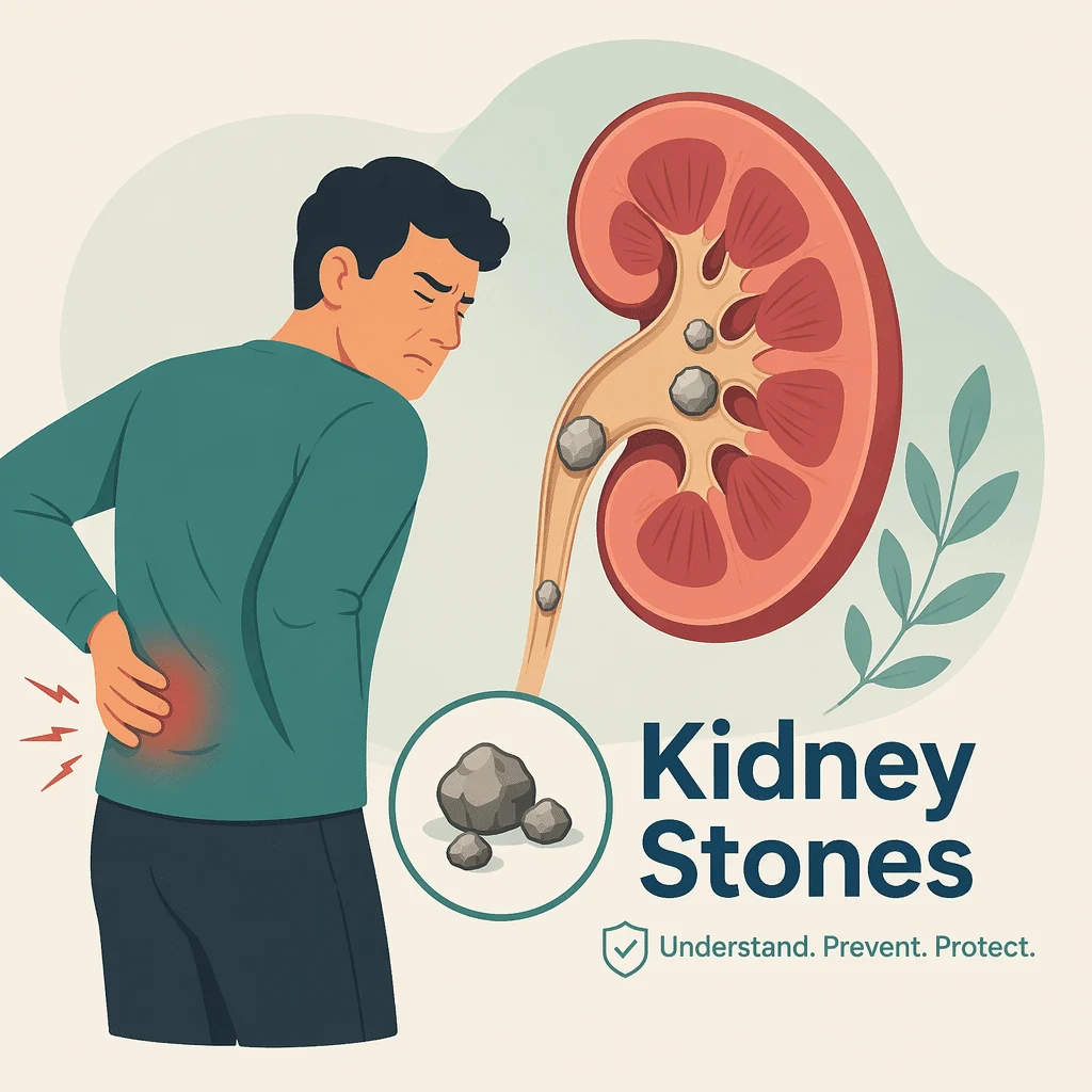
Batu ginjal adalah endapan kristal keras yang terbentuk di dalam ginjal, dan ketika salah satunya jatuh ke dalam saluran sempit yang mengalirkan ginjal, batu tersebut dapat menyebabkan salah satu rasa sakit paling hebat dalam dunia medis. Kondisi ini cukup umum: sekitar 11 persen pria dan 6 persen wanita di Amerika Serikat pernah mengalaminya, menurut National Institute of Diabetes and Digestive and Kidney Diseases. Batu ginjal juga cenderung berulang, karena satu batu membuat kemungkinan terbentuknya batu berikutnya jauh lebih besar.
Dalam artikel ini Anda akan mempelajari seperti apa rasanya serangan batu ginjal, termasuk ciri khas yang membedakannya dari nyeri perut hebat lainnya, jenis-jenis batu yang ada dan penyebabnya, cara memastikan diagnosis batu ginjal, cara penanganannya, serta langkah-langkah pencegahan yang benar-benar terbukti efektif. Artikel ini juga meluruskan satu saran keliru yang paling sering beredar soal batu ginjal: menghindari kalsium dari makanan Anda.
Baca kotak darurat di bawah ini terlebih dahulu: demam disertai nyeri pinggang dan dugaan batu ginjal adalah kondisi yang tidak boleh ditunggu di rumah.
Darurat: kapan batu ginjal menjadi berbahaya
Demam atau menggigil yang disertai nyeri pinggang dan dugaan adanya batu ginjal merupakan kondisi darurat urologi. Ginjal yang tersumbat sekaligus terinfeksi dapat berkembang menjadi sepsis yang mengancam jiwa dalam hitungan jam dan membutuhkan drainase segera; antibiotik saja tidak cukup. Segera pergi ke unit gawat darurat, atau hubungi layanan darurat jika Anda merasa sangat tidak sehat.
Segera cari pertolongan darurat jika:
- Anda sama sekali tidak bisa buang air kecil.
- Anda muntah terus-menerus hingga tidak bisa minum cairan sama sekali.
- Nyeri Anda sangat hebat dan tidak mereda dengan obat yang diresepkan dokter.
- Anda hanya memiliki satu ginjal atau ginjal transplantasi dan menduga ada sumbatan.
- Anda sedang hamil dan mengalami nyeri pinggang yang mungkin disebabkan oleh batu ginjal.
- Anda merasa bingung, menggigil, atau berkeringat dingin, atau jantung Anda berdebar kencang.
Apa itu batu ginjal, dan jenis-jenis yang perlu diketahui
Urine mengandung mineral dan zat sisa yang terlarut. Ketika urine menjadi terlalu pekat, atau keseimbangan antara zat pembentuk batu dan penghambat alami terganggu, mineral-mineral tersebut mengkristal dan saling menempel, tumbuh selama berminggu-minggu menjadi massa padat. Dokter menyebut kondisi ini nefrolitiasis.
Batu yang diam di dalam ginjal sering kali tidak menimbulkan gejala apa pun. Masalah mulai muncul ketika batu masuk ke ureter, yaitu saluran antara ginjal dan kandung kemih, di mana batu dapat menyumbat aliran urine dan memicu rasa nyeri. Jenis batu sangat penting karena menentukan cara pencegahannya: analisis laboratorium terhadap batu yang Anda keluarkan adalah informasi paling berharga yang bisa Anda dapatkan tentang risiko Anda sendiri.
Batu kalsium oksalat dan kalsium fosfat
Batu kalsium adalah jenis yang paling umum, dan batu kalsium oksalat lebih banyak dibandingkan kalsium fosfat. Batu ini terbentuk ketika urine mengandung terlalu banyak kalsium atau oksalat, atau terlalu sedikit sitrat — penghambat alami yang mencegah keduanya bergabung.
Batu kalsium fosfat lebih mungkin terbentuk ketika urine terus-menerus bersifat basa, dan lebih sering dikaitkan dengan asidosis tubular ginjal atau kelenjar paratiroid yang terlalu aktif. Panduan kami tentang pH urine normal membahas bagaimana tingkat keasaman berkaitan dengan jenis batu.
Batu asam urat, struvit, dan sistin
Batu asam urat terbentuk dalam urine yang terus-menerus bersifat asam dan berkaitan dengan asam urat, resistensi insulin, obesitas, serta pola makan yang banyak mengandung daging dan makanan laut. Jenis ini merupakan satu-satunya jenis batu yang umum dan kadang dapat dilarutkan dengan pengobatan yang diresepkan.
Batu struvit adalah batu infeksi: bakteri tertentu memecah urea dan membuat urine menjadi basa, sehingga magnesium amonium fosfat mengkristal. Batu ini tumbuh cepat dan mengikuti infeksi saluran kemih.
Batu sistin bersifat langka dan diturunkan secara genetik. Pada sistinuria, cacat transportasi yang diwariskan menyebabkan asam amino sistin bocor ke dalam urine, di mana ia sulit larut; batu ini biasanya mulai terbentuk sejak usia muda dan terus kambuh sepanjang hidup.
Seperti apa rasanya serangan batu ginjal
Kolik ginjal, yaitu nyeri akibat batu yang bergerak turun melalui ureter, muncul secara tiba-tiba, sering kali di malam hari, dan mencapai puncaknya dalam hitungan menit. Nyeri terasa di pinggang atau sisi punggung di bawah tulang rusuk, lalu menjalar ke depan dan ke bawah menuju selangkangan, buah zakar pada pria, atau labia pada wanita. Nyeri datang dalam gelombang, mencapai intensitas yang oleh banyak orang digambarkan lebih menyakitkan daripada melahirkan. Mual dan muntah sering terjadi, karena ginjal dan usus berbagi jalur saraf yang sama.
Inilah detail yang diketahui dokter tetapi jarang disampaikan kepada pasien: seseorang yang mengalami kolik ginjal tidak bisa diam. Mereka mondar-mandir, menggeliat, dan terus berganti posisi, tidak mampu berbaring tenang. Ini hampir kebalikan dari peritonitis — peradangan lapisan perut akibat usus buntu yang pecah — di mana gerakan justru memperparah nyeri dan penderita berbaring kaku tanpa berani bergerak. Tidak ada satu pola pun yang bisa memastikan diagnosis sendirian, tetapi kegelisahan disertai nyeri pinggang hebat di satu sisi sangat mengarah pada batu ginjal.
Urine mungkin tampak merah muda, merah, atau cokelat, dan darah mikroskopis bahkan lebih sering ditemukan; artikel kami tentang darah dalam urin menjelaskan alasannya. Begitu batu mendekati kandung kemih, rasa ingin buang air kecil yang terus-menerus adalah hal yang umum terjadi.
| Situasi | Apa artinya biasanya | Apa yang harus dilakukan? |
|---|---|---|
| Nyeri pinggang hebat yang tiba-tiba dan menjalar ke selangkangan | Kolik ginjal klasik akibat batu di ureter | Pemeriksaan segera di hari yang sama untuk penanganan nyeri dan pencitraan |
| Nyeri pinggang disertai demam atau menggigil | Kemungkinan ginjal tersumbat dan terinfeksi | Segera ke UGD; hubungi 119 jika kondisi sangat buruk |
| Batu sudah terdiagnosis dan sedang keluar sendiri di rumah | Perjalanan yang biasa terjadi pada banyak batu berukuran kecil | Ikuti rencana dokter Anda; saring urine dan simpan batunya |
| Dua batu atau lebih dari waktu ke waktu | Penyakit batu ginjal berulang dengan penyebab yang dapat diobati | Tanyakan tentang analisis batu dan pemeriksaan urine 24 jam |
| Dugaan batu ginjal saat kehamilan | Situasi berisiko lebih tinggi bagi ibu dan kehamilan | Pemeriksaan kebidanan dan urologi di hari yang sama; utamakan USG |
Siapa yang rentan terkena batu ginjal, dan mengapa
Faktor terbesar adalah kurang minum. Urine yang pekat membuat kristal lebih sulit larut karena kekurangan air — itulah mengapa batu ginjal lebih sering terjadi di iklim panas dan pada orang yang banyak berkeringat karena pekerjaannya, mulai dari koki dan pekerja atap hingga pekerja pabrik besi, serta mengapa serangan lebih sering terjadi di musim panas.
Pola makan berperan secara spesifik: natrium tinggi mendorong lebih banyak kalsium masuk ke urine, protein hewani berlebih meningkatkan beban asam dan kadar asam urat, serta minuman manis berhubungan dengan risiko yang lebih tinggi.
Beberapa kondisi meningkatkan risiko: obesitas, diabetes tipe 2, asam urat, dan hiperparatiroidisme primer, yaitu kondisi di mana kelenjar yang terlalu aktif mendorong kalsium masuk ke dalam darah lalu ke urine. Penyakit radang usus, diare kronis, dan operasi bariatrik dengan bypass usus menyebabkan hiperoksaluria enterik: penyerapan lemak yang terganggu menyisakan oksalat bebas di usus yang kemudian diserap dan dibuang melalui urine. Hormon paratiroid, kalsium darah dan asam urat adalah pemeriksaan yang biasa dilakukan.
Beberapa obat juga berkontribusi, termasuk topiramat, beberapa penghambat protease HIV, antasida berbasis kalsium dalam jumlah banyak, dan diuretik jangka panjang. Riwayat keluarga pun berperan. Sekitar setengah dari orang yang pernah mengalami satu batu ginjal akan mengalaminya lagi dalam sekitar sepuluh tahun — itulah mengapa pencegahan sangat penting.
Demam disertai batu ginjal: kondisi darurat yang tidak boleh ditunda
Sebagian besar serangan batu ginjal sangat menyakitkan, tetapi tidak berbahaya. Namun ada satu kondisi yang berbeda. Ketika batu menyumbat ureter dan urine yang tertahan di belakangnya menjadi terinfeksi, ginjal berubah menjadi kantong nanah yang tertutup dan bertekanan tinggi, memaksa bakteri dan racun masuk ke aliran darah. Ini disebut pielonefritis obstruktif, dan dapat berkembang menjadi syok septik dalam hitungan jam pada orang dewasa yang sebelumnya sehat. Demam atau menggigil hebat yang disertai nyeri pinggang adalah tanda peringatannya.
Solusinya bukan hanya antibiotik, karena antibiotik tidak dapat mencapai konsentrasi yang efektif di dalam sistem yang tersumbat. Ginjal harus segera didrainase, dengan memasang stent melalui ureter atau selang nefrostomi melalui punggung. Penanganan batu itu sendiri ditunda hingga infeksi mereda.
Jika Anda mengalami nyeri pinggang dan demam dalam bentuk apa pun, jangan menunggu untuk melihat apakah kondisi membaik sendiri. Segera periksakan diri pada hari yang sama.
Cara mendiagnosis batu ginjal
CT scan abdomen dan pelvis tanpa kontras adalah pemeriksaan standar. Pemeriksaan ini dapat mendeteksi hampir semua batu, menunjukkan ukuran dan posisinya, mengidentifikasi adanya sumbatan, serta menyingkirkan penyebab lain dari nyeri pinggang.
Ultrasonografi sering menjadi pilihan pertama pada ibu hamil dan pasien yang lebih muda karena tidak menggunakan radiasi. Pemeriksaan ini kurang akurat untuk batu ureter yang kecil, tetapi dapat mendeteksi pembengkakan ginjal akibat sumbatan dengan cukup andal — biasanya sudah cukup untuk menentukan langkah berikutnya.
Urinalisis dilakukan pada hampir setiap kasus, untuk mencari sel darah merah, tanda-tanda infeksi, dan kristal; panduan kami tentang kristal urine pada pemeriksaan mikroskopi menjelaskan apa yang ditunjukkan dan tidak ditunjukkan oleh temuan tersebut. Pemeriksaan darah biasanya mencakup fungsi ginjal, kalsium, dan asam urat, dan artikel kami tentang BUN dan kreatinin tinggi dapat membantu Anda memahaminya.
Jika batu berhasil ditangkap, kirimkan untuk dianalisis; biayanya terjangkau dan hasilnya sangat mengubah strategi pencegahan. Untuk batu yang berulang, batu pertama di usia muda, ginjal tunggal, atau riwayat keluarga yang kuat, dokter akan menambahkan evaluasi metabolik: pengumpulan urine 24 jam yang mengukur volume, kalsium, oksalat, sitrat, asam urat, natrium, pH, dan kreatinin urin, yang mengonfirmasi bahwa pengumpulan sudah lengkap.
Cara mengobati batu ginjal
Uraian berikut menjelaskan pilihan-pilihan yang tersedia; ini bukan rencana pengobatan, dan setiap keputusan ada di tangan dokter yang telah memeriksa Anda dan melihat hasil pencitraan Anda. Pengendalian nyeri adalah prioritas utama, dan pilihan obat bergantung pada fungsi ginjal Anda, obat-obatan lain yang sedang dikonsumsi, serta riwayat kesehatan Anda. Itulah mengapa tidak ada artikel yang seharusnya menyebutkan nama obat pereda nyeri atau dosisnya untuk Anda.
Sebagian besar batu kecil akan keluar sendiri seiring waktu dengan bantuan cairan dan pereda nyeri. Ukuran dan posisi batu menentukan peluangnya: semakin kecil batu dan semakin rendah letaknya, semakin besar kemungkinannya keluar. Dokter Anda mungkin menyarankan terapi ekspulsi medis, yaitu obat penghambat alfa yang merelaksasi ureter bagian bawah, yang umumnya dipertimbangkan untuk batu yang lebih besar di sepertiga bawah ureter, bukan untuk semua jenis batu.
Ketika batu tidak bisa keluar sendiri, ada tiga prosedur utama yang digunakan. Litotripsi gelombang kejut (shock wave lithotripsy) memusatkan energi akustik dari luar tubuh untuk memecah batu, tidak memerlukan sayatan, dan cocok untuk batu ginjal kecil serta batu di ureter bagian atas. Ureteroskopi memasukkan teleskop kecil melalui kandung kemih dan memecah batu menggunakan laser, menjangkau seluruh ureter dan ginjal dengan peluang terbaik untuk membersihkannya dalam satu sesi. Nefrolitotomi perkutan membuat jalur kecil melalui punggung menuju ginjal, digunakan untuk batu yang besar atau batu staghorn.
Ginjal yang tersumbat dan terinfeksi ditangani secara berbeda: drainase terlebih dahulu, batu kemudian.
Mencegah batu ginjal berikutnya
Pencegahan adalah bagian dari penanganan batu ginjal yang paling didukung bukti ilmiah, dan satu langkah menonjol di atas yang lain: minum cukup cairan sehingga urine yang dihasilkan banyak, encer, dan berwarna pucat sepanjang siang dan malam. Urine yang encer tidak mudah menjadi jenuh berlebih, dan kristal tidak dapat terbentuk jika urine tidak mencapai kondisi tersebut. Dokter Anda sebaiknya menentukan target volume cairan yang tepat untuk Anda, karena hal ini bergantung pada fungsi ginjal dan jantung Anda serta seberapa banyak cairan yang hilang melalui keringat.
Selain cairan, yang terbukti bermanfaat adalah mengurangi natrium, menjaga asupan protein hewani tetap sedang, menurunkan berat badan berlebih, dan mengurangi minuman manis. Membatasi oksalat hanya perlu dilakukan jika Anda membentuk batu kalsium oksalat dan hasil urine 24 jam menunjukkan kadar oksalat yang tinggi; menghindari bayam, kacang-kacangan, dan rhubarb secara menyeluruh tidak memberikan manfaat bagi orang lain.
Sebagian orang juga memerlukan pencegahan dengan obat yang disesuaikan dengan jenis batunya: diuretik tiazid untuk kadar kalsium urine yang tinggi, kalium sitrat untuk kadar sitrat rendah atau batu asam urat, dan allopurinol untuk kadar asam urat yang tinggi. Obat-obatan ini memiliki efek samping nyata dan dipilih berdasarkan hasil pemeriksaan urine Anda.
Kesalahpahaman tentang kalsium dalam makanan
Inilah koreksi yang paling sering disalahpahami dalam berbagai artikel: membatasi asupan kalsium dari makanan justru meningkatkan risiko batu kalsium oksalat, bukan menguranginya.
Alasannya bersifat mekanis. Kalsium yang dikonsumsi bersama makanan akan mengikat oksalat di dalam usus, membentuk senyawa yang tidak diserap dan dikeluarkan melalui tinja. Jika kalsium dihilangkan, oksalat tersebut justru akan diserap, mencapai ginjal, dan dikeluarkan ke dalam urine, sehingga bebas berpasangan dengan kalsium dan membentuk kristal. Seperti yang dinyatakan oleh National Institute of Diabetes and Digestive and Kidney Diseases, kalsium dari makanan tidak meningkatkan risiko terbentuknya batu kalsium oksalat, dan dalam jumlah yang tepat kalsium justru menghambat zat-zat lain di saluran pencernaan yang dapat memicu terbentuknya batu (NIDDK, Eating, Diet and Nutrition for Kidney Stones).
Suplemen kalsium adalah pertanyaan tersendiri. Jika dikonsumsi bersama makanan, efeknya mirip dengan kalsium dari makanan; jika dikonsumsi di antara waktu makan tanpa oksalat yang bisa diikat, kalsium ekstra tersebut diserap dan justru meningkatkan kadar kalsium dalam urine. Jika Anda mengonsumsi kalsium atau vitamin D suplemen dan pembentukan batu, diskusikan hal ini dengan dokter Anda.
Jus lemon, cuka sari apel, dan ramuan pembersih batu
Sitrat bukan sekadar mitos. Zat ini benar-benar menghambat pembentukan batu dengan cara mengikat kalsium dalam urine dan mengganggu pertumbuhan kristal — itulah mengapa kalium sitrat tersedia sebagai obat resep. Lemon kaya akan sitrat, dan beberapa penelitian kecil tentang terapi minuman lemon menunjukkan peningkatan sitrat urine yang cukup berarti pada sebagian pasien. Ini adalah pilihan yang masuk akal dan terjangkau untuk didiskusikan dengan dokter Anda, tidak lebih dari itu.
Yang tidak bisa dilakukan oleh air lemon adalah melarutkan batu kalsium. Tidak ada minuman apa pun yang bisa melakukannya. Satu-satunya batu yang dapat dilarutkan secara medis adalah batu asam urat dan beberapa batu sistin, dan itu dilakukan oleh dokter dengan cara mengalkalikan urine Anda menggunakan obat resep serta memantau hasilnya.
Cuka sari apel tidak memiliki bukti klinis dalam penanganan batu ginjal. Produk 'pembersih ginjal' dan protokol 'pembuang batu' yang dijual bebas pun tidak memiliki bukti, bahkan beberapa di antaranya mengandung bahan tinggi oksalat atau herbal diuretik yang justru memperburuk kondisi. Tinjauan bukti terbaru juga mencatat lebih banyak efek samping ringan dari jus lemon dibandingkan plasebo. Anggap semua ini sebagai hal yang belum terbukti, dan tangani dugaan batu ginjal sebagai masalah medis yang perlu ditangani dokter.
Kemajuan ilmiah terbaru dalam penanganan batu ginjal
Sebuah tinjauan bukti yang didanai pemerintah federal, diterbitkan dalam Annals of Internal Medicine pada tahun 2026 oleh Asher dan rekan-rekannya, menggabungkan 31 penelitian tentang pencegahan batu ginjal berulang. Hasilnya menunjukkan bahwa peningkatan asupan air, serta pola makan dengan kalsium normal hingga tinggi ditambah protein rendah dan natrium rendah, dapat mengurangi kekambuhan — bersama dengan terapi tiazid, alkali, dan allopurinol — meskipun tingkat kepastian buktinya masih rendah secara keseluruhan. Tinjauan ini juga mengaitkan jus lemon dengan lebih banyak efek samping ringan. Artinya bagi Anda: cairan dan pola makan tetap menjadi langkah yang paling didukung bukti, dan asupan kalsium normal adalah bagian dari anjuran tersebut, bukan pengecualian.
Uji klinis NOSTONE, yang diterbitkan dalam New England Journal of Medicine pada tahun 2023 oleh Dhayat dan rekan-rekannya, menguji hidroklortiaziid dibandingkan plasebo pada orang-orang dengan batu kalsium berulang. Dari tiga dosis dan hampir tiga tahun pemantauan, tingkat kekambuhan tidak berbeda secara bermakna dibandingkan plasebo, sementara kadar kalium rendah, gout baru, dan diabetes baru lebih sering terjadi pada kelompok yang mengonsumsi obat tersebut. Artinya bagi Anda: tiazid bukan lagi resep otomatis, dan wajar untuk menanyakan mengapa obat ini dipilih berdasarkan profil urine Anda.
Sebuah tinjauan tahun 2024 dalam Current Opinion in Nephrology and Hypertension oleh Bargagli dan rekan-rekannya menambahkan meta-analisis dari seluruh uji coba terkontrol plasebo untuk monoterapi tiazid, tidak menemukan keunggulan yang signifikan, dan menyarankan penggunaan yang lebih selektif. Artinya bagi Anda: pencegahan batu ginjal kini bergeser ke arah penanganan yang disesuaikan dengan profil kimia urine Anda secara individual, bukan diterapkan secara seragam untuk semua orang.
Sebuah tinjauan sistematis dan meta-analisis tahun 2025 dalam European Urology Focus oleh Nedbal dan rekan-rekan, dari seksi endourologi European Association of Urology, membandingkan ureteroskopi fleksibel dengan litotripsi gelombang kejut pada populasi gabungan yang sangat besar. Ureteroskopi lebih sering berhasil membersihkan batu dan lebih jarang memerlukan pengobatan ulang, dengan komplikasi, lama rawat inap, dan biaya yang sebanding. Artinya bagi Anda: jika keduanya ditawarkan, tanyakan seberapa besar kemungkinan masing-masing dapat membersihkan batu Anda dalam satu sesi, bukan sekadar mana yang terdengar lebih tidak invasif.
Terakhir, sebuah studi rumah sakit tahun 2025 dalam BMC Urology oleh Hsu dan rekan-rekan mengikuti orang-orang yang dirawat dengan sepsis akibat batu ureter atas yang menyumbat. Mereka yang mendapat drainase darurat lebih awal mencapai pengobatan definitif lebih cepat dan meninggalkan rumah sakit lebih cepat. Artinya bagi Anda: hal ini memperkuat kotak di bagian atas, bahwa ginjal yang terinfeksi dan tersumbat harus didrainase terlebih dahulu, baru kemudian batu ditangani.
Daftar istilah kunci
| Ketentuan | Definisi |
|---|---|
| Kolik ginjal | Nyeri seperti gelombang yang disebabkan oleh batu yang bergerak turun melalui ureter, dirasakan di pinggang dan menjalar ke selangkangan. |
| Ureter | Saluran sempit yang membawa urine dari setiap ginjal ke kandung kemih, dan tempat yang paling sering menjadi lokasi batu tersangkut. |
| Nefrolitiasis | Nama medis untuk penyakit batu ginjal. |
| Oksalat | Senyawa dalam beberapa makanan nabati yang mengikat kalsium; kadarnya yang terlalu tinggi dalam urine mendorong terbentuknya jenis batu yang paling umum. |
| Sitrat | Penghambat alami dalam urine yang mengikat kalsium dan memperlambat pertumbuhan kristal; kadar yang rendah meningkatkan risiko batu. |
| Hidronefrosis | Pembengkakan ginjal yang disebabkan oleh urine yang tertahan di belakang sumbatan. |
| Litotripsi gelombang kejut | Prosedur yang memecah batu menggunakan energi akustik terfokus yang diarahkan dari luar tubuh. |
| Ureteroskopi | Prosedur yang memasukkan teleskop kecil ke saluran kemih untuk memecah atau mengangkat batu menggunakan laser. |
| Nefrolitotomi perkutan | Pengangkatan batu berukuran besar melalui saluran kecil yang dibuat menembus punggung hingga ke ginjal. |
| Pengumpulan urine 24 jam | Urine yang dikumpulkan selama satu hari penuh dan dianalisis untuk menemukan penyebab metabolik dari batu yang berulang. |
Pertanyaan yang sering diajukan
Berapa lama waktu yang dibutuhkan untuk mengeluarkan batu ginjal?
Hal ini terutama bergantung pada ukuran dan posisi batu, dan tidak ada yang bisa menjamin batu akan keluar dengan sendirinya. Batu yang sangat kecil di bagian bawah ureter sering kali keluar dalam satu hingga dua minggu; batu yang lebih besar mungkin membutuhkan beberapa minggu, dan batu di atas ukuran tertentu biasanya memerlukan prosedur medis. Dokter biasanya menetapkan batas waktu untuk menunggu dan memantau, karena ureter yang tersumbat terlalu lama dapat merusak ginjal di belakangnya. Jika nyeri Anda memburuk, Anda mengalami demam, atau tidak ada kemajuan dalam jangka waktu yang diberikan dokter Anda, segera kembali lebih awal daripada menunggu.
Haruskah saya menghindari kalsium jika mengalami batu ginjal?
Tidak, dan ini adalah kesalahan yang paling umum terjadi. Diet rendah kalsium justru meningkatkan risiko batu kalsium oksalat, karena kalsium dari makanan mengikat oksalat di usus sehingga oksalat dikeluarkan melalui tinja dan tidak mencapai urine Anda. Jika asupan kalsium dikurangi, lebih banyak oksalat yang diserap dan dikeluarkan melalui urine — yang justru berlawanan dengan yang seharusnya. Asupan kalsium normal dari makanan bersifat protektif, dan juga melindungi tulang Anda. Suplemen kalsium adalah hal yang berbeda: jika dikonsumsi di luar waktu makan, suplemen ini dapat meningkatkan kadar kalsium dalam urine, jadi tanyakan kepada dokter Anda sebelum mulai atau berhenti mengonsumsinya.
Apakah jus lemon bisa melarutkan batu ginjal?
Tidak. Tidak ada minuman apa pun yang dapat melarutkan batu kalsium. Kebenarannya adalah bahwa sitrat, yang terkandung dalam jumlah banyak di lemon, memang benar-benar menghambat pembentukan batu, dan kalium sitrat merupakan obat pencegah yang nyata dan tersedia dengan resep dokter. Studi-studi kecil tentang terapi limun menunjukkan peningkatan moderat kadar sitrat dalam urine pada sebagian pasien, sehingga layak untuk didiskusikan dengan dokter Anda sebagai tambahan asupan cairan yang terjangkau. Ini bukan pengobatan untuk batu yang sudah ada, dan sebuah tinjauan bukti terbaru menemukan bahwa regimen air lemon menyebabkan lebih banyak efek samping ringan dibandingkan plasebo.
Apakah batu ginjal bisa merusak ginjal saya?
Biasanya tidak, jika ditangani dalam waktu yang wajar. Risiko muncul dari penyumbatan yang berkepanjangan dan dari infeksi. Ureter yang tersumbat selama berminggu-minggu dapat menyebabkan hilangnya fungsi ginjal secara permanen, dan ginjal yang tersumbat lalu terinfeksi merupakan kedaruratan medis. Orang dengan satu ginjal, ginjal transplantasi, atau penyakit ginjal yang sudah ada sebelumnya memiliki toleransi yang lebih kecil dan ditangani lebih segera. Itulah mengapa saran yang selalu diberikan adalah segera memeriksakan diri jika dicurigai ada batu, bukan mencoba bertahan menanggung sakitnya.
Apakah saya perlu menangkap batunya, dan apa yang terjadi setelahnya?
Ya. Saring urine Anda menggunakan filter yang disediakan klinik, atau kain saring halus, dan simpan semua benda padat yang tertangkap, meskipun terlihat seperti butiran pasir. Laboratorium akan menganalisis kandungannya, dan hasilnya menentukan seluruh rencana pencegahan Anda: saran pembatasan oksalat untuk satu jenis, pengalkalian urine untuk jenis lain, dan pengendalian infeksi untuk jenis ketiga. Bebas dari rasa sakit bukan berarti batu sudah keluar, sehingga pemeriksaan pencitraan lanjutan sering kali tetap dijadwalkan.
Apakah saya bisa memiliki batu ginjal jika hasil tes urine saya normal?
Ya. Sebagian besar orang dengan kolik ginjal memiliki darah dalam urine, tetapi sebagian kecil yang cukup berarti tidak, terutama ketika batu menyumbat ureter sepenuhnya. Oleh karena itu, hasil urinalisis yang normal tidak menyingkirkan kemungkinan adanya batu, dan pencitraan diperlukan untuk memastikannya. Sebaliknya juga berlaku: darah dalam urine memiliki banyak penyebab selain batu, sehingga temuan tersebut saja tidak dapat memastikan adanya batu.
Sumber
Detail studi dan kutipan diambil dari PubMed.
- National Institute of Diabetes and Digestive and Kidney Diseases (NIDDK) – Definisi & Fakta tentang Batu Ginjal
- National Institute of Diabetes and Digestive and Kidney Diseases (NIDDK) – Makan, Diet, & Nutrisi untuk Batu Ginjal
- MedlinePlus, U.S. National Library of Medicine – Batu ginjal
- American Urological Association – Panduan klinis Penanganan Medis Batu Ginjal
- Asher GN, Viprakasit DP, Aymes SE, Lusk JB, Ross S, Baker C, Rains C, Wright ST, Kahwati LC. Prevention of Recurrent Nephrolithiasis in Adults and Children: A Systematic Review. Ann Intern Med. 2026;179(5):696-707. https://doi.org/10.7326/ANNALS-25-04452
- Dhayat NA, Bonny O, Roth B, Christe A, Ritter A, Mohebbi N, et al. Hydrochlorothiazide and Prevention of Kidney-Stone Recurrence. N Engl J Med. 2023;388(9):781-791. https://doi.org/10.1056/NEJMoa2209275
- Bargagli M, Trelle S, Bonny O, Fuster DG. Thiazides for kidney stone recurrence prevention. Curr Opin Nephrol Hypertens. 2024;33(4):427-432. https://doi.org/10.1097/MNH.0000000000000990
- Nedbal C, Geraghty R, Tramanzoli P, Sessa D, Gauhar V, Galosi AB, Gregori A, Somani BK. Cost and Effectiveness of Flexible Ureteroscopy Versus Shockwave Lithotripsy for Urolithiasis: A Systematic Review and Meta-analysis by the European Association of Urology Endourology Section. Eur Urol Focus. 2025;11(6):1012-1022. https://doi.org/10.1016/j.euf.2025.06.004
- Hsu JT, Lin CM, Hung SF, Chung SD, Cheng PY. Optimizing antibiotic duration and percutaneous nephrostomy timing in urosepsis complicated by upper ureteral stones: a retrospective study. BMC Urol. 2025;25(1):182. https://doi.org/10.1186/s12894-025-01868-7
Bacaan lebih lanjut
- Urine berbusa: penyebab dan risikonya
- Kekurangan magnesium: gejala, penyebab, dan penanganannya
- Elektrolit dalam urin: menafsirkan hasil tes Anda
- Panel fungsi ginjal: apa yang diukurnya
- Warna urine: memahami penyebab dan perubahannya
Pemeriksaan batu ginjal biasanya menghasilkan beberapa angka sekaligus: urinalisis, kreatinin dan fungsi ginjal, kalsium darah dan asam urat, serta pengumpulan urine 24 jam. AI DiagMe membaca hasil-hasil tersebut dan menjelaskan dalam bahasa yang mudah dipahami apa yang diukur oleh masing-masing nilai dan mengapa dokter Anda memintanya. Ini membantu Anda memahami hasil Anda dan mempersiapkan pertanyaan yang lebih baik. Layanan ini tidak mendiagnosis batu ginjal dan tidak menggantikan dokter Anda.
Pahami hasil lab Anda dengan AI DiagMe.
Dapatkan interpretasi hasil Anda dalam hitungan menit.