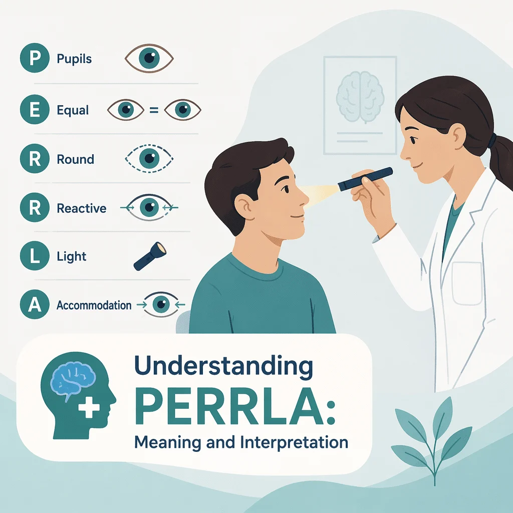
The PERRLA meaning is straightforward once you know what the letters stand for: Pupils Equal, Round, Reactive to Light and Accommodation. It is shorthand that a clinician writes after checking your pupils with a small flashlight, and it usually sits in the eye or neurological part of a visit summary. Most people who look the term up have just spotted it in their own medical record and want to know whether it points to a problem. In this article you will learn what each letter describes, how the pupil exam is carried out, what a normal note does and does not rule out, which pupil patterns deserve attention, and when a change in your pupils is a true emergency. You will also see where blood work fits into a wider evaluation.
The PERRLA meaning, letter by letter
PERRLA is an acronym, not a measurement with a number attached. It records what the examiner saw during a few seconds of looking at your pupils, the black openings at the center of each iris that widen and narrow to control how much light reaches the back of the eye. Because it is written in shorthand, it can look more technical than it is. Our team also maintains a guide to common medical lab test abbreviations.
P and E: pupils equal
The examiner compares the size of the two pupils. In ordinary room light, adult pupils usually measure between two and four millimeters, and they widen in the dark. Equal means the two are the same size, or close enough that the difference is not considered meaningful. A visible difference between the two is called anisocoria, and on its own it is not automatically abnormal.
R: round
A healthy pupil is a smooth circle. An oval, teardrop or notched pupil can follow eye surgery, an injury to the iris, or inflammation inside the eye, so the shape is worth recording even when everything else looks normal.
R: reactive to light
When light reaches one eye, that pupil should shrink quickly. This is the pupillary light reflex. The signal travels along the optic nerve into the brainstem, then back out along the third cranial nerve to the small muscle that closes the pupil. Because the circuit passes through the brainstem, it is one of the few brain pathways a clinician can watch directly from the bedside.
L and A: light and accommodation
Accommodation is what your eyes do when you shift focus from something far away to something close. The lens changes shape, the eyes turn slightly inward, and the pupils get smaller. A clinician tests it by asking you to look at a distant point and then at a finger held near your nose.
You will also meet shorter and longer variants of the acronym. PERRL stops at the light reaction because accommodation was not tested, which is common in a busy clinic. PERRLA/EOMI adds extraocular movements intact, meaning the eyes moved normally in every direction. Some notes add a number, such as 3 mm, which is simply the measured pupil size.
How the pupil exam is actually performed
The whole examination takes under a minute and needs only a penlight and a dimmed room. Knowing the steps makes the note in your chart far easier to read.
The direct and consensual light response
The examiner lowers the light, asks you to look at a distant target so your eyes are not focusing up close, then brings a beam onto one eye from the side. The pupil in the lit eye should constrict, which is the direct response. The pupil in the other eye should constrict at the same moment, which is the consensual response. Both happen because the two halves of the reflex are connected inside the brainstem. Comparing them tells the examiner whether a problem sits on the incoming side of the circuit or the outgoing side.
The near response
You are then asked to look at a far target and switch to a finger or pen held about six inches from your nose. The pupils should tighten as the eyes converge. A pupil that reacts poorly to light but constricts well to near focus is described as light-near dissociation, a pattern that sends the examiner looking for specific causes rather than dismissing it.
The swinging-flashlight test
The examiner moves the light back and forth between the two eyes every few seconds. Normally each pupil stays small as the beam arrives. If one pupil widens instead of holding steady when the light reaches it, that eye is sending a weaker signal to the brain. This pattern is called a relative afferent pupillary defect, and it points to a problem in the optic nerve or the retina rather than in the pupil muscle itself. It is one of the few bedside findings that can reveal optic nerve damage before vision loss becomes obvious. Optic nerve inflammation has many possible causes, and our team explains the warning signs of multiple sclerosis.
What the PERRLA meaning tells you, and what it does not
A normal note is reassuring about one specific circuit at one specific moment. It is not a clean bill of health for your eyes or your brain, and reading it that way is the most common mistake patients make.
- It suggests the light reflex pathway was working on both sides when the examination was done.
- It makes a large pupil-involving third nerve problem unlikely at that moment.
- It does not test how well you see. Visual acuity, color vision and visual fields all need separate testing.
- It does not exclude a stroke. Many strokes leave the pupils untouched, and our team describes the warning signs of a stroke.
- It does not measure eye pressure, so it says nothing about glaucoma. Our team also outlines the symptoms and testing for glaucoma.
- It does not exclude a slow-growing mass inside the skull, which can spare the pupils for a long time. Another article reviews the symptoms of a brain tumor.
- It is a snapshot. Pupils can be normal at one visit and abnormal a few hours later, which is exactly why the check is repeated in hospital.
Pupil findings and what they can point to
The table below groups the patterns clinicians look for, what each one can suggest, and how quickly it usually needs attention. It is a reading aid for your own chart, not a way to diagnose yourself.
| Pupil finding | What it can point to | How urgent |
|---|---|---|
| Both pupils equal, round and brisk to light | An intact pupil pathway at the time of the exam | Routine, no action needed |
| A small, long-standing size difference, both pupils reacting normally | Physiologic anisocoria, a harmless lifelong variation | Routine, mention it at your next visit |
| One widely dilated pupil that barely reacts, with a drooping eyelid and double vision | Pressure on the third cranial nerve, which can come from an aneurysm | Emergency, call 911 |
| One small pupil with a drooping eyelid on the same side | Horner syndrome, affecting a nerve pathway that runs from the brain to the eye | Urgent, same-day medical assessment |
| One pupil that widens when the light swings onto it | A relative afferent pupillary defect, suggesting optic nerve or retinal disease | Urgent, eye or neurology assessment within days |
| A large pupil that reacts slowly to light but better to near focus | Adie tonic pupil, usually benign and often found in young adults | Non-urgent, confirm with an eye specialist |
| Both pupils very small or very large after a medication or eye drop | A drug or eye-drop effect rather than nerve damage | Routine, check with your pharmacist or prescriber |
| A newly unequal pupil after a head injury, or with sudden severe headache, weakness or confusion | Possible bleeding or swelling inside the skull | Emergency, call 911 |
Common and harmless reasons pupils differ
Unequal pupils frighten people, and most of the time the explanation is ordinary. The reassuring pattern is a difference that has been there for years, stays the same in bright and dim light, and comes with normal eyelid position and normal eye movements.
- Physiologic anisocoria: a small built-in difference in pupil size that many healthy people carry all their lives. Old photographs often settle the question.
- Dilating drops: after a routine eye examination the treated pupil stays wide for several hours.
- Accidental contact with a medicine: touching a motion-sickness patch and then rubbing one eye, or breathing in a nebulized medication that leaks around the mask, can widen a single pupil.
- Prescribed medicines: opioids tend to make both pupils very small, while some antihistamines, decongestants, antidepressants and stimulants tend to widen them.
- Past eye surgery, trauma or inflammation: these can leave a pupil irregular in shape or slow to react, permanently and harmlessly.
Alcohol, cannabis and recreational stimulants also change pupil size, which is one reason clinicians ask about them before ordering scans. None of these explanations should be assumed when the change is new and sudden.
When to seek care immediately
Call 911 or go to an emergency department now if a pupil has become unequal, unusually large or unresponsive and it comes with any of the following:
- A sudden, severe headache, especially the worst headache of your life.
- Any head injury, even one that seemed minor at the time.
- A drooping eyelid, double vision or a sudden loss of vision.
- Weakness or numbness on one side of the body, a facial droop, slurred speech or trouble understanding speech.
- Confusion, unusual drowsiness, vomiting, a seizure or a stiff neck with fever.
Do not wait to see whether it improves, do not drive yourself, and do not call a routine appointment line instead. A newly dilated pupil with a drooping eyelid and double vision can signal an aneurysm pressing on a nerve, and a newly unequal pupil after a head injury can signal bleeding inside the skull. Both are time-critical. A first-ever severe headache is not the same as a familiar one, and our team covers the causes and treatments of migraine.
Book a non-urgent appointment, rather than emergency care, if you notice a small difference in pupil size that has been stable for years, a pupil that is slow to react without any other symptom, or a shape irregularity you have had since an old eye operation.
Where blood tests fit into a pupil work-up
No blood test measures PERRLA, and no laboratory result can confirm or exclude a pupil abnormality. The pupil exam is answered by the examination itself, by an eye specialist, and when needed by imaging of the brain, the eye socket and the blood vessels of the neck.
Blood work enters the picture one step later. When pupil or nerve findings are part of a wider evaluation, clinicians frequently order laboratory tests to look for conditions that can contribute to nerve or eye problems, or that need to be managed alongside them. Typical requests include blood glucose and hemoglobin A1c for diabetes, thyroid function tests, vitamin B12, and inflammatory markers such as C-reactive protein and the erythrocyte sedimentation rate when giant cell arteritis is a concern in an older adult with headache and visual symptoms.
Reading those results is where a clear explanation helps. Our team publishes an A1C conversion chart for translating a percentage into an average glucose value, details the symptoms and causes of low vitamin B12, explains the normal ranges for thyroid levels, and reviews the causes of high CRP levels. None of these tests diagnoses a pupil problem. They describe the background against which a clinician interprets one.
Latest scientific advances in pupil assessment
Research since 2023 has concentrated on one known weakness of the classic exam: the penlight check depends on the examiner’s eye and judgment, so two clinicians can record different findings in the same patient. Here is what recent work found, and what it means for you.
Machines now measure what the penlight only estimates
A 2025 review of hospital practice concluded that manual penlight assessment is imprecise compared with quantitative pupillometry, a handheld device that films the pupil and reports its size and speed of reaction as numbers rather than impressions (Campos and colleagues). A large international intensive care study known as ORANGE, which followed patients with acute brain injury, reported that standard penlight findings are interpreted subjectively, while the automated device produces a reproducible score called the neurological pupil index (Oddo and colleagues, 2023).
What this means for you: if you or a relative are in hospital after a brain injury or a stroke, staff may check the pupils with a small camera-based device instead of a flashlight. It is not a different test. It is the same reflex, measured more consistently and recorded so that the next nurse can compare like with like.
Telling a worrying small pupil from a harmless one
A 2024 study used automated pupillometry to separate Horner syndrome from physiologic anisocoria, the harmless size difference described earlier (Disse and colleagues). The device tracked how slowly the smaller pupil re-opens once the lights go down, a feature called dilation lag, and the authors reported that this measurement helped tell the two situations apart.
What this means for you: an eye clinic may eventually settle that question with a short recording rather than the eye drop test used today, which means waiting in the clinic while drops take effect. This is a research finding on a limited number of patients, not yet standard care.
Smartphone cameras are being tested, with caution
A pilot study published in 2024 combined a smartphone camera with a machine learning model to detect mild traumatic brain injury from the pupil reaction (Maxin and colleagues). The number of participants was small and the authors present the work as early. A scoping review the same year, meaning a study that maps what is already known instead of pooling results, examined pupillometry after head trauma and found it promising for predicting recovery while calling for larger and better standardized studies (Kiani and colleagues).
What this means for you: a phone app is not a substitute for an examination, and no consumer app available today can tell you whether your pupils are normal. Treat these results as a sign of where the field is heading, not as something to try at home.
Glossary
| Term | Definition |
|---|---|
| Accommodation | The automatic adjustment your eyes make to focus on a nearby object. The lens changes shape, the eyes turn inward and the pupils narrow. |
| Adie tonic pupil | A usually harmless condition in which one pupil is larger, reacts slowly to light and constricts better when focusing up close. |
| Anisocoria | A difference in size between the two pupils. It can be a harmless lifelong trait or a sign of a nerve problem, depending on the other findings. |
| Consensual light response | The narrowing of the pupil in the eye that is not being lit, when light is shone into the other eye. |
| EOMI | Extraocular movements intact. A note meaning the eyes moved normally in all directions when the examiner tested them. |
| Horner syndrome | A combination of a small pupil, a drooping upper eyelid and sometimes reduced sweating on one side of the face, caused by interruption of a nerve pathway to the eye. |
| PERRLA | Pupils Equal, Round, Reactive to Light and Accommodation. A shorthand note recording a normal bedside pupil examination. |
| Ptosis | A drooping upper eyelid. When it appears suddenly alongside a change in pupil size, it needs prompt medical attention. |
| Pupillometry | Measurement of pupil size and reaction with a device rather than by eye. Quantitative pupillometry produces numbers that different staff can compare. |
| Relative afferent pupillary defect | A weaker light signal from one eye, seen as that pupil widening when the light swings onto it. It suggests optic nerve or retinal disease. |
Frequently asked questions
What is the difference between PERRL and PERRLA?
The two notes describe the same examination, stopped at different points. PERRL records that the pupils are equal, round and reactive to light. PERRLA adds that the accommodation, or near focus, response was also tested and was normal. In many settings the near response is skipped because it takes an extra step and rarely changes the immediate decision, so PERRL is often simply a shorter version of the same reassuring finding rather than a sign that something was abnormal.
What does PERRLA and EOMI mean together?
EOMI stands for extraocular movements intact. Written together, PERRLA and EOMI mean that the pupils looked and reacted normally, and that the eyes moved normally in all directions when the examiner asked you to follow a finger. The combination is a quick screen of several cranial nerves at once, and it is one of the most frequent lines in a routine physical examination note.
What does a note saying my pupils are 3 mm mean?
It is the measured diameter of the pupils, in millimeters. In ordinary room light adult pupils commonly sit between two and four millimeters, so 3 mm is a typical reading. The number matters mainly for comparison: a nurse who records 3 mm now and 5 mm two hours later has captured a change worth investigating, even though either figure on its own might look unremarkable.
Can I ask for a PERRLA test at the laboratory?
No. PERRLA is a physical examination finding, not a laboratory test, so there is nothing to collect or analyze. Anyone with a penlight and the right training can perform it, and it costs nothing. If you are worried about your pupils, ask your primary care clinician or an eye specialist to examine you rather than requesting a test. Blood work may be ordered afterward to investigate contributing conditions, but never to assess the pupils themselves.
Can caffeine, stress or tiredness change my pupil size?
Yes, and this is normal. Pupils widen with stress, excitement, pain and stimulants including caffeine, and they narrow with drowsiness and in bright light. These changes affect both eyes together, which is the key point. Something that makes only one pupil change size is a different situation and deserves attention, particularly if it appears suddenly or comes with an eyelid or vision change.
What does it mean if my chart says the pupils are equal but sluggish?
Sluggish means the pupils did react to light, but more slowly or less completely than expected. It can simply reflect a bright examination room, older age, tiredness or a medication effect. It can also be an early sign that the reflex pathway is under strain, which is why a sluggish reaction in someone who is unwell, injured or drowsy is usually rechecked shortly afterward rather than ignored.
Sources
- MedlinePlus, National Library of Medicine — Anisocoria, Medical Encyclopedia — medlineplus.gov
- Belliveau AP, Somani AN, Dossani RH — Pupillary Light Reflex — StatPearls, National Library of Medicine — ncbi.nlm.nih.gov
- Mayo Clinic — Horner syndrome, symptoms and causes — mayoclinic.org
- Campos E, Tejerina Alvarez EE, Lorente Balanza JA — Clinical utility of quantitative pupillometric monitoring in patients with acute brain injury — Medicina Clinica, 2025 — doi.org
- Oddo M, Taccone FS, Citerio G, et al. — The Neurological Pupil index for outcome prognostication in people with acute brain injury (ORANGE): a prospective, observational, multicentre cohort study — The Lancet Neurology, 2023 — doi.org
- Disse LR, Bockisch CJ, Weber KP, Fierz FC — Differentiation of Horner Syndrome and Physiological Anisocoria by Automated Pupillometry — Journal of Neuro-Ophthalmology, 2024 — doi.org
- Maxin AJ, et al. — Smartphone Pupillometry and Machine Learning for Detection of Acute Mild Traumatic Brain Injury: Cohort Study — JMIR Neurotechnology, 2024 — doi.org
- Kiani I, et al. — Prognostic role of quantitative pupillometry in traumatic brain injury: a scoping review — Neurological Sciences, 2024 — doi.org
Further reading
- The causes and treatments of black spots in vision
- Conjunctivitis and uveitis: causes and symptoms compared
- The symptoms, signs and testing of meningitis
- Diabetes blood tests: glucose, HbA1c and insulin explained
- The complete blood count and how to read your results
Understand your lab results with AI DiagMe
A pupil examination answers one question, and the blood tests ordered around it answer others. If you are holding a report that mentions hemoglobin A1c, thyroid hormones, vitamin B12 or C-reactive protein, AI DiagMe turns those numbers into plain language and shows you which values sit outside the expected range. It is built to help you understand your results and prepare better questions for your appointment. It does not diagnose, and it does not replace your doctor.
Get your results interpreted in minutes