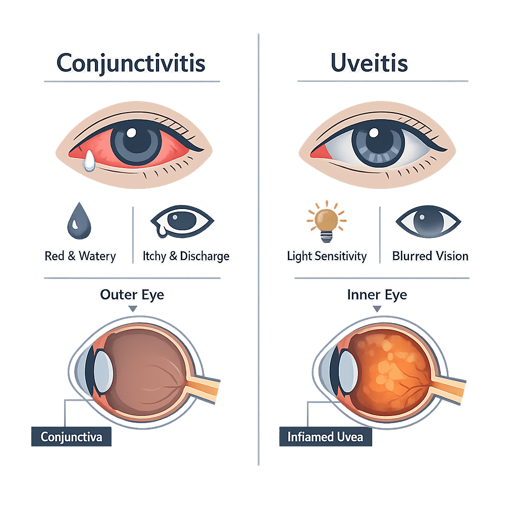
Să deosebești conjunctivita de uveită contează mai mult decât se crede, pentru că una este de obicei o neplăcere trecătoare, iar cealaltă îți poate amenința vederea. Ambele înroșesc ochiul. Ambele pot provoca lăcrimare. Privind în oglindă, seamănă suficient de mult încât oamenii presupun de obicei varianta mai ușoară și așteaptă să treacă. Diferența nu stă în roșeața în sine, ci în ce o însoțește: mâncărimea și secreția lipicioasă indică o direcție, durerea profundă, sensibilitatea reală la lumină și vederea încețoșată indică alta. În acest articol vei afla unde se situează fiecare afecțiune în interiorul ochiului, ce semne poți verifica singur, semnalele de alarmă care necesită consultație în aceeași zi, de ce uveita indică adesea o afecțiune din altă parte a corpului și ce analize de sânge îi ajută pe medici să găsească cauza.
Conjunctivită vs uveită: diferența în un minut
Conjunctivita este inflamația peliculei de suprafață a ochiului. Uveita este inflamația din interiorul ochiului. Această singură distincție explică aproape tot ce urmează.
Deoarece conjunctivita se află la exterior, irită: ochiul mâncărime, se simte ca și cum ar avea nisip în el, lăcrimează și produce adesea secreții care lipesc genele peste noapte. Vederea rămâne normală după ce clipești și îndepărtezi pelicula. Majoritatea cazurilor sunt virale, se rezolvă de la sine în una până la două săptămâni, iar formele comune se transmit ușor între persoane. Echipa noastră explică și tipurile, simptomele și tratamentul conjunctivitei pentru cititorii care știu deja că aceasta este problema lor.
Uveita se află în profunzimea ochiului, într-un țesut bogat în vase de sânge și nervi. Doare mai degrabă decât provoacă mâncărime, lumina puternică devine cu adevărat dureroasă, iar vederea se încețoșează într-un mod pe care clipitul nu îl rezolvă. Nu este contagioasă. Nu se vindecă de la sine, iar inflamația netratată din interiorul ochiului poate cicatriza structuri delicate, poate crește presiunea intraoculară sau poate deteriora retina. De aceea uveita este afecțiunea care necesită urgență.
Unde se află fiecare afecțiune în interiorul ochiului
Conjunctiva: stratul de suprafață
Conjunctiva este o membrană subțire și transparentă care acoperă albul ochiului și căptușește interiorul pleoapelor. Conține vase de sânge fine, de obicei invizibile. Când este iritată de un virus, bacterii sau un alergen, aceste vase se dilată și întreaga suprafață capătă o nuanță difuză roz sau roșie, care se extinde până în colțurile ochiului și sub pleoape.
Uveea: stratul de dedesubt
Uveea este stratul pigmentat din mijlocul ochiului. Are trei componente: irisul, inelul colorat pe care îl vedem; corpul ciliar, aflat imediat în spatele irisului, care focalizează cristalinul și produce lichidul din interiorul ochiului; și coroida, un strat vascular care hrănește retina. Inflamația secțiunii anterioare, numită uveită anterioară sau irită, este cea mai frecventă formă și cea mai susceptibilă de a fi confundată cu conjunctivita. Când se declanșează, vasele care se congestionează sunt cele profunde, dispuse în jurul marginii irisului, producând un inel concentrat de roșeață cunoscut sub numele de flush ciliar, nu o nuanță roz uniformă.
Conjunctivită vs uveită: semnele pe care le poți observa singur
Tabelul de mai jos prezintă ce poate evalua o persoană în mod rezonabil acasă. Ajută la estimarea probabilităților, dar nu înlocuiește un examen medical, iar niciun rând din el nu constituie o dovadă în sine.
| Ce observi | Indică conjunctivita | Indică uveita |
|---|---|---|
| Unde se află roșeața | O roșeață difuză, roz, pe albul ochiului și pe fața interioară a pleoapelor | Un inel de roșeață mai profundă în jurul marginii irisului colorat |
| Descărcare | Apoasă, filantă sau groasă și lipicioasă; pleoapele se pot lipi în timpul nopții | Puțină sau deloc; ochiul lăcrimează fără a produce puroi |
| Cum se simte | Mâncărime, arsură, senzație de nisip, senzația că ai ceva sub pleoapă | O durere surdă, profundă, în ochi sau în spatele ochiului, adesea mai intensă la focalizare |
| Vederea | Normală, cu excepția unei încețoșări scurte care dispare la clipit | Cu adevărat înceșoșată sau diminuată, și nu se limpezește |
| Sensibilitate la lumină | Cel mult ușoară | Pronunțată; lumina puternică poate fi activ dureroasă |
| Pupila | Dimensiune și formă normale, reacționează normal | Poate părea mai mică, neregulată ca formă sau lentă în reacție |
| Cum începe | De obicei un ochi, extinzându-se la celălalt în câteva zile | De obicei un singur ochi, cu debut în câteva ore până la câteva zile |
| Contagios | Formele virale și bacteriene se transmit ușor de la o persoană la alta | Nu este contagioasă |
| Evoluție obișnuită | Majoritatea cazurilor se rezolvă în una până la două săptămâni | Necesită tratament de specialitate urgent pentru a proteja vederea |
Un indiciu practic merită reținut. Mâncărime cu secreție și vedere normală este un semn liniștitor. Durere cu sensibilitate la lumină și vedere redusă nu este, indiferent de ce se întâmplă cu secreția.
Semne de alarmă: când un ochi roșu necesită îngrijire în aceeași zi
Oricare dintre următoarele transformă un ochi roșu dintr-o problemă de rezolvat în cursul săptămânii într-una care necesită atenție chiar în ziua respectivă. Contactează un specialist oftalmolog sau un serviciu de urgență în loc să aștepți să vezi dacă trece.
- Vedere încețoșată, estompată sau redusă, care nu se limpezește la clipit
- Durere reală în ochi sau în jurul ochiului, spre deosebire de mâncărime sau senzație de nisip
- Disconfort pronunțat la lumină puternică, suficient de intens încât să te facă să clipești des sau să cauți o cameră întunecată
- Roșeață concentrată într-un inel în jurul irisului (partea colorată a ochiului)
- O pupilă care pare diferită ca mărime sau formă față de cealaltă
- Apariția bruscă de muște volante, flash-uri de lumină sau o umbră care se deplasează în câmpul vizual
- Un ochi roșu și dureros la orice persoană care poartă lentile de contact, a suferit o intervenție chirurgicală oculară sau un traumatism ocular, ori are o afecțiune autoimună cunoscută
- Un ochi roșu la un nou-născut sau la un sugar foarte mic
Muștele volante merită o mențiune separată, deoarece pot însoți o inflamație mai profundă în ochi și pot semnala și probleme la nivelul retinei. Un alt ghid descrie cauzele petelor negre în câmpul vizual. Durerea însoțită de vedere redusă și cornee tulbure poate indica și o creștere bruscă a presiunii intraoculare, de aceea această bibliotecă acoperă cauzele și investigarea glaucomului .
Ce cauzează conjunctivita și ce cauzează uveita
Cauzele conjunctivitei
Centrele pentru Controlul și Prevenirea Bolilor grupează cauzele frecvente în trei categorii: virusuri, bacterii și alergeni. Conjunctivita virală este cea mai frecventă, apare de obicei însoțind o răceală, produce secreții apoase și se vindecă fără tratament specific. Conjunctivita bacteriană tinde să producă secreții mai groase și mai persistente. Conjunctivita alergică afectează ambii ochi simultan, provoacă mâncărime intensă și este însoțită de strănut sau nas înfundat. Substanțele iritante, precum clorul, fumul și cosmeticele, provoacă un al patrulea tip, neinfecțios. Când factorul declanșator pare sezonier sau de mediu, echipa noastră explică rezultatele unui test de sânge pentru alergii, care poate identifica alergenii specifici implicați.
Cauzele uveitei
Uveita reprezintă o categorie diferită de problemă. În aproximativ jumătate din cazuri, nu se identifică nicio cauză. În restul cazurilor, inflamația este provocată fie de sistemul imunitar care atacă ochiul, fie de o infecție care îl afectează. Grupul autoimun include spondilita anchilozantă și afecțiunile coloanei vertebrale asociate, sarcoidoza, bolile inflamatorii intestinale, artrita idiopatică juvenilă, artrita psoriazică și boala Behcet. Grupul infecțios include virusurile herpetice și herpesul zoster, toxoplasmoza, sifilisul, tuberculoza și boala Lyme. Traumatismele și unele medicamente reprezintă o proporție mai mică.
De ce uveita semnalează adesea o afecțiune în altă parte a corpului
Acesta este aspectul despre care pacienții sunt cel mai rar informați și tocmai de aceea un prim episod de uveită duce de obicei la analize de sânge, nu doar la picături pentru ochi.
Coloana vertebrală și ochiul
Un marker genetic numit HLA-B27, prezent pe suprafața globulelor albe, leagă o familie de afecțiuni inflamatorii care afectează coloana vertebrală și articulațiile mari de inflamația recurentă din partea anterioară a ochiului. Persoanele care îl poartă au un risc mult mai mare de a face atacuri repetate de uveită anterioară, iar la unele dintre ele ochiul se inflamează înainte ca durerile de spate să fie vreodată recunoscute. Rigiditatea matinală în zona lombară care durează mai mult de jumătate de oră, la o persoană cu episoade recurente de ochi roșu și dureros, este o combinație care merită menționată medicului.
Sarcoidoza, bolile intestinale și bolile articulare
Sarcoidoza, în care mici grupuri de celule inflamatorii se formează în plămâni, ganglioni limfatici, piele și ochi, se poate manifesta prima dată prin simptome oculare. Boala Crohn și colita ulcerativă prezintă un risc recunoscut de uveită, iar această bibliotecă acoperă simptomele și diagnosticul bolii Crohn’s pentru cititorii cu simptome digestive alături de inflamație oculară.
Infecții care ajung la ochi
Mai multe infecții pot inflama uveea, uneori la ani după expunerea inițială, motiv pentru care investigațiile pot părea surprinzător de extinse. Echipa noastră explică semnificația unui test RPR pozitiv pentru sifilis, un articol complementar descrie interpretarea rezultatelor IgG și IgM pentru toxoplasmoză, iar un ghid suplimentar acoperă tipurile și rezultatele testului pentru tuberculoză.
Analizele de sânge pe care medicii le recomandă frecvent
Nu există un singur test pentru uveită. Diagnosticul ocular se stabilește prin examinare; analizele de sânge caută cauza din spatele acesteia, ghidate de simptomele tale, nu ca un set standard de investigații. Primele sunt markerii generali ai inflamației, iar echipa noastră explică cum să citești o analiză de sânge pentru CRP, în timp ce un articol complementar acoperă semnificația rezultatelor testului VSH din sânge. Când se suspectează o cauză autoimună, această bibliotecă detaliază testele incluse într-un panel autoimun, iar un alt ghid explică semnificația testului pentru anticorpi antinucleari. Tipizarea HLA-B27, imagistica toracică și screeningul pentru infecții sunt adăugate în funcție de tabloul clinic.
Cum diagnostichează și tratează medicii fiecare afecțiune
Conjunctivita este de obicei diagnosticată pe baza simptomelor și a examinării ochiului. Recoltarea de probe se face doar în cazurile severe, persistente sau la nou-născuți. Conjunctivita virală se tratează cu comprese reci, picături lubrifiante și măsuri de igienă pentru a preveni răspândirea. Conjunctivita alergică răspunde la picături antihistaminice sau stabilizatoare ale mastocitelor. Picăturile cu antibiotice scurtează modest evoluția cazurilor bacteriene, dar sunt prescrise mult mai des decât susțin dovezile.
Uveita este diagnosticată cu o lampă cu fantă, un microscop cu un fascicul îngust și luminos care permite unui specialist să vadă celulele inflamatorii plutind în lichidul din partea anterioară a ochiului. Presiunea din interiorul ochiului este măsurată, iar retina este examinată prin pupila dilatată. Tratamentul începe de obicei cu picături oftalmice cu corticosteroizi pentru a calma inflamația, plus picături care dilată pupila pentru a ușura durerea și a preveni lipirea irisului inflamat de cristalin. Boala mai severă sau recurentă poate necesita injecții cu corticosteroizi în jurul ochiului, tratament oral sau medicamente care suprimă sistemul imunitar pe termen lung. Timpul contează: cu cât inflamația este controlată mai devreme, cu atât mai puține cicatrici rămân.
Cele mai recente progrese științifice
Cercetările publicate după 2023 au rafinat modul în care medicii decid cine are nevoie de ce analiză și cât de atent trebuie triată un ochi roșu. Nimic din acestea nu schimbă semnalele de alarmă menționate mai sus.
Un prim episod de uveită poate dezvălui o boală spinală ascunsă
O analiză sistematică și meta-analiză publicată în 2023 a reunit studii despre persoane prezentate cu inflamație la nivelul segmentului anterior al ochiului și a căutat spondiloartrita — o familie de afecțiuni inflamatorii care afectează coloana vertebrală — care nu fusese încă diagnosticată. O minoritate semnificativă s-a dovedit a o avea. Ce înseamnă asta pentru tine: dacă ai avut uveită și ai și dureri de spate sau rigiditate matinală, menționează ambele lucruri aceluiași medic, în loc să le tratezi ca probleme separate.
Testarea HLA-B27 a intrat în investigațiile standard
Un consens de experți publicat în 2024 a stabilit cum ar trebui evaluată și tratată inflamația non-infecțioasă la nivelul segmentului anterior al ochiului, cu și fără markerul HLA-B27. Testul este poziționat ca parte a evaluării de rutină, nu ca un extra de specialitate, deoarece prezența markerului influențează probabilitatea recurenței atacurilor și cât de atent trebuie urmărit pacientul. Ce înseamnă asta pentru tine: faptul că ți se recomandă o analiză de sânge după o problemă oculară este o practică normală, nu un semn că se suspectează ceva mai rar.
Testarea de laborator pentru cauze infecțioase a devenit mai țintită
O analiză din 2023 privind investigațiile de laborator în uveita infecțioasă a susținut că testele ar trebui alese în funcție de tabloul clinic, nu trimise în bloc pentru fiecare pacient. Ce înseamnă asta pentru tine: dacă medicul tău comandă trei analize în loc de cincisprezece, aceasta este practica actuală recomandată, iar o listă mai scurtă duce adesea mai repede la răspuns.
Ochiul poate fi primul semn al sarcoidozei
O analiză din 2023 privind uveita asociată sarcoidozei a descris cât de frecvent inflamația oculară este primul semn al bolii, înainte să apară orice manifestare pulmonară sau cutanată, și cum se combină imagistica cu analizele de sânge pentru a confirma diagnosticul. Ce înseamnă asta pentru tine: o radiografie toracică după un episod de uveită nu este o reacție exagerată; este una dintre cele mai rapide metode de a identifica o cauză tratabilă.
Picăturile cu antibiotice sunt încă folosite în exces în conjunctivita la copii
Un studiu amplu publicat într-o revistă de oftalmologie de top în 2024 a urmărit copii și adolescenți tratați pentru conjunctivită. Majorității li s-au prescris picături oftalmice cu antibiotice, însă cei care le-au primit nu au avut mai puține șanse să necesite îngrijiri suplimentare ulterior. Ce înseamnă asta pentru tine: un ochi lipicios nu înseamnă automat că sunt necesare antibiotice, iar un medic care recomandă igienă și supraveghere atentă urmează dovezile științifice.
Semnele de alarmă sunt ratate mai des decât ar trebui
Un proiect de îmbunătățire a calității din 2023 în medicina de familie a analizat modul în care erau documentate consultațiile pentru ochi roșu acut și a constatat că semnele de avertizare cheie erau consemnate doar într-o minoritate dintre ele; utilizarea unor ghiduri structurate a îmbunătățit situația. O analiză din 2024 pentru medicii de familie a prezentat aceleași semne de alarmă pentru practica de zi cu zi. Ce înseamnă asta pentru tine: să cunoști singur semnele de alarmă este cu adevărat util, iar faptul că spui clar că vederea îți este afectată sau că lumina te deranjează schimbă urgența cu care ești consultat.
Glosar de termeni cheie
| Termen | Definiție |
|---|---|
| Conjunctiva | Membrana subțire și transparentă care acoperă albul ochiului și căptușește interiorul pleoapelor. |
| Uvea | Stratul mijlociu pigmentat al ochiului, alcătuit din iris, corpul ciliar și coroidă. |
| Uveita anterioară | Inflamația părții anterioare a uveei, în principal a irisului. Numită și irită, este cea mai frecventă formă. |
| Flush ciliar | O zonă de roșeață profundă în jurul irisului, acolo unde albul ochiului se întâlnește cu irisul colorat, tipică pentru inflamația din interiorul ochiului. |
| Fotofobie | Disconfort sau durere cauzate de lumina puternică. |
| Lampă cu fantă | Un microscop cu un fascicul îngust și luminos care permite unui specialist oftalmolog să examineze structurile din partea anterioară a ochiului. |
| HLA-B27 | Un marker genetic prezent pe suprafața globulelor albe, asociat cu anumite forme de artrită a coloanei vertebrale și cu atacuri repetate de uveită anterioară. |
| Spondiloartrita | Un grup de afecțiuni inflamatorii care afectează coloana vertebrală și articulațiile mari, cauzând de obicei dureri de spate și redoare matinală. |
| Sarcoidoză | O afecțiune în care grupuri mici de celule inflamatorii se formează în organe precum plămânii, ganglionii limfatici, pielea și ochii. |
| Sinechii | Aderențe care se formează atunci când țesutul irisului inflamat se lipește de structurile din jur, putând lăsa pupila cu o formă neregulată. |
Întrebări frecvente
Poate conjunctivita să evolueze spre uveită?
Conjunctivita obișnuită nu progresează spre uveită; ele reprezintă inflamații ale unor straturi diferite ale ochiului. Există totuși excepții de reținut. Unele virusuri, în special cele din familia herpesului și virusul care provoacă zona zoster, pot afecta atât suprafața ochiului, cât și interiorul acestuia, fie simultan, fie unul după celălalt. O infecție severă sau netratată a corneei poate, de asemenea, să propage inflamația spre interior. În practică, dacă un ochi roșu care a început cu mâncărime și secreții dezvoltă o durere surdă, sensibilitate la lumină sau vedere încețoșată după câteva zile, această schimbare este semnalul că trebuie să mergi la un consult, nu să mai aștepți.
Există un tratament curativ pentru uveită?
Un episod unic cu o cauză identificată poate fi adesea tratat și să nu mai revină niciodată, ceea ce reprezintă cel mai aproape de vindecare în sensul deplin al cuvântului. Uveita legată de o afecțiune autoimună cronică este mai bine descrisă ca putând fi ținută sub control: tratamentul calmează fiecare puseu și reduce frecvența acestora, însă tendința de fond rămâne. Ceea ce influențează cel mai mult evoluția este cât de repede este tratat primul episod, deoarece cea mai mare parte a leziunilor permanente provine din inflamația lăsată să evolueze necontrolat, nu din afecțiunea în sine.
Ce picături oftalmice se folosesc pentru uveită?
Două categorii de picături fac cea mai mare parte a muncii. Picăturile cu corticosteroizi reduc inflamația în sine și sunt administrate de obicei frecvent la început, apoi reduse treptat sub supraveghere medicală. Picăturile midiatrice lărgesc pupila, ceea ce ameliorează durerea profundă cauzată de spasmul mușchiului de focalizare și previne lipirea irisului inflamat de cristalin. Deoarece picăturile cu corticosteroizi pot crește presiunea din interiorul ochiului și, la utilizare îndelungată, pot contribui la apariția cataractei, ele sunt prescrise și monitorizate de un specialist oftalmolog, nu folosite dintr-un flacon vechi de acasă.
Este uveita contagioasă?
Nu. Uveita este o inflamație a unui strat din interiorul propriului tău ochi și nu poate fi transmisă nimănui altcuiva, chiar și atunci când o infecție din altă parte a corpului a declanșat-o. Conjunctivita este diferită: formele virale și bacteriene se transmit ușor prin mâini, prosoape și perne folosite în comun. Dacă nu ești sigur(ă) de care dintre ele ai, spălatul pe mâini nu costă nimic și acoperă ambele posibilități cât timp mergi să îți examinezi ochiul.
Cât de repede poate uveita să afecteze vederea?
Vederea se poate înceța în una sau două zile de la debutul unui episod, iar o inflamație severă netratată poate începe să provoace leziuni permanente în câteva zile sau săptămâni, nu luni. Acesta este motivul pentru care urgența este atât de importantă. Complicațiile includ creșterea presiunii intraoculare, opacifierea cristalinului, edem la nivelul centrului retinei și aderențe care deformează pupila. Tratat prompt, majoritatea oamenilor își recuperează vederea complet. De aceea, un ochi care doare, este sensibil la lumină și vede mai puțin clar trebuie evaluat în aceeași zi, nu lăsat o săptămână să se amelioreze de la sine.
Am nevoie de antibiotice pentru un ochi roșu și lipicios?
De obicei, nu. Majoritatea conjunctivitelor infecțioase sunt virale, iar picăturile cu antibiotice nu au niciun efect împotriva unui virus. Chiar și în cazurile bacteriene, cele mai multe se vindecă fără tratament, iar picăturile reduc simptomele doar în mică măsură. Medicii le prescriu în situații specifice: nou-născuți, purtători de lentile de contact, secreție abundentă sau persistentă și persoane cu sistemul imunitar slăbit. Dacă ochiul te doare, nu suportă lumina puternică sau vederea ți s-a înceșoșat, întrebarea nu mai este ce antibiotic să folosești, ci dacă inflamația se află în interiorul ochiului — ceea ce necesită o altă examinare și un alt tratament.
Surse
- National Eye Institute, National Institutes of Health — Uveitis — nei.nih.gov
- Centers for Disease Control and Prevention — About Pink Eye (conjunctivitis) — cdc.gov
- Mayo Clinic — Uveitis: symptoms and causes — mayoclinic.org
- Lee SC, Lee JY, Kim SH, et al. — The effect of uveitis and undiagnosed spondyloarthritis: a systematic review and meta-analysis — Scientific Reports, 2023 — pubmed.ncbi.nlm.nih.gov/37679498
- Chao YJ, Hwang DK, Chen SN, et al. — Diagnosticul, tratamentul și prevenirea uveitei anterioare acute neinfecțioase cu sau fără antigenul leucocitar uman B27 la adulți: consens de experți în Taiwan — Ocular Immunology and Inflammation, 2024 — pubmed.ncbi.nlm.nih.gov/36701640
- Dutta Majumder P, Ghosh A, Biswas J, et al. — Investigații de laborator în uveita infecțioasă — Ocular Immunology and Inflammation, 2023 — pubmed.ncbi.nlm.nih.gov/36698066
- Giorgiutti S, Jacquot R, El Jammal T, et al. — Uveita asociată sarcoidozei: o trecere în revistă — Journal of Clinical Medicine, 2023 — pubmed.ncbi.nlm.nih.gov/37176633
- Shapiro DJ, Hersh AL, Kronman MP, et al. — Tratamentul antibiotic și utilizarea serviciilor medicale la copii și adolescenți cu conjunctivită — JAMA Ophthalmology, 2024 — pubmed.ncbi.nlm.nih.gov/38935389
- Winters S, Bernstein E, Mitchell A — Conjunctivitis: Diagnosis and Management — American Family Physician, 2024 — pubmed.ncbi.nlm.nih.gov/39172671
- Hayre A, Ahmed I, Patel N, et al. — Un proiect de îmbunătățire a calității consultațiilor pentru ochiul roșu acut în medicina primară: îmbunătățirea identificării semnalelor de alarmă — Cureus, 2023 — pubmed.ncbi.nlm.nih.gov/38205501
Lectură suplimentară
- Simptomele, testele și tratamentele bolilor autoimune
- Artrita reumatoidă: simptome, diagnostic și tratament
- Psoriazis: simptome, tipuri, cauze și factori declanșatori
- Boala Lyme: simptome, stadii, testare și tratament
- Cum să-ți citești rezultatele analizelor de sânge
Înțelege-ți rezultatele analizelor cu AI DiagMe
Ochiul roșu este examinat de un specialist oftalmolog, însă cauza inflamațiilor repetate este adesea descoperită în analizele de sânge. AI DiagMe citește rapoartele pe care le ai deja — de la CRP și VSH până la testul anticorpilor antinucleari, un panel autoimun sau screening pentru infecții — și îți explică fiecare valoare pe înțelesul tău, ca să poți vedea ce s-a urmărit și ce a ieșit. Te ajută să-ți înțelegi rezultatele și să pregătești întrebări mai bune pentru medic. Nu pune diagnostice și nu înlocuiește medicul tău.
Obține interpretarea rezultatelor în câteva minute