

































Understanding your blood analyses through quality publications
Welcome to our resource center, your comprehensive portal for health information and medical analyses.Specifically, this section is designed to offer you easy access to reliable and educational publications.In turn, these resources will help you better understand your analysis results and deepen your medical knowledge.
Categories
Most popular
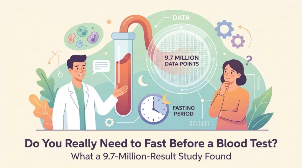
Do You Really Need to Fast Before a Blood Test? What a 9.7-Million-Result Study Found
Most of us have been told the same thing for years: no food after dinner,…
Retatrutide Side Effects: What the 2026 Phase 3 Trials Show
Retatrutide side effects are back in the headlines after Eli Lilly reported new phase 3…
Silent Atherosclerosis in Young Adults: What 16,808 People Revealed
Silent atherosclerosis, the build-up of fatty plaque inside artery walls before any symptom appears, is…
Unexplained Weight Loss and Blood Tests: What a 2026 Oxford Study Found
Losing weight without trying is a symptom that is easy to brush off and hard…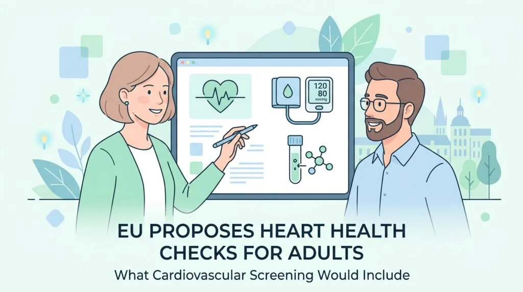
EU Proposes Heart Health Checks for Adults: What Cardiovascular Screening Would Include
The European Commission has put forward a plan to make heart health checks a normal…
mRNA Flu Vaccine: What the First FDA-Approved Shot Means for You
The first mRNA flu vaccine has been approved in the United States, and it changes…
Blood Proteins and 10-Year Mortality: What a 38,000-Person Study Shows
Could one blood sample say something about your risk of dying in the next five…
Sepsis Blood Tests in 2026: What a New Biomarker Could Change
September is Sepsis Awareness Month in the United States, and this year the spotlight includes…
Breast Cancer Screening Guidelines 2026: What to Know This Awareness Month
Every October, pink ribbons bring one question back into focus: when should you actually start…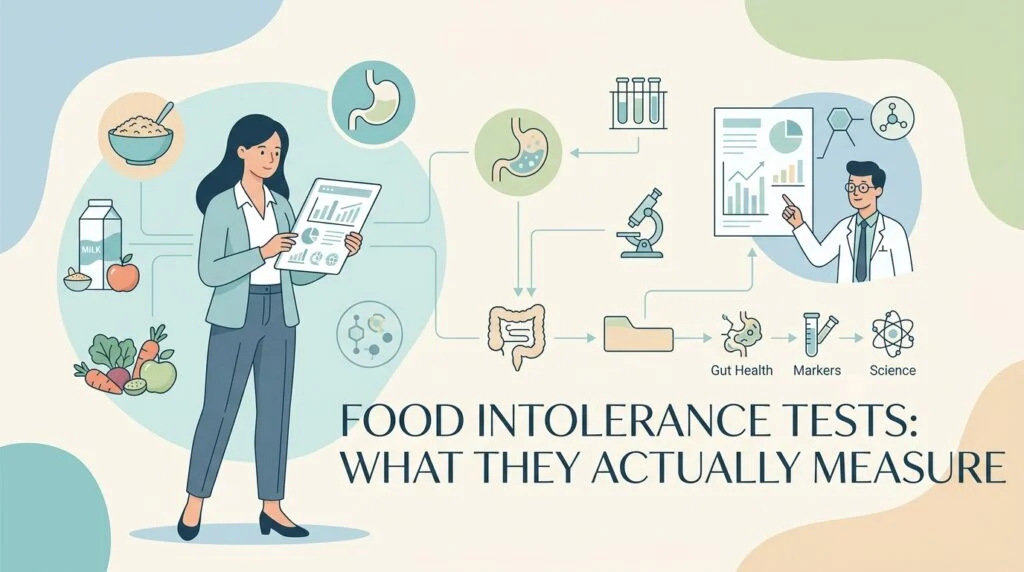
Food Intolerance Tests: What They Actually Measure
Most at-home food intolerance and sensitivity tests measure IgG antibodies, which reflect what you eat…
New 10-Minute Troponin Test Could Speed Up Heart Attack Diagnosis
A French diagnostics company has just launched a point-of-care blood test that can measure a…
FDA Advisory Panel Backs Grail’s Galleri Blood Test: What the Vote Changes Now
On September 23, 2026, an FDA advisory panel did something no expert committee had done…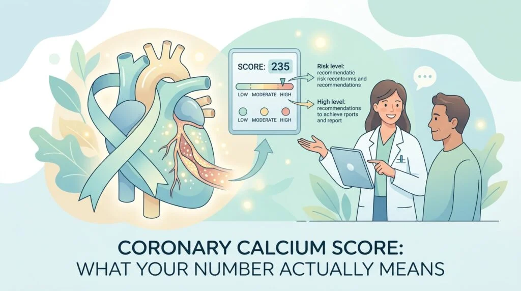
Coronary Calcium Score: What Your Number Actually Means
A coronary calcium score measures calcified plaque, not blockage. This guide explains the two ways…
TORCH Test and Autism: What a Study of 3.7 Million Births Found
A TORCH test is a group of blood tests that looks for infections a pregnant…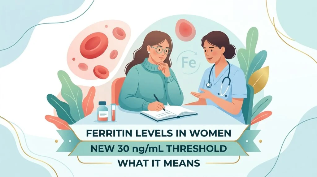
Ferritin Levels in Women: What the New 30 ng/mL Threshold Means
Ferritin levels in women have just been redefined. On September 16, 2026, the American Society…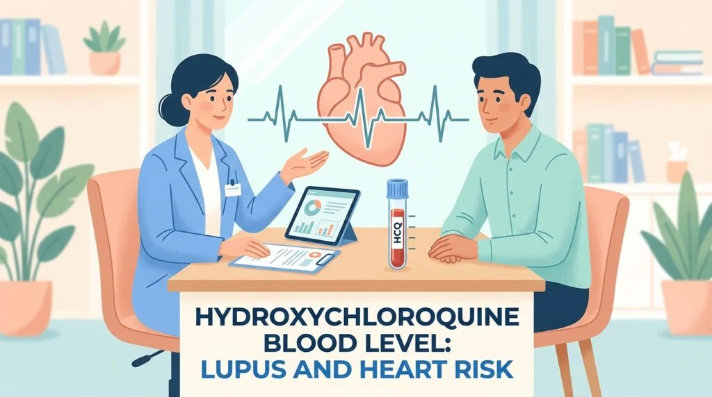
Hydroxychloroquine Blood Level: Lupus and Heart Risk
A Yale-led study covered on 21 September 2026 found that the amount of hydroxychloroquine actually…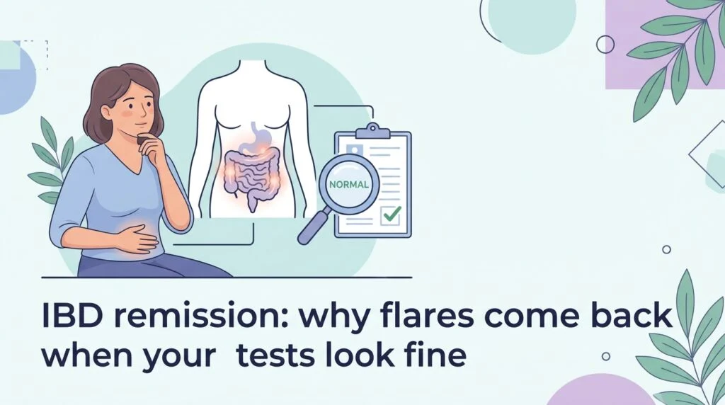
IBD remission: why flares come back when your tests look fine
Being in remission from inflammatory bowel disease usually means the symptoms have gone quiet and…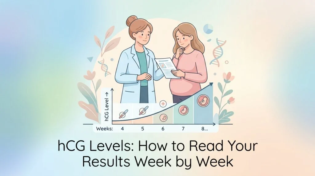
hCG Levels: How to Read Your Results Week by Week
hCG reference ranges by week are so wide that a single value rarely settles anything.…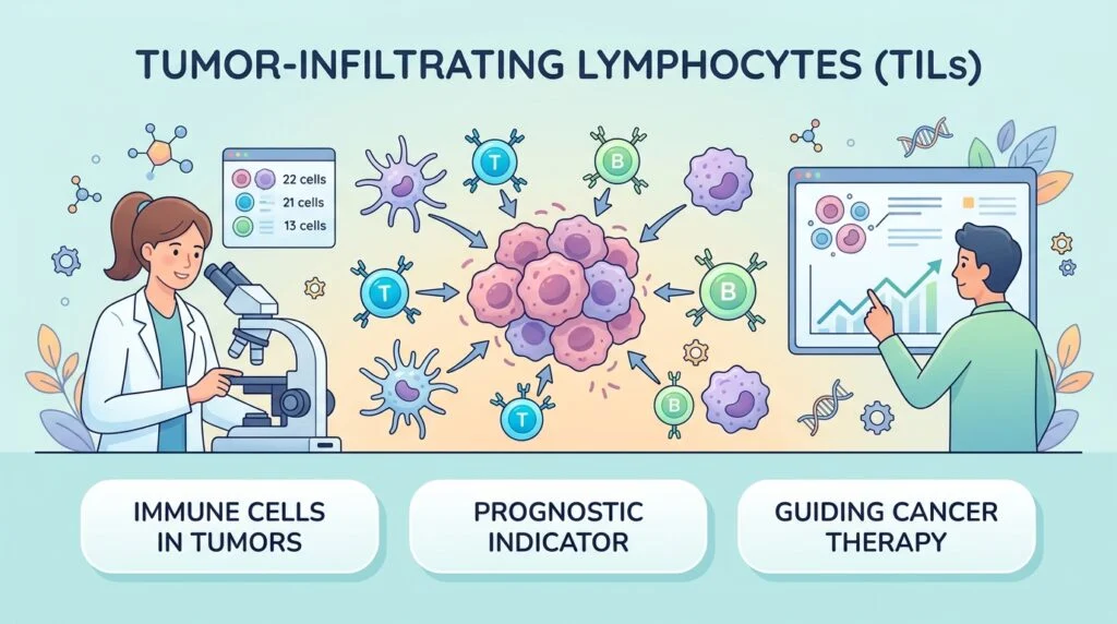
Tumor-Infiltrating Lymphocytes (TILs): What They Mean in Cancer
Tumor-infiltrating lymphocytes are immune cells found inside tumor tissue. This guide explains what brisk, non-brisk…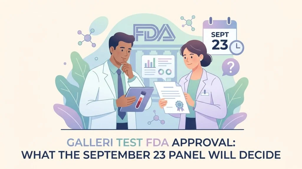
Galleri Test FDA Approval: What the September 23 Panel Will Decide
A US Food and Drug Administration advisory panel meets on September 23, 2026 to vote…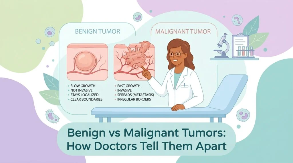
Benign vs Malignant Tumors: How Doctors Tell Them Apart
What really separates benign from malignant tumors, why imaging and blood markers cannot settle the…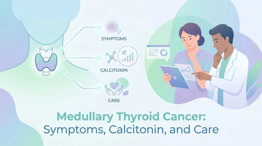
Medullary Thyroid Cancer: Symptoms, Calcitonin, and Care
A plain-language guide to medullary thyroid cancer: how it differs from papillary and follicular disease,…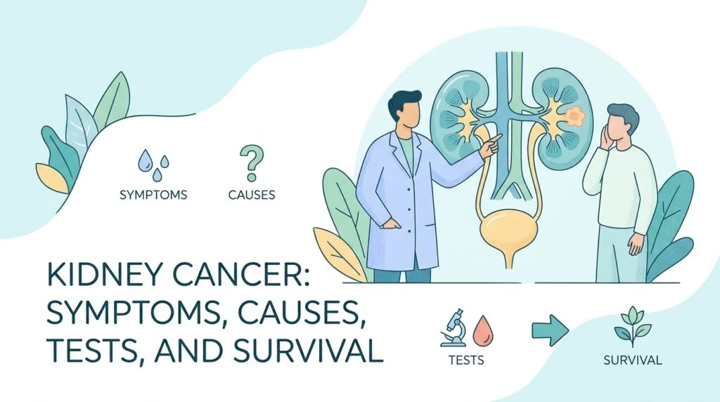
Kidney Cancer: Symptoms, Causes, Tests, and Survival
A plain-language guide to kidney cancer: the types, why two thirds of cases are now…
How Much Whole Grain Per Day? What 87 Trials Found
On 16 September 2026 the European Heart Journal published something dietary advice rarely offers: a…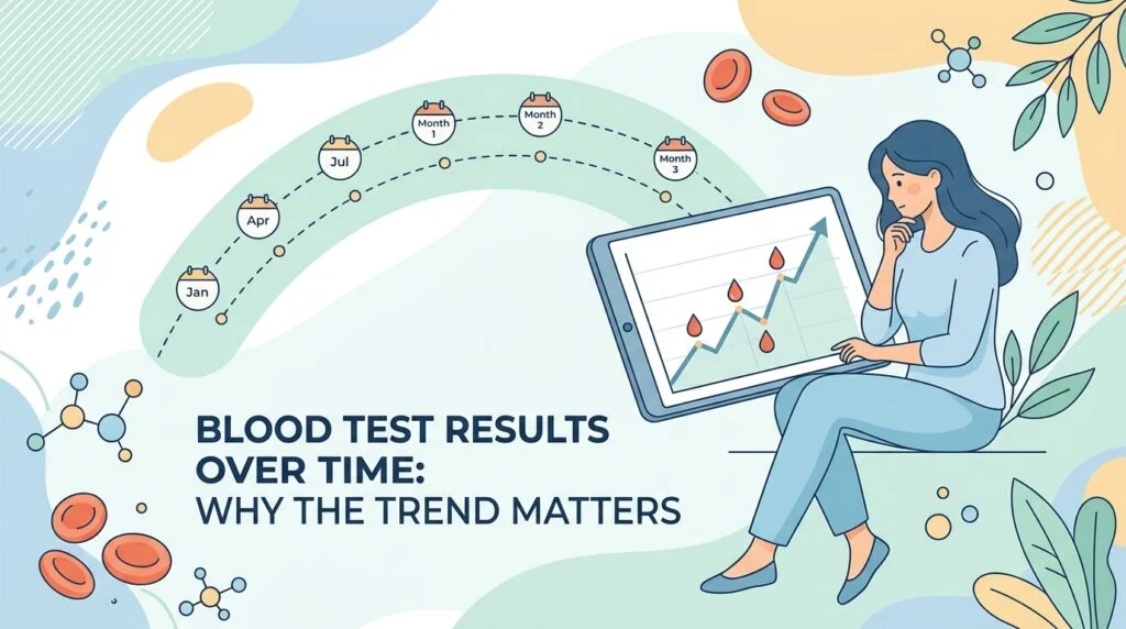
Blood Test Results Over Time: Why the Trend Matters
On September 17, 2026, a research team at the University of Oxford published a study…
Epstein-Barr Virus and Multiple Sclerosis: What the New Relapse Study Changes
Almost every adult carries the Epstein-Barr virus, and almost nobody develops multiple sclerosis. That gap…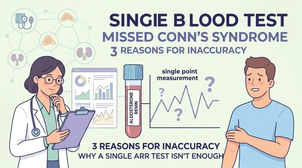
Aldosterone Renin Ratio: 3 Reasons a Single Blood Test Misses Conn’s Syndrome
On September 14 and 15, 2026, research from the University of Bristol and the University…
Pharmacogenomic Testing: 6 Facts About Gene-Guided Prescribing
Two people can swallow the same pill, at the same dose, and end up in…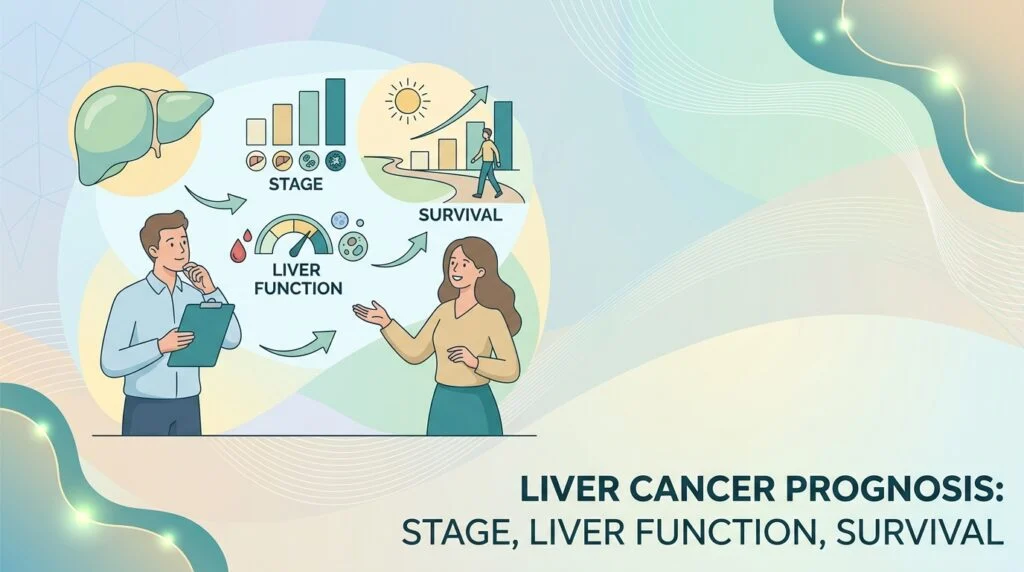
Liver Cancer Prognosis: Stage, Liver Function, Survival
How liver cancer prognosis is actually assessed: BCLC staging, Child-Pugh liver function, performance status, survival…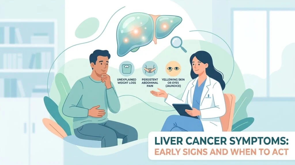
Liver Cancer Symptoms: Early Signs and When to Act
A plain-language guide to liver cancer symptoms: why they appear late, what early and advanced…
The kind of publications you will find here
Blood markers – Discover detailed explanations of the different markers present in your blood analyses. Each article in our publications explains the marker’s role in the body. It also discusses its clinical significance and reasons why it might be elevated or decreased.
Pathologies and diseases – Browse our library of publications on various medical conditions, their symptoms, links to lab analyses, and common therapeutic approaches.
Company news – Stay informed about the latest news concerning AI DiagMe, our partnerships, technological innovations, and upcoming events.
Medical news – Follow recent advancements in the medical field, relevant scientific discoveries, and changes in health practices, with a particular focus on diagnosis and interpretation of analyses. Indeed, our publications aim to provide comprehensive insights into these areas.
Medical glossary – Consult our comprehensive glossary of medical terms to help you understand the specialized vocabulary encountered in your analysis results or during medical consultations.
Our team, composed of healthcare professionals and artificial intelligence experts, regularly updates this resource center to guarantee accurate and up-to-date information. Ultimately, our goal is to support you in understanding your health. To that end, we aim to help you have more informed discussions with your healthcare professionals.
We invite you to explore these publications as a supplement to your medical consultations. Remember that this information does not replace the advice of a qualified healthcare professional.
Going further
Any questions, check out our FAQ or contact us.
If you want to learn more about blood markers in an engaging way, feel free to subscribe and listen to AI DiagMe Podcasts.
Want to take control of understanding your blood tests? Understand your laboratory analysis results in minutes with our aidiagme.com platform; your health deserves this special attention!