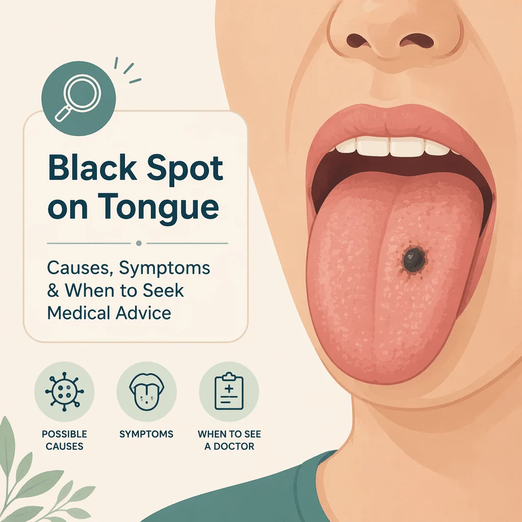
O pată neagră pe țesutul limbii este unul dintre acele semne care aproape întotdeauna arată mai grav decât este în realitate. Majoritatea petelor închise la culoare de pe limbă sunt inofensive, iar multe nu sunt leziuni propriu-zise, ci colorații care dispar în câteva zile odată ce cauza este eliminată.
Cu toate acestea, gura este unul dintre locurile unde o pată pigmentată cu adevărat îngrijorătoare poate fi ignorată luni de zile, considerată “doar o pată”. De aceea, acest articol îndeplinește ambele roluri: mai întâi explică cauzele benigne, apoi prezintă puținele caracteristici care indică faptul că o pată ar trebui examinată în curând.
În acest articol vei afla de ce limba reține atât de ușor culoarea, ce este limba păroasă neagră, care medicamente și alimente colorează limba, cum arată o maculă melanotică și un tatuaj de amalgam, și ce modificări ar trebui să te trimită la stomatolog. Caseta cu semnele de alarmă de mai jos este partea pe care trebuie s-o citești dacă nu citești nimic altceva.
De ce suprafața limbii prinde culoarea atât de ușor
Partea superioară a limbii nu este netedă. Este acoperită de mii de mici proeminențe numite papile, cele mai numeroase fiind papilele filiforme: subțiri, asemănătoare unor fire, ușor keratinizate, cam ca niște peri foarte scurți.
Acești peri sunt uzați în permanență de mâncare, băutură și de frecarea produsă de vorbit și înghițit. Când descuamarea ține pasul cu creșterea, limba rămâne roz și uniformă. Când încetinește, papilele se alungesc și rețin tot ce trece peste ele: cafea, ceai, vin roșu, gudron de tutun, coloranți alimentari, reziduuri de apă de gură și bacterii producătoare de pigment.
Asta explică de ce atât de multe modificări ale limbii de culoare închisă sunt superficiale. Ceva aflat pe suprafața papilelor reprezintă un depozit. Ceva aflat în interiorul mucoasei bucale reprezintă o leziune. A face distincția între acestea două este cel mai important lucru pe care îl face un medic. Modificările reliefate sau cu textură aparte, cum ar fi cele observate în cazul HPV pe limbă, se simt diferit sub vârful limbii față de o pată plată.
Limba păroasă neagră: cel mai frecvent motiv pentru care limba se înnegrește
Limba păroasă neagră, cunoscută medical sub numele de lingua villosa nigra, este același proces dus la extrem. Papilele filiforme nu se mai descuamează, se alungesc și capătă un aspect împâslit, asemănător unui covor, pe mijlocul și spatele limbii. Pigmentul acumulat și bacteriile colorează apoi acel „covor" în maro, verde închis sau negru.
Este benignă și nu evoluează spre altceva. De obicei nu provoacă durere, deși unii oameni semnalează respirație urât mirositoare, senzație de greață sau o schimbare în percepția gustului alimentelor. Dacă gustul este principala ta preocupare, cauzele unui gust neplăcut în gură și gât se suprapun cu factorii declanșatori de mai jos.
Ce tinde să o declanșeze
Studiile identifică în mod constant aceiași factori predispozanți: fumatul, consumul excesiv de cafea sau ceai negru, igiena orală precară, gura uscată, debilitatea generală sau boala și anumite medicamente. Antibioticele sunt un factor declanșator bine documentat, la fel și apele de gură oxidante, inclusiv cele care conțin compuși cu peroxid. Firul comun este orice lucru care încetinește reînnoirea suprafeței limbii sau perturbă echilibrul microbilor rezidenți. De aceea apare adesea în timpul tratamentului pentru altceva și dispare când acel tratament se încheie.
Ce o rezolvă de obicei
Studiile publicate descriu tratamentul ca eliminarea, pe cât posibil, a factorului cauzator, asigurarea pacientului că afecțiunea este benignă și menținerea igienei orale prin debridare blândă, astfel încât papilele să se descuameze normal. În practică: perierea blândă a limbii cu o periuță moale, hidratare corespunzătoare și reducerea consumului de tutun.
Blândețea este cuvântul-cheie și se aplică în mod specific limbii păroase negre. Nu este un motiv să freci energic, să răzuiești cu forță sau să aplici agenți de albire ori apă oxigenată pe orice zonă a limbii tale. Îngrijirea orală de rutină contează aici, la fel cum contează și pentru depunerile întărite din jurul lucrărilor dentare: constanța contează mai mult decât forța. O colorație închisă care nu dispare după câteva săptămâni de curățare blândă merită arătată unui stomatolog, deoarece s-ar putea să nu fie o simplă colorație.
Colorarea: cauza pe care oamenii aproape niciodată nu o asociază
Bismutul și de ce medicamentele pentru stomac îți înnegresc limba
Subsalicilatul de bismut este substanța activă din mai multe remedii gastrice fără prescripție medicală, cel mai cunoscut fiind Pepto-Bismol. În gură și în intestin, bismutul reacționează cu cantități mici de compuși cu sulf produși de bacteriile obișnuite, formând sulfură de bismut, care este neagră.
Rezultatul este o decolorare neagră tranzitorie a limbii, adesea însoțită în același timp de scaune de culoare închisă. Este complet inofensivă și reversibilă. Culoarea revine de obicei la normal în câteva zile de la oprirea medicamentului, iar limba rămâne netedă, fără papilele alungite specifice limbii negre păroase.
Acest lucru contează deoarece o limbă neagră însoțită de scaune negre sună suficient de înfricoșător încât să îi trimită pe oameni la urgențe. Dacă ai luat un remediu care conține bismut în zilele anterioare, acesta este adesea tot răspunsul. Dacă nu ai luat, scaunele negre reprezintă o problemă separată de discutat cu un medic.
Alimente, băuturi, tutun și alți agenți
O mulțime de lucruri obișnuite colorează temporar limba: lemn-dulce, fructe de pădure închise la culoare, sfeclă roșie, coloranți alimentari din dulciuri și băuturi reci, cafea tare și ceai negru, vin roșu și suplimente de fier dacă o pastilă se dizolvă pe limbă. Tutunul contribuie atât prin petele de gudron, cât și prin melanoza fumătorului, în care mucoasa bucală produce mai mult pigment ca răspuns la expunerea cronică la fum. Studiile de specialitate menționează și o listă lungă de medicamente, printre care unele antibiotice, antidepresive și medicamente pentru tensiunea arterială.
Colorarea are o caracteristică utilă: apare destul de brusc, este difuză sau în pete, nu formează o zonă bine delimitată, nu are o componentă reliefată sau nodulară și se estompează odată ce cauza dispare.
Pete plate care aparțin mucoasei în sine
Unele pete închise la culoare nu se află pe limbă, ci în interiorul ei. Acestea persistă pe termen nelimitat fără a cauza vreodată probleme.
Macula melanotică orală este cea mai frecventă leziune pigmentată a gurii: o pată maro sau neagră, plată, bine delimitată, de obicei sub un centimetru, cel mai adesea pe buză sau pe mucoasa obrazului, dar poate apărea și pe limbă. Rezultă dintr-o creștere locală a melaninei, pigmentul care colorează pielea și părul, fără o înmulțire a celulelor producătoare de pigment. Este benignă și, de obicei, rămâne la aceeași dimensiune și culoare ani de zile.
Tatuajul cu amalgam este și mai simplu de explicat. Atunci când o plombă de culoare argintie este frezată sau plasată, fragmente microscopice de metal se pot încrusta în gingia sau mucoasa adiacentă. Organismul le izolează, lăsând o pată permanentă, plată, de culoare albastru-cenușie sau neagră. Este inofensivă, nu se modifică și nu trebuie îndepărtată. Indiciul este localizarea: o pată închisă la culoare situată lângă sau în dreptul unei plombe vechi are o explicație evidentă.
Ambele pot semăna cu ceva mai grav, motiv pentru care medicii întreabă de cât timp există pata și dacă s-a modificat.
Pigmentarea fiziologică normală
Pigmentarea fiziologică, numită uneori pigmentare rasială, este o variantă normală, nu o boală. Este frecventă la persoanele cu ten mai închis și apare sub forma unei colorații maronii sau brun-negricioase a gingiilor, a mucoasei obrajilor și, uneori, a limbii.
De obicei este simetrică, afectează zone largi mai degrabă decât un singur punct bine delimitat, este prezentă din copilărie și nu se modifică. Nimic din toate acestea nu descrie o leziune care necesită investigații. Merită menționată, deoarece persoanele cu acest aspect sunt uneori îndemnate să se îngrijoreze de felul în care arată dintotdeauna gura lor.
Ce vezi și ce înseamnă de obicei
Tabelul de mai jos este un instrument orientativ, nu un diagnostic. Există pentru a te ajuta să descrii cu precizie ceea ce observi.
| Ce observi | Ce este de obicei | Ce să faci |
|---|---|---|
| Un strat întunecat, păros sau împâslit, pe mijlocul și spatele limbii | Limba păroasă neagră, o supracreștere benignă a papilelor de pe suprafața limbii | Periaj ușor zilnic al limbii, hidratare corespunzătoare, eliminarea factorilor declanșatori precum fumatul sau un medicament recent. Menționează acest lucru la următoarea vizită la stomatolog |
| O înnegrire bruscă a întregii limbi după administrarea unui remediu pentru stomac, adesea însoțită de scaune închise la culoare | Colorarea cu bismut, o reacție chimică inofensivă în gură și în tractul digestiv | Este de așteptat să dispară în câteva zile de la oprirea medicamentului. Dacă scaunele închise la culoare persistă fără o explicație legată de bismut, consultă un medic |
| O singură pată plată, brun-neagră, cu aspect neschimbat de ani de zile | Cel mai adesea este o maculă melanotică orală, cea mai frecventă leziune pigmentată benignă a cavității bucale | Menționează-l la un control stomatologic de rutină, pentru a fi documentat și comparat în timp |
| O pată albastru-cenușie lângă sau în dreptul unei plombe vechi | Tatuaj amalgam, fragmente de material de obturaţie dentară încorporate în ţesut | Inofensiv și permanent. Menționează-l pentru a fi consemnat în fișa ta dentară |
| O pată pigmentată nouă care crește, își schimbă culoarea sau forma, ulcerează sau sângerează | Necesită evaluare. Majoritatea unor astfel de pete se dovedesc a fi benigne, dar acesta este tiparul care nu trebuie presupus a fi o simplă colorare | Programează-te la un consult stomatologic sau medical cât mai curând, în loc să o urmărești luni de zile |
Când o pată pigmentată trebuie examinată
Melanomul mucoasei orale este rar, iar marea majoritate a petelor închise la culoare din gură nu sunt nimic de acest fel. Merită totuși un capitol aparte, dintr-un singur motiv: gura este un loc unde melanomul este diagnosticat târziu, de obicei pentru că pata nu durea.
Leziunile pigmentate timpurii din gură sunt nedureroase. Fără durere nu există niciun semnal de alarmă, astfel că pata este observată, considerată o simplă colorare și lăsată în pace. Până când produce o umflătură, o ulcerație care nu se vindecă, sângerare sau dinți care se clatină, de obicei există deja de mult timp. Acest tipar se repetă în toată literatura de specialitate.
Regula practică nu este “să presupui ce e mai rău”. Este regula folosită pentru petele pigmentate de oriunde de pe corp, de la un pată închisă la culoare sub unghie până la a spune o creștere cutanată inofensivă provenită dintr-un melanom: ceea ce contează nu este cum arată o pată într-o anumită zi, ci dacă se schimbă.
Semne de alarmă: consultă un stomatolog sau medic cât mai curând
Programează-te fără întârziere dacă o pată pigmentată din gura ta prezintă oricare dintre următoarele:
- Este nouă sau o pată existentă a început să se schimbe
- Crește sau se extinde la margini
- Marginile sunt neregulate, zimțate sau asimetrice
- Culoarea variază în interiorul petei sau include zone roșii, albastre sau cenușii alături de negru
- A devenit proeminentă sau există o umflătură sub ea sau lângă ea
- Este ulcerată, sângerează sau nu se vindecă
- Dinții din apropiere s-au clătinat sau proteza nu mai potrivește
- Există amorțeală la nivelul limbii sau gurii, sau un nodul la nivelul gâtului
Și regula generală, valabilă chiar și fără niciunul dintre semnele de mai sus: orice pată pigmentată din gură care persistă aproximativ două săptămâni după ce ai eliminat cauza evidentă trebuie examinată. Dacă ai oprit medicamentul, ai redus cafeaua, ți-ai curățat ușor limba și pata nu a dispărut, programează-te la un consult în loc să continui să o urmărești.
Cauze sistemice: când gura este primul semn
Câteva afecțiuni din alte zone ale corpului se anunță prin pigmentare la nivelul gurii, iar în ambele cazuri de mai jos modelul este difuz, nu o singură pată neagră.
Boala Addison, sau insuficiența suprarenală primară, reprezintă incapacitatea glandelor suprarenale de a produce suficient cortizol. O caracteristică recunoscută este pigmentarea brună în pete a mucoasei bucale și a gingiilor, adesea însoțită de pigmentarea pliurilor cutanate și a cicatricilor. Rareori apare izolat: tabloul clinic mai larg include de obicei oboseală persistentă, scădere în greutate, poftă de sare, amețeli la ridicarea în picioare și tensiune arterială scăzută. Investigațiile se concentrează pe hormonii suprarenali, iar ghidurile noastre despre analiză de sânge pentru cortizol, cortizolul scăzut dimineața, și analiza de sânge pentru ACTH explică acele rezultate.
Sindromul Peutz-Jeghers este o afecțiune ereditară în care apar pete mici, întunecate, asemănătoare pistruilor, în jurul buzelor, în interiorul gurii și uneori pe degete, de obicei din copilărie, însoțite de polipi în intestin. Modificările de la nivelul gurii nu reprezintă problema în sine; implicarea intestinului este cea care contează, motiv pentru care acest tablou clinic impune trimiterea la specialist, nu simpla liniștire a pacientului.
Cauzele nutriționale arată diferit. Deficiențele precum vitamina B12 scăzută sau feritină scăzută tind să facă limba dureroasă, netedă și roșie, mai degrabă decât pigmentată.
La cine să mergi și ce face medicul de fapt
Pentru orice problemă din interiorul gurii, medicul stomatolog este primul la care ar trebui să mergi. Mulți oameni sunt surprinși de acest lucru, crezând că stomatologii se ocupă doar de dinți, însă examinarea țesuturilor moi este o procedură de rutină la un control de bilanț, iar stomatologii văd mult mai multe leziuni orale decât majoritatea medicilor de familie.
Perioada Institutul Național de Cercetare Dentară și Craniofacială subliniază același lucru, recomandând ca orice rană, umflătură, pată sau zonă cu sângerare în gură care persistă mai mult de două săptămâni să fie consultată de un stomatolog sau de un medic, și precizând că examinarea țesuturilor moi este nedureroasă și durează doar câteva minute.
Medicul te întreabă când ai observat prima dată pata, dacă s-a schimbat, ce medicamente iei, dacă fumezi și dacă există lucrări dentare în apropiere, apoi o examinează la lumină bună: plată sau reliefată, cu margini clare sau difuze, unică sau multiplă.
Dacă aspectul și istoricul corespund clar unei cauze benigne, rezultatul este de obicei liniștirea pacientului și o notă în fișa medicală, astfel încât modificarea să poată fi comparată ulterior. Dacă există o incertitudine reală, sau leziunea este nouă sau în schimbare, pasul definitiv este biopsia, în care un mic fragment de țesut este examinat la microscop. Studiile privind leziunile pigmentate orale sunt consecvente în această privință: aspectul clinic restrânge lista de posibilități, iar histologia o tranșează.
Un lucru pe care nimeni nu îl poate face, inclusiv această pagină, este să evalueze o leziune pigmentată dintr-o fotografie sau o descriere scrisă. Culoarea se reproduce nesigur, adâncimea și textura nu se fotografiază, iar istoricul contează la fel de mult ca aspectul vizual. Privitul imaginilor online te ajută să înțelegi categoriile; nu îți poate spune în care dintre ele te încadrezi.
Ce spun studiile despre petele pigmentate de pe limbă
Baza de dovezi este inegală, și merită menționat acest lucru. Leziunile pigmentate benigne sunt bine descrise în recenzii. Melanomul oral este suficient de rar încât cea mai mare parte a cunoștințelor provin din rapoarte de caz și serii de cazuri, adică grupuri mici de pacienți descriși împreună, nu din studii clinice.
O analiză din 2025 publicată în American Journal of Clinical Dermatology de Wolk și colaboratori a acoperit întregul spectru al leziunilor pigmentate orale, de la pigmentarea normală și tatuajul de amalgam până la melanomul mucoasei orale. Concluzia: aceste leziuni arată similar din punct de vedere clinic, în ciuda unor biologii foarte diferite, și, deși majoritatea sunt benigne, necesită o evaluare atentă, cu biopsie atunci când este indicat. O analiză narativă din 2025 publicată în Journal of Oral Pathology and Medicine de Maldonado-Mendoza a adăugat că leziunile cu celule pigmentare de orice tip reprezintă mult sub un procent din totalul leziunilor orale. Ce înseamnă asta pentru tine: dacă un medic dorește să examineze mai atent sau să recolteze o probă, nu înseamnă că suspectează ceva grav. Acest lucru reflectă o suprapunere reală în aspectul clinic.
O analiză din 2014 publicată în World Journal of Gastroenterology de Gurvits și Tan rămâne cel mai complet studiu despre limba neagră păroasă, citat deoarece nimic ulterior nu l-a înlocuit. Concluzia: afecțiunea este benignă, prevalența variază considerabil între populații, iar factorii declanșatori sunt fumatul, consumul excesiv de cafea sau ceai, igiena orală precară, gura uscată, debilitatea și utilizarea anumitor medicamente. Tratamentul constă în eliminarea factorului declanșator, reasigurarea pacientului și debridarea blândă. Ce înseamnă asta pentru tine: dacă limba ta închisă la culoare este pufoasă și difuză, nu o pată distinctă, te poți aștepta la o rezolvare completă prin măsuri blânde.
O meta-analiză din 2025, publicată în Journal of Dental Research, Dental Clinics, Dental Prospects de Rodriguez-Archilla și Valverde-Martinez, a combinat zeci de studii care acopereau câteva mii de persoane cu melanom oral. O meta-analiză reunește studii separate într-o singură estimare. Ce s-a descoperit: supraviețuirea a fost scăzută și a scăzut constant după diagnostic, ceea ce autorii au atribuit răspândirii precoce combinate cu diagnosticarea tardivă. Ce înseamnă asta pentru tine: motivul pentru care o pată care se schimbă trebuie examinată ține de momentul intervenției, nu de probabilitatea cancerului. Întârzierea este cea care agravează cazul rar.
În final, un raport de caz din 2009 și o analiză din Journal of Drugs in Dermatology, semnat de Cohen, a documentat apariția limbii negre cauzate de subsalicilat de bismut, confirmată atunci când decolorarea a reapărut la reluarea medicamentului. Ce s-a constatat: limba rămâne netedă, iar culoarea revine la normal odată ce substanța este oprită. Ce înseamnă asta pentru tine: o limbă neagră și netedă după un remediu pentru stomac are o explicație benignă, autolimitantă. Acesta este un singur caz cu o analiză a literaturii de specialitate, nu un studiu amplu, deci tratează-l ca pe o ilustrare, nu ca pe o dovadă a frecvenței.
Concluzia sinceră: cauzele benigne sunt bine caracterizate, cea gravă nu este, deoarece este prea rară pentru a fi studiată corespunzător. Această asimetrie explică de ce există regula leziunii în schimbare.
Întrebări frecvente
O pată neagră pe limbă înseamnă cancer?
Aproape sigur că nu. Melanomul oral este rar, iar marea majoritate a petelor închise la culoare de pe limbă sunt cauzate de colorare, limbă păroasă neagră, o macule melanotică benignă sau pigmentare normală. Motivul pentru care cancerul este menționat deloc nu este că ar fi probabil, ci că este ușor de trecut cu vederea: cancerele pigmentate în stadiu incipient din cavitatea bucală nu dor, așa că sunt ignorate. De aceea, recomandările se concentrează pe schimbare, nu pe aspect. O pată care arată la fel de ani de zile este liniștitoare. O pată nouă sau una care crește, se ulcerează, sângerează sau își schimbă culoarea ar trebui examinată.
Poate Pepto-Bismol să îmi coloreze limba în negru?
Da. Subsalicilatul de bismut, substanța activă, reacționează cu compușii de sulf produși de bacteriile normale din gură și intestin, formând sulfură de bismut neagră. Aceasta colorează frecvent atât limba, cât și scaunul în același timp. Este inofensivă, nu afectează limba, iar culoarea revine la normal în câteva zile de la oprirea medicamentului. Limba rămâne netedă în aceste cazuri. Dacă culoarea neagră persistă mult timp după ce ai oprit medicamentul sau dacă scaunele închise la culoare continuă fără o explicație legată de bismut, merită să menționezi acest lucru medicului.
Ar trebui să merg la medic sau la dentist pentru o pată închisă la culoare pe limbă?
Mai întâi un stomatolog, pentru orice apare în interiorul gurii. Stomatologii examinează țesuturile moi orale ca parte de rutină a unui control, văd mult mai multe leziuni orale decât majoritatea medicilor de familie și pot îndruma direct către specialiști în medicină orală sau chirurgie oro-maxilo-facială. Medicul de familie este alegerea mai potrivită dacă pata este însoțită de simptome în altă parte a corpului, cum ar fi oboseală persistentă, scădere în greutate, amețeli la ridicarea în picioare sau scaune închise la culoare, deoarece acestea indică o cauză sistemică, nu una locală.
Cât timp ar trebui să aștept înainte de a merge la un control?
Aproximativ două săptămâni după eliminarea cauzei evidente este pragul utilizat pe scară largă. Dacă ai renunțat la bismut, ai redus consumul de cafea, ai încetat sau ai redus fumatul și ți-ai curățat ușor limba, cele mai multe colorări și cele mai multe cazuri de limbă păroasă neagră se vor fi îmbunătățit vizibil în acest interval. O pată care nu s-a modificat deloc merită o consultație. Nu aștepta două săptămâni dacă există semne de alarmă: o pată nouă care crește, își schimbă culoarea, se ulcerează, sângerează sau este asociată cu o umflătură sau cu dinți care se mișcă trebuie văzută fără întârziere.
Pot să periez sau să rachez pata?
Perierea ușoară zilnică a limbii cu o periuță moale face parte din recomandările standard pentru limba păroasă neagră și funcționează deoarece problema constă într-un strat care nu s-a descuamat. Nu este potrivită pentru o pată pigmentată plată de pe mucoasa bucală, unde frecarea nu realizează nimic și irită țesutul. Nu raca niciodată cu forță și nu aplica niciodată înălbitor, apă oxigenată sau orice alt produs de albire sau oxidant pe o leziune din gură. Dacă curățarea ușoară nu a îndepărtat o zonă închisă la culoare, aceasta este o informație diagnostică pe care trebuie să o aduci la cunoștința medicului stomatolog, nu un motiv să încerci mai insistent.
De ce am mai multe puncte negre mici în loc de o singură pată?
Mai multe puncte mici închise la culoare răspândite pe suprafața limbii sunt de obicei pigment prins în papilele individuale, ceea ce este frecvent în cazul cafelei sau ceaiului tari, al tutunului, al alimentelor de culoare închisă și al limbii păroase negre în stadiu incipient. Resturile alimentare prinse arată similar. Uneori, aspectul reflectă papile fungiforme pigmentate, o variantă benignă în care papilele în formă de ciupercă de la vârful limbii sunt în mod natural mai închise la culoare. Mai multe pete închise apărute în același timp și care au rămas stabile sunt mult mai puțin îngrijorătoare decât o singură pată care se mărește, dar aceeași regulă de două săptămâni se aplică dacă persistă.
Glosar
| Termen | Definiție |
|---|---|
| Papile filiforme | Proiecțiile subțiri, asemănătoare unor fire, care acoperă cea mai mare parte a suprafeței superioare a limbii și îi conferă textura |
| Limba păroasă neagră (lingua villosa nigra) | O afecțiune benignă în care papilele filiforme nu se descuamează, se alungesc și rețin pigment și bacterii, dând un aspect întunecat și păros |
| Macule melanotică orală | O pată plată, bine delimitată, maro sau neagră, cauzată de melanină în exces în mucoasa bucală; cea mai frecventă leziune pigmentată benignă din această zonă |
| Tatuaj de amalgam | O pată permanentă albastru-cenușie sau neagră cauzată de fragmente microscopice de material de obturaţie dentară încorporate în mucoasă |
| Melanomul mucoasei orale | Un cancer rar care apare din celulele producătoare de pigment din mucoasa gurii, remarcat prin faptul că este nedureros în stadiile timpurii și, prin urmare, diagnosticat târziu |
| Pigmentare fiziologică | Colorare maro normală, de obicei simetrică, a gingiilor și a mucoasei bucale, mai frecventă la persoanele cu ten mai închis |
| Melanina | Pigmentul natural care dă culoarea pielii, părului și unor părți ale mucoasei bucale |
| Bismut subsalicilat | Substanța activă din mai multe remedii gastrice fără prescripție medicală; formează un compus negru inofensiv în gură și în tractul digestiv |
| Xerostomia | Termenul medical pentru gura uscată persistentă, un factor declanșator recunoscut al limbii negre păroase |
| Biopsie | Prelevarea unui mic fragment de țesut pentru a fi examinat la microscop; testul definitiv pentru o leziune orală incertă |
Surse
- Institutul Național de Cercetare Dentară și Craniofacială. Cancerul oral.
- MedlinePlus, Biblioteca Națională de Medicină. Afecțiuni ale limbii.
- Institutul Național al Cancerului. Tratamentul cancerului de buze și al cavității orale (PDQ) – Versiunea pentru pacienți.
- Wolk R, Massi D, Trochesset D. Pigmented Lesions of the Oral Mucosa: Clinical Presentation, Histology, and Recommendations for Management. American Journal of Clinical Dermatology, 2025. doi:10.1007/s40257-025-00950-y. Accesat prin PubMed.
- Gurvits GE, Tan A. Black hairy tongue syndrome. World Journal of Gastroenterology, 2014. doi:10.3748/wjg.v20.i31.10845. Accesat prin PubMed.
- Rodriguez-Archilla A, Valverde-Martinez P. Survival and prognostic factors in mucosal melanomas of the oral cavity: A meta-analysis. Journal of Dental Research, Dental Clinics, Dental Prospects, 2025. doi:10.34172/joddd.025.41492. Accesat prin PubMed.
- Maldonado-Mendoza J. Oral Melanocytic Neoplasms: A Narrative Review. Journal of Oral Pathology and Medicine, 2025. doi:10.1111/jop.70094. Accesat prin PubMed.
- Cohen PR. Black tongue secondary to bismuth subsalicylate: case report and review of exogenous causes of macular lingual pigmentation. Journal of Drugs in Dermatology, 2009. PubMed 20027942.
Lectură suplimentară
- HPV pe limbă: simptome, riscuri și tratamente
- Melanom: Simptome, Diagnostic, Tratamente
- Keratoză Seboreică vs Melanom: Ghid de Simptome
- Cortizol scăzut dimineața: cauze, simptome, tratamente
- Hemoleucograma completă: înțelege-ți rezultatele
Înțelege-ți rezultatele analizelor cu AI DiagMe
O pată neagră pe limbă se diagnostichează prin examinare vizuală, nu printr-un test de sânge, deci acesta este un subiect în care analizele de laborator nu oferă răspunsul. AI DiagMe nu poate vedea limba ta și nu poate evalua o leziune pigmentată dintr-o descriere sau o fotografie. Doar o examinare față în față, și uneori o biopsie, pot face asta.
Unde putem ajuta este după aceea. Dacă un medic investighează o cauză sistemică, precum boala Addison, poate solicita analize care includ cortizolul, ACTH sau un hemoleucogramă completă, iar acele rezultate sunt adesea mai greu de înțeles decât scrisoarea în care sosesc. AI DiagMe le transformă în limbaj simplu pe care îl poți prezenta medicului tău. Nu pune diagnostice.
Obține interpretarea rezultatelor în câteva minute