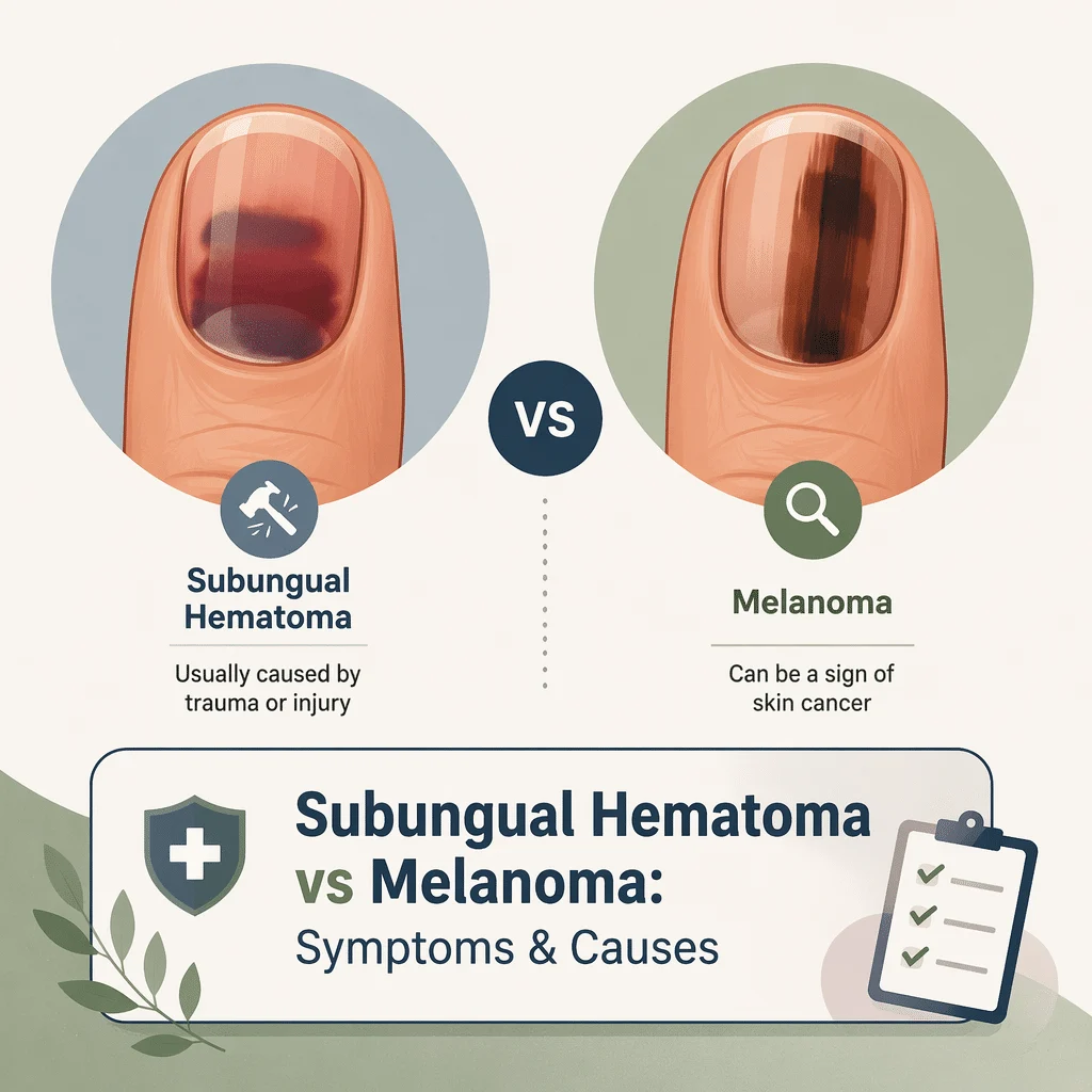
The question of subungual hematoma vs melanoma comes up whenever a dark mark appears under a nail, and the honest answer is that the two can look almost identical in the first days. A subungual hematoma is trapped blood under the nail plate, almost always after an injury. Nail melanoma is a rare cancer that begins in the pigment-producing cells of the nail unit. Both can leave a brown or black patch in someone who remembers a minor knock, which is why a remembered injury settles nothing on its own. In this article you’ll learn which features clinicians actually use, why the way a mark behaves over several weeks tells you more than the way it looks today, which harmless look-alikes are common, and what happens at a dermatology appointment. Nothing here replaces an in-person exam.
What each condition actually is
Blood trapped under the nail
A subungual hematoma is a bruise in a place where the blood has nowhere to go. When the small vessels in the nail bed are damaged, blood collects in the narrow space between the bed and the hard plate above it. Because the plate cannot expand, the pooled blood presses outward, which is why a fresh hematoma can throb even when the injury looked trivial. The Cleveland Clinic describes the typical trigger as direct trauma, such as something crushing or slamming the nail.
Melanoma in the nail unit
Nail melanoma, also called subungual melanoma or nail unit melanoma, starts in the melanocytes, the cells that make pigment. Most sit in the nail matrix, the growth zone tucked under the cuticle that manufactures the nail plate. When abnormal melanocytes there produce pigment, it is baked into the plate as it forms and emerges as a lengthwise stripe. According to a 2023 review of nail unit melanoma published in the journal Cells, roughly two in three nail melanomas first show up as exactly this: a pigmented band running down the nail. That band has a long list of harmless causes too, which is the heart of the problem.
Subungual hematoma vs melanoma: a side-by-side comparison
The table below sets out the features clinicians weigh. Read it as a set of probabilities, not a verdict.
| Feature | More typical of a subungual hematoma | More typical of nail melanoma |
|---|---|---|
| Remembered injury | Often present, sometimes dramatic and dated to the hour | Frequently also reported, because most people can recall knocking a nail at some point |
| Color | Starts red or purple, turns dark brown or black, then often fades toward brown, green or yellow as it resolves | Brown to black pigment that does not pass through the bruise color sequence |
| Behavior over weeks | Moves toward the fingertip as the nail grows, leaving clear nail behind at the cuticle | Stays anchored at the cuticle, because the matrix keeps producing pigment |
| Shape | Blotch, pool or rounded patch, often with a sharp leading edge | Lengthwise band running from cuticle to free edge |
| Band width over time | Not applicable; the patch shrinks rather than widens | May widen, and classically widens more at the cuticle end than at the tip |
| Colors within the mark | Fairly uniform for its stage of healing | Several shades of brown, gray and black side by side within one band |
| Side borders | Defined edge that follows the pooled blood | Blurred or irregular side borders, band not parallel-sided |
| Pigment on surrounding skin | Absent, although bruising of the finger itself can occur | Pigment spilling onto the cuticle or side nail folds, known as the Hutchinson sign |
| Number of nails | Usually one, matching the injured digit | Usually one, most often a thumb, index finger or big toe; bands on many nails point away from melanoma |
| Nail surface | Smooth, unless the injury also damaged the bed | Splitting, thinning, ridging or crumbling of the plate along the band |
| Pain | Common early, from pressure; settles as the blood is absorbed or released | Usually painless in early disease |
A remembered injury is the least reliable line in the table, because a knock is such an ordinary event that people recall one whether or not it explains the mark. And a dark nail with none of the melanoma features still deserves an opinion if it has not changed after a couple of months.
The most useful sign is whether the mark grows out
If you take one idea from this page, take this one. Blood trapped under the plate becomes stuck to its underside, and the nail keeps growing forward from the matrix, so the trapped blood travels with it like a mark on a conveyor belt. Week by week, a strip of clear nail appears behind it at the cuticle, and the dark patch drifts toward the fingertip until it is trimmed away.
A melanocytic band behaves in the opposite way. The pigment is not stuck to the plate: it is manufactured continuously by cells in the matrix and laid into every new millimeter of nail. The band therefore stays anchored at the cuticle no matter how long you watch it, because it is being made afresh.
The Cleveland Clinic notes that it may take six to nine months for a nail to grow out completely and return to its normal color, and fingernails clear faster than toenails. What matters here is not the total time but the direction of travel: after four to eight weeks you should be able to see whether clear nail is appearing behind the mark.
Watching is reasonable when you had an unmistakable injury, the mark is behaving like a bruise, and you have a plan to get it checked if it does not move. It is not reasonable when there was no injury, when the mark is a band rather than a blotch, or when any feature from the right-hand column is present.
Warning signs that mean a nail should be examined
The ABCDEF checklist for nail pigment
Dermatologists use a nail-specific memory aid modeled on the ABCDE rule for moles. It is a prompt to seek an opinion, not a test you can pass at home.
- A, for age and ancestry: nail pigment appearing in adulthood, and higher-risk ancestry groups in whom nail melanoma is more often diagnosed late.
- B, for the band: a brown-to-black band that is wide, has blurred edges, or contains several different shades.
- C, for change: a band that is widening, darkening, or altering its shape, or a mark that fails to grow out.
- D, for digit: a single thumb, index finger or big toe is the most common site.
- E, for extension: pigment extending onto the cuticle or the skin at the sides of the nail, the Hutchinson sign.
- F, for family or personal history of melanoma or atypical moles.
When to see a doctor
- A dark band under a nail that appeared without any injury you can identify.
- A mark that has not started to move toward the fingertip after roughly two months.
- A band that is getting wider, especially if it is wider at the cuticle end.
- Pigment on the cuticle or the skin beside the nail.
- Splitting, thinning or crumbling of the nail plate along the pigmented strip.
- A nail that bleeds, oozes, or develops a lump or ulcer.
- Severe throbbing pain after a crush injury, or a nail injury with a deep cut, a deformed fingertip, or a nail torn from its bed.
- Repeated dark marks under nails with no trauma to explain them, especially alongside easy bruising elsewhere.
Nail melanoma is uncommon, and most dark nails are not cancer. But it is diagnosed later than melanoma on ordinary skin, largely because a bruise is such a convincing explanation. Our team also covers the symptoms and diagnosis of melanoma in a separate guide, and another article explains the link between low iron and bruising.
How a subungual hematoma happens and how it is managed
Crushes, stubs and running shoes
The classic causes are a hammer blow, a car door, a dropped weight, or a toe stubbed hard against furniture. Less obvious is the repetitive kind. When a running or hiking shoe is a little too short, the toe strikes the front of the shoe on every downhill stride, and thousands of small impacts do what one big impact does. The result is the dark toenail runners call runner’s toe, most often on the second toe when it is longer than the big toe. Blood thinners and clotting disorders also let a minor knock produce a large collection, and our team also covers the clotting tests in a coagulation panel.
Drainage, and when it is considered
Trephination means making a small opening in the nail plate to let trapped blood escape and relieve pressure. A 2025 review of fingertip injuries in American Family Physician describes subungual hematoma as diagnosed clinically and managed with either observation or nail trephination. It is usually considered when pressure pain is significant and the injury is recent, since older blood has often clotted and will not drain.
The procedure belongs in a clinic. The Cleveland Clinic is explicit that you should never attempt drainage at home: it risks infection, damage to the nail bed, and a permanently deformed nail. Draining also does not answer the melanoma question, because it only removes blood that was already there.
When imaging matters
A hard crush to a fingertip or toe can fracture the bone beneath. The Cleveland Clinic notes that imaging is used to check for broken bones or other injuries not visible on the surface of the skin. A large hematoma after a significant crush, a fingertip that looks bent or shortened, or a nail lifted off its bed are reasons to consider an X-ray. Deep cuts in the bed may need repair to prevent a permanently split nail.
Harmless look-alikes worth knowing about
Longitudinal melanonychia
Longitudinal melanonychia is simply the descriptive term for a lengthwise pigmented band, and most of the time it is benign. It is common and often entirely normal in people with darker skin tones, where the pigment cells in the matrix are more readily activated. Bands on multiple nails, present for years, with straight parallel edges and a uniform color, are the reassuring pattern. Nail melanoma still occurs across all skin tones, so a band that is new, single, widening or changing deserves an in-person opinion.
Medicines and treatments
Some drugs darken nails, usually across several nails at once and with a clear starting point in time. Certain chemotherapy agents, antimalarials, antibiotics and antiretrovirals are recognized causes. Bringing a list of your medicines to the appointment can settle the question quickly.
Infections and other stains
Fungal nail infection can look dark when debris or old blood collects under a thickened plate; the nail is usually crumbly and discolored across its width rather than in a clean stripe. Pseudomonas bacteria produce a distinctive green-black stain, most often under a nail that has lifted away from its bed. Diabetes changes nail growth and healing too, and our team also describes the nail changes seen in diabetes. Inflammatory skin disease can affect nails as well, and this guide explains the symptoms and triggers of psoriasis. For pigmented spots on the skin rather than the nail, another article compares the differences between seborrheic keratosis and melanoma.
What happens at the dermatologist
The visit starts with history and a look at all twenty nails, because a pattern across several nails changes the interpretation of any one of them. Expect questions about when the mark appeared, whether it has changed, and what medicines you take.
Next comes dermoscopy, an examination with a handheld magnifier and a strong light. It lets the clinician see the internal structure of a band: whether the lines within it are regular and parallel, whether the colors are uniform, and whether pigment truly extends onto the cuticle or is only showing through translucent skin. That last distinction matters, because a false Hutchinson sign is common in benign bands.
If uncertainty remains, the answer comes from a nail matrix biopsy. A small sample of the growth zone is removed under local anesthetic and examined under a microscope. It is the only test that gives a definitive answer, and the 2023 nail unit melanoma review is unambiguous that biopsy with histopathology is necessary to confirm the diagnosis. A biopsy can leave a permanent line in the nail, which is why clinicians weigh it carefully rather than performing one on every band.
Latest scientific advances
Research from the last three years has focused on making this judgment more accurate before anyone reaches for a scalpel.
Telling a real Hutchinson sign from a false one
Pigment spreading onto the cuticle has long been treated as a red flag, but plenty of harmless bands show something similar. A 2025 study in the Journal of the American Academy of Dermatology compared the two directly. The pigment around benign bands tended to have a straight side border and to fade gradually toward the cuticle. The pigment around melanomas was more often patchy, wider than the band itself, and spread further across the nail fold. What this means for you: pigment on the cuticle is a reason to be seen, not a diagnosis, and an experienced clinician can often separate the two in the room.
Watching a band instead of biopsying it straight away
A 2025 study in a European dermatology journal followed adults with a single pigmented band, photographing and re-examining them over time rather than removing them immediately. The changes that mattered were the appearance of new colors, a grainy pattern of pigment, and pigment that grew visibly more intense. Bands that stayed stable stayed benign. What this means for you: structured follow-up is a legitimate plan for an uncertain band, but only with photographs and a booked review date rather than a vague intention to keep an eye on it.
Reassurance for pigmented bands in children
Parents are often alarmed by a dark stripe on a child’s nail, and the features that alarm dermatologists in adults turn up frequently in children without meaning the same thing. A 2024 systematic review and meta-analysis, which pools many studies, gathered data on more than a thousand children with pigmented nail bands. Harmless moles were by far the most common explanation, worrying-looking features such as wide, dark or multi-colored bands were common, and invasive melanoma was not identified in the pooled data. What this means for you: a child’s band still needs a proper look, but the odds are strongly reassuring, and careful monitoring is often preferred to immediate surgery.
A simpler check during surgery
A 2026 study in the International Journal of Dermatology tested whether simply looking directly at the nail matrix during a biopsy, without magnifying equipment, helps identify the earliest melanomas. An irregular, variegated pattern seen with the naked eye picked out early cases more reliably than examining the nail plate alone. What this means for you: nothing you can do at home, but surgeons can extract more information from a procedure you are already having.
Glossary of key terms
| Term | Definition |
|---|---|
| Subungual hematoma | A collection of blood trapped between the nail bed and the nail plate, usually after an injury. Subungual means under the nail. |
| Nail unit | The whole structure of the nail: the hard plate, the bed underneath it, the matrix that produces it, the cuticle and the skin folds at the sides. |
| Nail matrix | The growth zone hidden under the cuticle where the nail plate is manufactured. Pigment made here is built into the nail as it grows. |
| Longitudinal melanonychia | A brown or black band running lengthwise along the nail. It is a description, not a diagnosis, and it has many benign causes. |
| Melanocyte | A pigment-producing cell. Melanocytes in the nail matrix are the source of a pigmented band, and melanoma is a cancer of these cells. |
| Hutchinson sign | Pigment extending from the nail onto the cuticle or the skin folds at the sides. It raises concern for nail melanoma. |
| Pseudo-Hutchinson sign | Pigment that only appears to spill onto the skin, often because it is showing through a translucent cuticle. It is seen with benign bands. |
| Dermoscopy | Examination of the skin or nail with a handheld magnifier and light, which reveals structures and colors invisible to the naked eye. |
| Trephination | A small opening made in the nail plate by a clinician to release trapped blood and relieve pressure. |
| Nail matrix biopsy | Removal of a small sample of the nail growth zone under local anesthetic, examined under a microscope. It is the only definitive test. |
Frequently asked questions
How long does a subungual hematoma take to go away?
The pain usually eases within a few days as the pressure settles, but the visible mark lasts far longer, because it has to travel the full length of the nail before it can be trimmed off. The Cleveland Clinic gives a figure of six to nine months for a nail to grow out completely and return to its normal color. Fingernails clear sooner than toenails, and a big toenail sits at the slower end. The useful checkpoint is much earlier: within about four to eight weeks you should be able to see clear nail appearing behind the mark at the cuticle. If that has not happened, the mark needs an in-person assessment rather than more waiting.
Does a subungual hematoma always need to be drained?
No. Many are managed with observation alone, particularly when the pain is mild and the collection is small. Drainage is aimed at relieving pressure, so it is most relevant when a recent injury is causing significant throbbing. It also becomes less useful as time passes, because clotted blood does not drain readily. The decision belongs to a clinician who can examine the finger or toe, check for an underlying fracture and look at the nail bed, none of which you can assess yourself.
Should I go to the doctor for a subungual hematoma?
Go if the pain is severe or worsening, if the injury was a hard crush, if the fingertip looks deformed, if the nail has been torn or lifted from its bed, if there is a deep cut, or if you have diabetes or poor circulation. Go regardless of pain if you cannot account for the mark with an injury, or if it has not begun to grow out after a couple of months. Never attempt to drain a nail at home.
Can a doctor tell subungual hematoma vs melanoma just by looking?
Often, but not always, and this is the honest limitation at the center of the topic. History, a careful look at all twenty nails and dermoscopy resolve most cases confidently. When they do not, no amount of further looking will settle it, and a nail matrix biopsy is the only test that gives a definitive answer. That is also why photographs sent to a friend, an app, or an online forum cannot rule anything out. If a clinician recommends a biopsy for a band, it is because the visual assessment has reached its limit, not because they think you have cancer.
What does an early nail melanoma look like?
Most often it is an unremarkable brown or black stripe on a single nail, most commonly a thumb, index finger or big toe. Early on it may be narrow, painless and easy to dismiss. The features that raise concern are change over time, a band that widens toward the cuticle, several shades within one band, blurred side borders, pigment on the surrounding skin, and splitting or thinning of the nail along the stripe. A minority of nail melanomas produce no pigment at all and appear as a persistent lump, ulcer or unexplained nail destruction, which is another reason not to rely on color alone.
Is a dark line under a child’s nail dangerous?
It is usually not. Nail melanoma is exceedingly rare in children, and pooled research on more than a thousand children with pigmented nail bands found harmless moles to be by far the most common cause, with no invasive melanomas identified. Children’s bands also often look alarming, with wide, dark or multi-colored pigment, without that carrying the same meaning as it would in an adult. A child’s band should still be examined by a clinician and followed over time, but immediate surgery is frequently avoidable.
Sources
- Cleveland Clinic — Subungual Hematoma: causes, drainage and healing time. my.clevelandclinic.org
- National Cancer Institute — Melanoma Treatment (PDQ), patient version: signs of melanoma and how it is diagnosed. cancer.gov
- Johns Hopkins Medicine — Melanoma: warning signs and sites where melanoma can start, including under a fingernail or toenail. hopkinsmedicine.org
- MedlinePlus Medical Encyclopedia (National Library of Medicine) — Nail abnormalities: dark streaks and the Hutchinson sign. medlineplus.gov
- Conway J, Bellet JS, Rubin AI, Lipner SR — Adult and Pediatric Nail Unit Melanoma: Epidemiology, Diagnosis, and Treatment — Cells, 2023. doi.org/10.3390/cells12060964
- Tsai SY, Hamilton CE, Mologousis MA, Hawryluk EB — Melanoma-like features in pediatric longitudinal melanonychia: a systematic review and meta-analysis — Pediatric Dermatology, 2024. doi.org/10.1111/pde.15597
- Bang JS, Kim JY — Clinical differences between Hutchinson’s sign in subungual melanoma and pseudo-Hutchinson’s sign in benign longitudinal melanonychia — Journal of the American Academy of Dermatology, 2025. doi.org/10.1016/j.jaad.2025.10.089
- Moscarella E, Brancaccio G, Apalla Z, et al. — Digital dermoscopy follow-up for acquired longitudinal melanonychia — Journal of the European Academy of Dermatology and Venereology, 2025. doi.org/10.1111/jdv.20822
- Galili E, Kotek O, Shemer A, et al. — Intraoperative Nail Matrix Assessment in Longitudinal Melanonychia — International Journal of Dermatology, 2026. doi.org/10.1111/ijd.70205
- Hilgefort J, Becker J, Chu J — Fingertip Injuries — American Family Physician, 2025. pubmed.ncbi.nlm.nih.gov
Further reading
- Anemia: Symptoms, Causes, Types, and How It’s Diagnosed
- Low Ferritin: Causes, Symptoms, and Treatment
- Iron Studies Panel: Ferritin, Serum Iron, TIBC and Transferrin Saturation Explained
- CBC vs CMP: Understanding the Tests
- Skin Rash: Causes, Symptoms, and Treatments
Understand your lab results with AI DiagMe
Nail melanoma is diagnosed by biopsy, not by a blood test, and no laboratory panel can tell a bruise from a cancer under a nail. Blood work still has a place at the edges of this topic: dark marks under several nails with no injury to explain them, or bruising out of proportion to a knock, can prompt a doctor to order a complete blood count, a platelet count or clotting times. If you already have those numbers, this guide explains how to read a complete blood count. AI DiagMe turns lab results into plain language so you can arrive at your appointment with better questions; it helps you understand your results, it does not diagnose, and it does not replace your doctor.
Get your results interpreted in minutes