


Seborrheic Keratosis vs. Melanoma describes two very different skin findings that can look similar at first glance. In this article I explain what each one is, how to tell them apart, when to seek medical care, what tests doctors use, and what treatments the two conditions require. You will learn clear signs to watch for, simple self-check steps, and practical prevention tips.
What seborrheic keratosis vs. melanoma means
Seborrheic keratosis refers to a common, noncancerous skin growth that often appears as a waxy or “stuck-on” patch. Melanoma refers to a cancer that starts in pigment-producing cells. Both can appear as dark spots, but melanoma can be life threatening if not treated early. In this section I define each condition in plain language and outline the main differences you should know.
Seborrheic keratosis vs. melanoma: appearance and texture
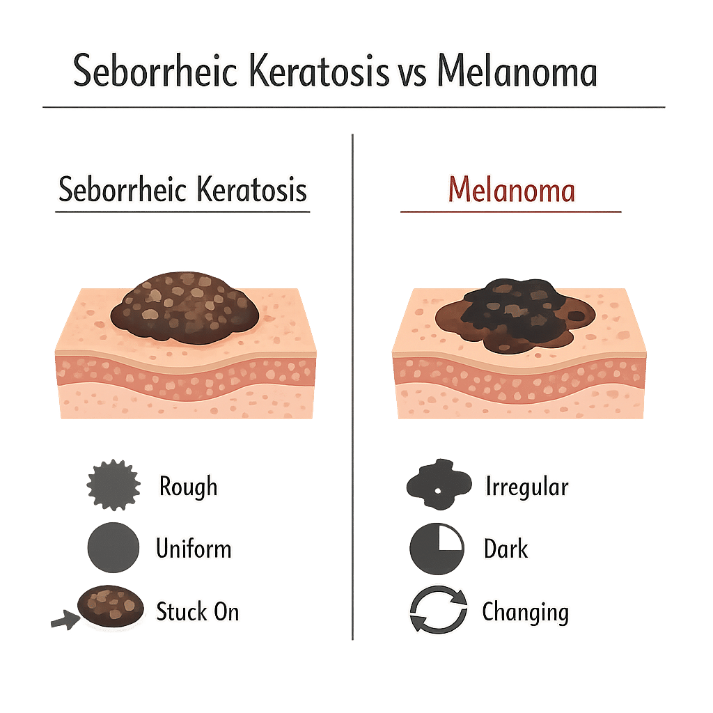
Seborrheic keratoses usually look like round or oval patches. They often have a rough, scaly, or waxy surface. Colors range from light tan to dark brown or black. Many people describe them as feeling like a small crust or stuck-on sticker. They rarely change quickly.
Melanoma commonly appears with irregular color and shape. It may show multiple colors in one spot. The edge often looks uneven or scalloped. A new dark spot that grows, changes shape, or bleeds needs prompt attention. Melanoma often feels flat or slightly raised, and it can develop a firm lump.
How they look and feel
Seborrheic keratoses usually cause no pain. They may itch occasionally. They rarely bleed unless scraped or irritated. Melanoma may itch, bleed, or form an open sore. It can grow steadily over weeks or months. Pay attention when a spot changes noticeably. Also, compare the spot to nearby moles or patches for differences.
Causes and risk factors for melanoma and seborrheic keratosis
Seborrheic keratoses develop most often with aging and family tendency. Sun exposure does not strongly cause them, though sun damage may coexist. Melanoma links strongly to ultraviolet (UV) light from sun or tanning beds. Other melanoma risks include fair skin, many moles, family history, and a history of severe sunburns. In short, one condition mainly reflects age and genetics, while the other links to UV damage and cancer risk.
Seborrheic keratosis vs. melanoma: how doctors make a diagnosis
Doctors start with a careful skin exam. They use a dermatoscope, a handheld tool that magnifies skin features. That tool helps reveal patterns beneath the surface. If a spot looks suspicious, the doctor will recommend a biopsy. During a biopsy the doctor removes part or all of the spot and sends it to a lab for analysis. Pathology gives a definitive diagnosis.
When to see a doctor for seborrheic keratosis vs. melanoma
See a doctor if a spot changes size, shape, or color. Also seek care if a lesion itches intensely, bleeds, or forms an open sore. If you find a new dark spot after age 30, mention it at your next checkup. If you or a family member has melanoma, ask for a skin exam sooner. When in doubt, get professional evaluation rather than assuming a spot is harmless.
Tests to distinguish seborrheic keratosis vs. melanoma
Clinicians use visual exam and dermoscopy first. When needed, they perform a biopsy. A shave biopsy removes the top layers. An excisional biopsy removes the whole lump and a margin of normal skin. Lab specialists then examine tissue under a microscope. They look at cell shape, pattern, and activity to confirm melanoma or a benign growth. Genetic or molecular tests sometimes help in complex cases.
Treatment options for seborrheic keratosis and melanoma
For seborrheic keratosis doctors commonly offer simple removal for comfort or cosmetic reasons. They may freeze the spot with liquid nitrogen, scrape it away, or use laser treatment. Those options remove the growth quickly and typically heal well.
For melanoma treatment depends on stage. Early melanoma usually requires a surgical excision with a margin of normal skin. If the tumor has spread, doctors may recommend lymph node testing, immunotherapy, targeted drugs, or radiation. Specialists design a plan based on tumor thickness and spread. Always follow specialist advice for melanoma care.
Prevention and skin monitoring tips
Protect your skin from UV light by using sunscreen, wearing protective clothing, and avoiding tanning beds. Reapply sunscreen every two hours when outdoors. Perform a monthly skin self-check in good light. Use photos to track new or changing spots. At least once a year, ask a clinician for a full-body skin exam if you have risk factors. Early detection makes treatment more effective.
Seborrheic keratosis vs. melanoma: common mistakes and red flags
People sometimes assume that dark, old-looking spots are harmless. That mistake can delay diagnosis. Also, DIY removal attempts risk infection and hide important features from a doctor. Red flags include rapid growth, color changes, bleeding, and irregular borders. If a spot looks different from your other marks, call a clinician for advice. Trust professional evaluation for anything suspicious.
Frequently Asked Questions (FAQ)
Q: Can seborrheic keratosis turn into melanoma?
A: No. Seborrheic keratoses do not turn into melanoma. However, a melanoma can appear near or next to other skin growths. Always get new or changing spots checked.
Q: How fast does melanoma grow?
A: Growth varies. Some melanomas grow over weeks, while others change more slowly. Any noticeable change over a short period warrants evaluation.
Q: Will a biopsy hurt?
A: A biopsy uses local anesthetic, so you should feel little to no pain during the procedure. You may feel mild soreness afterward, which usually improves in a few days.
Q: Are home remedies safe to remove seborrheic keratosis?
A: No. Home remedies can cause burns and infection. They can also remove evidence needed for diagnosis. See a clinician for safe removal options.
Q: How often should I check my skin?
A: Check once a month. Also see a clinician yearly if you have risk factors. If you notice changes, seek care immediately.
Q: Can sunscreen prevent melanoma completely?
A: No. Sunscreen reduces risk but cannot eliminate it. Combine sunscreen with protective clothing and safe sun habits.
Glossary of Key Terms
- Biopsy: The removal of a small skin sample for lab testing.
- Dermatoscope: A handheld device that magnifies and lights the skin for detailed inspection.
- Immunotherapy: Medicines that help the immune system attack cancer.
- Melanoma: A type of skin cancer that arises from pigment cells.
- Seborrheic keratosis: A common, benign (noncancerous) skin growth.
- UV light: Ultraviolet rays from the sun or tanning beds that can damage skin.
Understand Your Lab Test Results with AI DiagMe
Understanding your skin biopsy or lab results can feel overwhelming. AI DiagMe helps translate lab values and pathology terms into plain language, and it highlights what actions you can discuss with your clinician. Use it to gain clarity before your next visit and to prepare informed questions for your care team.
➡️ Analyze Your Lab Results with AI DiagMe Now
