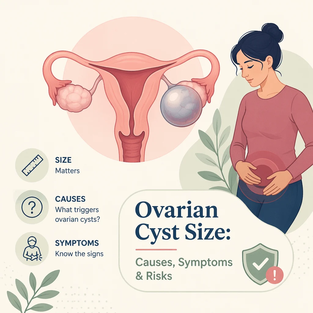
The ovarian cyst size written on an ultrasound report is usually the first number a woman reads, and it is rarely the number that decides anything. A measurement in centimeters tells your doctor how much room a cyst takes up. It does not say what the cyst is made of, whether it is likely to disappear on its own, or whether it carries any risk at all. Two women can both be told they have a 4 cm cyst and be given completely different advice.
In this article you will learn how ovarian cyst size is measured, why the words simple and complex on your report carry more weight than the centimeters, how size thresholds are actually used to choose between watching and operating, why menopausal status changes the reading, which blood tests belong in the work-up, and which symptoms mean you should be seen the same day.
What ovarian cyst size actually tells you
An ovarian cyst is a sac, usually filled with fluid, sitting in or on an ovary. Mayo Clinic describes them exactly that way, and MedlinePlus adds that most women have one at some point in their lives and that they are usually harmless and go away by themselves.
On ultrasound, the sonographer measures the cyst in three planes and reports the largest diameter, generally in centimeters, sometimes in millimeters. Ten millimeters equals one centimeter, so a cyst reported as 35 mm and one reported as 3.5 cm are the same cyst. The measurement is reproducible and easy to compare between scans, which is exactly why it ends up in the headline of the report.
What the ovarian cyst size cannot do is tell you what you are dealing with. A large, entirely fluid-filled sac with thin walls behaves very differently from a small one containing solid tissue. That is why radiologists give at least as much attention to the description of the cyst as to its diameter, and why a bigger measurement is not automatically worse news.
Why functional cysts form, and why most of them go away
Most ovarian cysts are a by-product of a working menstrual cycle rather than a disease. MedlinePlus notes that cysts usually form during ovulation, when the ovary releases an egg. These are called functional cysts, and they come in two flavors.
Follicular cysts
Every month, a fluid-filled follicle grows on the ovary to carry the maturing egg. Mayo Clinic explains that a follicular cyst begins when the follicle does not rupture: it fails to release its egg and simply keeps growing. Nothing has gone wrong with your body; a normal step has just been skipped.
Corpus luteum cysts
After the egg is released, the empty follicle becomes a small hormone-producing structure called the corpus luteum. Cleveland Clinic explains that instead of dissolving, it can fill with fluid and form a cyst. These are the cysts most often found by chance in early pregnancy.
The timelines are reassuring. Mayo Clinic states that functional cysts are usually harmless, rarely cause pain, and often disappear on their own within two to three menstrual cycles. Cleveland Clinic puts it in days rather than cycles: functional cysts generally shrink over time, usually within 60 days, without treatment. This is the single most useful fact for anyone staring at a fresh ultrasound report, and it explains why the most common medical response to a cyst is a repeat scan rather than an operation.
Simple versus complex: the words that outrank the measurement
If you read only one line of your report, read the description. A simple cyst is a single-chamber sac filled with clear fluid, with thin, smooth walls and nothing solid inside. A complex cyst contains something else: internal walls dividing it into chambers, thickened walls, debris, or solid areas and small projections growing into the cavity.
That distinction drives almost everything that follows. The StatPearls chapter on ovarian cysts hosted on the NCBI Bookshelf states that simple cysts smaller than 6 cm carry less than a 1 percent risk of being cancerous, and that single-chamber cysts under 10 cm are usually benign regardless of the patient’s age. The same chapter lists the features that push toward surgery: a cyst larger than 10 cm, a complex multi-chamber mass, or papillary projections and solid components. Notice that only one of those four is a measurement.
Endometriomas
Sometimes called chocolate cysts, these form when tissue similar to the uterine lining grows on the ovary and bleeds into a pocket month after month. They have a characteristic ground-glass look on ultrasound. Our library covers endometriosis, how it is diagnosed and how it is treated.
Dermoid cysts
Also called mature teratomas, these are present from birth and contain tissue such as fat, hair or even teeth. They look alarming on a report and are almost always benign, but they do not shrink on their own and they carry a higher risk of twisting.
Cystadenomas
These grow from the surface of the ovary and fill with watery or mucous fluid. They can become very large and are usually benign, but because they keep growing they are more often removed than watched.
Polycystic ovaries are not ovarian cysts
This is the most common confusion in the whole topic. A report saying an ovary has a polycystic appearance describes many small follicles arranged around the edge of the ovary. Those are immature follicles, not cysts, and they are not measured or followed the way a cyst is. Our library explains polycystic ovary syndrome, its symptoms and its diagnostic tests.
How ovarian cyst size guides follow-up and surgery
Size does count, but it acts as a modifier on top of the appearance rather than as a verdict of its own. The table below shows how a typical report line translates into a plan. Every threshold in it comes from a published source, and your own doctor may reasonably decide differently based on your symptoms and history.
| What the report says | What it usually means | What typically happens next |
|---|---|---|
| Simple cyst under 6 cm, before menopause | Almost certainly a functional cyst; StatPearls puts the risk of cancer for simple cysts under 6 cm at under 1 percent | Watchful waiting, often with no repeat scan at all; most shrink without treatment |
| Simple cyst 6 to 10 cm, before menopause | Still usually benign; StatPearls notes single-chamber cysts under 10 cm are usually benign at any age | A repeat ultrasound in a few weeks or months to confirm it is shrinking or gone |
| Simple cyst larger than 10 cm | Benign in appearance, but bulky enough to press on neighboring organs | Removal is commonly discussed; Cleveland Clinic and StatPearls both cite 10 cm as a threshold for considering surgery |
| Any cyst found after menopause | Ovulation has stopped, so a functional cyst is no longer the expected explanation | Follow-up imaging and often blood tests before anything is assumed |
| Complex cyst with solid areas, thick walls or papillary growths | The description, not the measurement, is what raises concern | Specialist review with structured risk scoring; StatPearls lists these features among surgical indications |
Two other things push toward surgery independently of the ovarian cyst size: symptoms you cannot live with, and a cyst that refuses to disappear. MedlinePlus puts it simply, saying you may need surgery if you have pain, are past menopause, or if the cyst does not go away.
Why menopausal status changes how your ovarian cyst size is read
The same 4 cm simple cyst is a routine finding at 32 and a reason for a proper work-up at 62. The logic is straightforward. Before menopause, ovulation happens every month, so the most likely explanation for a new fluid-filled sac is the cycle itself. After menopause, ovulation has stopped, so the ordinary explanation no longer applies and the cyst has to be accounted for another way.
That does not make a postmenopausal cyst dangerous. Simple fluid-filled cysts are found fairly often after menopause and most are still benign. What changes is the threshold for looking more closely: repeat imaging is more likely to be arranged, and blood work is more likely to be added. MedlinePlus is direct about the background risk, stating that ovarian cysts can rarely become cancerous and that this risk increases as you get older. Our library describes the symptoms and stages of menopause in more detail.
When to seek care immediately
Most cysts cause nothing worse than a dull ache. A small number cause an emergency, and the warning signs are the same whatever the ovarian cyst size on your last report.
Go to an emergency department the same day, without waiting for an appointment, if you have sudden severe pain low in the abdomen or on one side, especially when it comes with any of the following:
- nausea or vomiting
- fever
- dizziness, faintness or actually fainting
- rapid breathing or a racing heart
- cold, clammy skin
Mayo Clinic’s instruction is to get immediate medical help for sudden severe abdominal or pelvic pain, or pain with fever or vomiting. Cleveland Clinic uses almost identical wording for severe abdominal pain that comes on suddenly with vomiting or fever.
Two complications explain why. Ovarian torsion is the ovary twisting on the ligaments that hold it, which cuts off its own blood supply; a heavy cyst makes this more likely, and the ovary can be lost if it is not untwisted quickly. A ruptured cyst spills its contents into the pelvis, and if it tears a blood vessel it can bleed internally. MedlinePlus is unambiguous: if a cyst bursts or causes bleeding, you should get medical help right away. Milder one-sided pain that builds gradually is a different situation, and our library covers ovary pain after sex and its usual causes.
Which blood tests belong in the work-up
No blood test can tell you whether a cyst is harmless. That sentence is worth reading twice, because the internet is full of the opposite claim. Blood work supports the picture the ultrasound has already drawn; it does not replace it.
CA-125 is not a screening test
CA-125 is a protein measured in blood, and it is the test most often misunderstood in this whole subject. MedlinePlus states plainly that a provider cannot use CA-125 to screen for ovarian cancer if you are not at high risk, because non-cancerous conditions raise it too. The list of benign causes is long and ordinary: endometriosis, pelvic inflammatory disease, uterine fibroids, liver disease, and simply having your period or being pregnant. A raised CA-125 in a 30-year-old with endometriosis usually means endometriosis.
Where the test earns its place is in a postmenopausal woman with a mass on ultrasound, and in monitoring a cancer that has already been diagnosed. Our library explains the CA-125 blood marker and how it is interpreted, and also covers the wider family of tumor markers and their limits.
The other tests your doctor may order
A pregnancy test comes first in anyone who could be pregnant, because a corpus luteum cyst is a normal early-pregnancy finding and because ectopic pregnancy is on the list of things that cause sudden pelvic pain. Our library explains how a pregnancy test works and how to read it. A complete blood count checks for blood loss and for signs of infection, and our library covers the complete blood count and how to read it.
In younger women with a solid or partly solid mass, doctors may add germ cell tumor markers, since the rare tumors seen in that age group tend to release specific proteins. Our library describes the alpha-fetoprotein blood test. Hormone testing has no role in sizing a cyst, though it may be requested if the underlying question is about cycles or fertility. If the concern extends beyond a cyst, our library covers ovarian cancer, its symptoms and its testing pathway.
Latest scientific advances in reading ovarian cysts
The research of the past few years has moved away from the measurement and toward structured ways of describing what a cyst looks like. Here is what has changed, in plain terms.
The first shift is a shared vocabulary. O-RADS is a scoring system that sorts a cyst into a risk group, roughly from almost certainly harmless up to high concern, based on its appearance rather than its diameter. A large international study published in JAMA Oncology in 2023 tested it against what those cysts actually turned out to be in thousands of women, and found that the categories did separate low-risk from high-risk lesions as intended. What this means for you: the risk category on your report is not one radiologist’s impression, it comes from a system that has been checked against real outcomes.
The second is that no single system has won. A 2025 pooled analysis in the Journal of Clinical Ultrasound compared O-RADS head to head with two tools from the IOTA group, the Simple Rules and the ADNEX model, and found no meaningful difference between them. A 2026 review in the same journal went further and asked whether the Simple Rules still work when a general gynecologist rather than a subspecialist applies them; the drop in accuracy was small. What this means for you: if your first scan is done at a community clinic rather than a referral center, the assessment is still likely to be sound.
The third is early automation. A team at the University of Chicago published work in 2024 training software to trace the outline of a cyst on ultrasound automatically and separate its internal parts, as a step toward computer-assisted classification. This is a research result, not a tool in clinics, and it should be read as a direction of travel rather than a change to your care.
The fourth is combination. A 2025 study in Cancer Imaging built a score merging the O-RADS category, contrast-enhanced ultrasound and the CA-125 result, and reported fewer false alarms than ultrasound scoring alone. It was done at a single center and needs confirmation elsewhere before anyone changes practice, but it points at where the field is heading: imaging and blood work read together, neither one trusted on its own.
Running underneath all of it is a steady move toward doing less. The StatPearls guidance that a single-chamber cyst under 10 cm can be monitored with repeat ultrasound rather than removed reflects a broad shift away from operating on findings that would have resolved by themselves.
Glossary
| Term | Definition |
|---|---|
| Adnexa | The structures beside the uterus: the ovaries, the fallopian tubes and their supporting tissue. An adnexal mass is a lump found in that area. |
| Corpus luteum | The small hormone-producing structure left behind on the ovary after an egg is released. It normally dissolves; if it fills with fluid it becomes a cyst. |
| Cystadenoma | A benign growth arising from the surface of the ovary and filling with watery or mucous fluid. It can become large and does not resolve on its own. |
| Dermoid cyst | Also called a mature teratoma. A benign cyst present from birth that contains tissue such as fat, skin or hair. |
| Endometrioma | A cyst formed when tissue similar to the lining of the uterus grows on the ovary and bleeds into a pocket. Sometimes called a chocolate cyst. |
| Follicle | The small fluid-filled sac on the ovary that holds a maturing egg. A follicle that grows without releasing its egg becomes a follicular cyst. |
| O-RADS | Ovarian-Adnexal Reporting and Data System. A standardized way of scoring how likely an ovarian mass is to be benign, based on its appearance on imaging. |
| Ovarian torsion | The ovary twisting on its supporting ligaments and cutting off its own blood supply. A medical emergency causing sudden severe one-sided pain. |
| Simple cyst | A single-chamber sac containing only clear fluid, with thin smooth walls and nothing solid inside. The lowest-concern appearance on a report. |
| Transvaginal ultrasound | A scan performed with a slim probe placed in the vagina, giving a much closer view of the ovaries than a scan through the abdomen. |
Frequently asked questions
What size of ovarian cyst is dangerous?
There is no single number that turns a cyst dangerous, which is why no reputable source publishes one. Appearance matters more: StatPearls reports that simple cysts under 6 cm carry less than a 1 percent risk of being cancerous, and that single-chamber cysts under 10 cm are usually benign at any age. Above 10 cm, both Cleveland Clinic and StatPearls describe surgery as something to discuss, mainly because large cysts press on other organs and are more prone to twisting. A complex cyst with solid parts is taken more seriously than a large simple one, whatever the centimeters say.
Can a 3 cm ovarian cyst be cancerous?
It is very unlikely if the cyst is described as simple. StatPearls places simple cysts under 6 cm in a group with less than a 1 percent risk of cancer, and a 3 cm cyst before menopause is the classic profile of an ordinary functional cyst. The picture is different if a 3 cm cyst is described as complex, or if it is found after menopause; in those situations the size is reassuring but the context is not, and follow-up is arranged on the strength of the description rather than the measurement.
How fast do ovarian cysts grow?
There is no reliable published growth rate, and any specific figure you find should be treated with suspicion. Functional cysts follow the rhythm of the cycle, appearing and often resolving within weeks: Mayo Clinic gives two to three menstrual cycles and Cleveland Clinic gives around 60 days. Dermoid cysts and cystadenomas grow slowly over years. Because growth cannot be predicted from one scan, doctors measure change directly by repeating the ultrasound after an interval and comparing.
What is a normal ovarian cyst size in millimeters?
Small fluid-filled structures on the ovary are normal rather than abnormal. Developing follicles are present every cycle and are typically reported in millimeters; a mature follicle before ovulation is a normal finding, not a cyst. Reports often start using the word cyst above about 30 mm, or 3 cm. Ten millimeters is one centimeter, so a 45 mm cyst and a 4.5 cm cyst are identical, and the unit change on a report does not mean anything has changed.
Will birth control pills shrink a cyst I already have?
They are not described that way. MedlinePlus states that birth control pills can help prevent new cysts, which is a different claim from shrinking an existing one. By suppressing ovulation they stop the monthly process that creates functional cysts in the first place, which is useful for someone who keeps getting them. A cyst that is already present is generally left to resolve on its own or reassessed on a repeat scan.
Can a large cyst affect pregnancy?
Most pregnancy-related cysts settle by themselves. StatPearls reports that most pregnancy-associated cysts, both corpus luteum and follicular, resolve spontaneously by 14 to 16 weeks of gestation, and that resolution is less likely when a cyst is larger than 5 cm or has a complex appearance. A cyst found in early pregnancy is therefore usually watched rather than treated. Persistent, large or complex cysts are discussed individually with an obstetrician, since the balance of risks in pregnancy is not the same as outside it.
Sources
- MedlinePlus, U.S. National Library of Medicine — Ovarian Cysts, 2026 — medlineplus.gov/ovariancysts.html
- Mayo Clinic — Ovarian cysts: symptoms and causes, 2026 — mayoclinic.org
- Cleveland Clinic — Ovarian Cysts, 2026 — my.clevelandclinic.org
- StatPearls, NCBI Bookshelf, National Library of Medicine — Ovarian Cyst — ncbi.nlm.nih.gov/books/NBK560541
- MedlinePlus Medical Tests — CA-125 Blood Test (Ovarian Cancer), 2026 — medlineplus.gov
- Timmerman S, Valentin L, Ceusters J, et al. — External Validation of the Ovarian-Adnexal Reporting and Data System (O-RADS) Lexicon and the International Ovarian Tumor Analysis 2-Step Strategy — JAMA Oncology, 2023 — doi.org/10.1001/jamaoncol.2022.5969
- Almeida G, Bort M, Alcazar JL — Comparison of the Diagnostic Performance of O-RADS With IOTA Simple Rules and ADNEX Model for Classifying Adnexal Masses: A Head-To-Head Meta-Analysis — Journal of Clinical Ultrasound, 2025 — doi.org/10.1002/jcu.24048
- Hierard D, Padilla-Prieto C, Arego C, et al. — Have IOTA Simple Rules the Same Diagnostic Performance When Used by Nonexpert Examiners as Compared to Expert Examiners? Systematic Review and Meta-Analysis — Journal of Clinical Ultrasound, 2026 — doi.org/10.1002/jcu.70248
- Whitney HM, Yoeli-Bik R, Abramowicz JS, et al. — AI-based automated segmentation for ovarian/adnexal masses and their internal components on ultrasound imaging — Journal of Medical Imaging, 2024 — doi.org/10.1117/1.JMI.11.4.044505
- Jiang Z, Pu W, Luo X, et al. — Integrating O-RADS US v2022, CEUS, and CA125 to enhance the diagnostic differentiation of ovarian masses: development of the OCC-US model — Cancer Imaging, 2025 — doi.org/10.1186/s40644-025-00918-5
Further reading
- Our library explains how to read your blood test results.
- Our library covers the follicle-stimulating hormone blood test.
- Our library describes luteinizing hormone and how to read its results.
- Our library explains estradiol, the main estrogen marker.
- Our library covers testosterone as a blood marker in women and men.
Understand your lab results with AI DiagMe
A cyst on an ultrasound rarely travels alone: your doctor may order a pregnancy test, a complete blood count, or a CA-125 measurement alongside it, and those results arrive as numbers with no explanation attached. AI DiagMe turns a lab report into plain language, showing you what each value measures, where it sits relative to its reference range, and which questions are worth raising at your next appointment. It helps you understand your results. It does not diagnose you, and it does not replace your doctor.
Get your results interpreted in minutes