A gallbladder rupture is a surgical emergency: the wall of the gallbladder tears and bile leaks out into the abdomen, where it does not belong. It almost never happens out of nowhere. In the great majority of cases it is the end point of an inflamed, infected gallbladder that has gone untreated for too long, and it carries a real risk of severe infection and death. That is why the only safe response to the warning signs described below is to seek emergency medical care immediately, rather than waiting to see whether the pain settles on its own. In this article you’ll learn how a gallbladder reaches the point of tearing, which symptoms should send you to an emergency room, how doctors confirm the diagnosis, what treatment involves, and what recent research says about timing and outcomes.
What a gallbladder rupture actually is
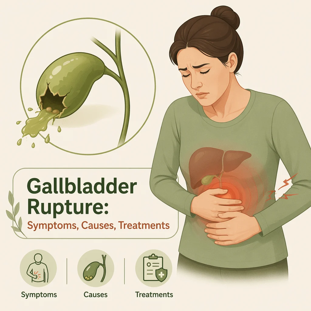
The gallbladder is a small pear-shaped pouch tucked under the liver on the right side of the abdomen. Its only job is to store bile, the greenish digestive fluid the liver makes to help you absorb fat, and to squeeze it into the small intestine after a meal. A gallbladder rupture, also called gallbladder perforation, means that this pouch has developed a hole or tear.
Bile is not a harmless fluid once it escapes. Inside the digestive tract it is useful; loose in the abdominal cavity it is chemically irritating and frequently contaminated with bacteria. The result is either a walled-off pocket of infected fluid next to the liver, or a widespread inflammation of the abdominal lining known as bile peritonitis. Both need hospital treatment. Neither improves with rest, painkillers, or dietary changes.
The three patterns doctors describe
Surgeons group perforations using a system named after the physician who first described them, the Niemeier classification. Stripped of the technical vocabulary, it describes three very different situations.
- Free perforation: the tear opens directly into the abdominal cavity and bile spreads widely. This is the least common pattern and the most dangerous, because it produces bile peritonitis quickly.
- Contained perforation: the body walls the leak off with inflamed tissue and forms an abscess, a pocket of pus, around the gallbladder. This is the pattern reported most often in hospital series.
- Chronic perforation with a fistula: the leak slowly carves an abnormal channel between the gallbladder and a neighboring structure, usually a loop of bowel. It develops over weeks rather than hours.
The distinction matters because it changes the treatment plan. A free perforation is an operating-room emergency. A contained perforation can sometimes be drained first and operated on later, once the infection is controlled.
How a gallbladder gets to the point of tearing
There is a fairly consistent chain of events behind most cases. It usually starts with gallstones. When a stone lodges in the neck of the gallbladder or in the cystic duct, bile can no longer drain. Pressure builds inside the pouch, the wall stretches and becomes inflamed, and acute cholecystitis begins. The National Institute of Diabetes and Digestive and Kidney Diseases describes this blockage as the trigger for a gallbladder attack, and notes that untreated gallstone disease can lead to complications.
If the pressure is not relieved, the stretched wall starts to lose its blood supply. Tissue begins to die, a stage called gangrenous cholecystitis, and dead tissue tears easily. Mayo Clinic describes exactly this sequence: untreated inflammation can cause death of gallbladder tissue, which may in turn cause the gallbladder to burst or develop a tear. The table below shows how the pain typically changes as that chain progresses.
| Stage | How the pain behaves | Typical duration | What else you may notice |
|---|---|---|---|
| Gallstone attack (biliary colic) | Steady, intense pain high in the right side or center of the abdomen, often after a fatty meal; it fades on its own | Minutes to a few hours | Nausea and vomiting; no fever between attacks; you feel normal afterward |
| Acute cholecystitis | The pain no longer settles; it stays, worsens, and often spreads toward the right shoulder blade | More than six hours, then days | Fever, marked tenderness when the area is pressed, rising white cell count and CRP |
| Gangrenous cholecystitis | Pain may become oddly less sharp as the dying wall loses its nerve supply, while the person looks visibly worse | Days | High fever, fast pulse, drowsiness or confusion, especially in older adults |
| Perforation | Pain can ease briefly as the built-up pressure is released, then returns, spreads across the whole abdomen and stays | Hours to days after the wall gives way | Rigid, board-like abdomen, shaking chills, rapid heartbeat, low blood pressure, jaundice |
When there are no gallstones at all
A minority of cases begin without a single stone. In people who are already critically ill after major surgery, severe burns, or a long stay in intensive care, blood flow to the gallbladder can fall so low that the wall becomes inflamed and ischemic, meaning starved of oxygen. Researchers reviewing this pattern in eGastroenterology in 2023 argued that it deserves its own label, ischemic cholecystitis, because it behaves differently from stone-related disease and carries a heavier burden of complications. Severe infections such as typhoid and, in some parts of the world, parasitic infestation of the bile ducts can also inflame a stone-free gallbladder to the point of perforation.
Warning signs that call for emergency care
Gallbladder rupture does not announce itself with one unmistakable sign. In one hospital series of 72 patients published in 2025, pain in the upper right abdomen was present in roughly nine out of ten cases, but the rest of the picture varied widely. What matters is the combination and the direction of travel: pain that is escalating rather than settling, in someone who has been unwell for days.
Call emergency services now, or go straight to an emergency room, if you have any of the following.
- Severe upper right abdominal pain that keeps getting worse over hours instead of easing
- Pain that spreads to the right shoulder, the shoulder blade, or across the whole abdomen
- An abdomen that becomes hard and board-like, painful to touch anywhere
- Fever with shaking chills, sometimes called rigors
- Yellowing of the skin or the whites of the eyes, or urine that turns dark
- Repeated vomiting, particularly when nothing stays down
- A racing heartbeat, clammy skin, dizziness on standing, or very little urine
- New confusion, drowsiness, or slurred speech in an older adult
The deceptive lull
One pattern deserves a warning of its own. A gallbladder under high internal pressure hurts intensely. When the wall finally gives way, that pressure is released, and the pain can briefly improve. People understandably read this as recovery. It is not. Within hours the pain usually returns, spreads, and becomes constant as bile irritates the lining of the abdomen. A sudden improvement after days of severe biliary pain is a reason to be checked urgently, not a reason to stay home.
Who is most at risk
Perforation is a complication of delay more than anything else. The single most consistent risk factor across published series is a long interval between the first symptoms and arriving at a hospital; in the 2025 series mentioned above, patients had on average been unwell for more than two weeks before presenting. Other recognized risk factors include:
- Older age, which blunts pain and fever and can mask how serious the illness is
- Diabetes, which impairs the immune response and dulls abdominal pain through nerve damage
- A weakened immune system, whether from medication, chemotherapy, or chronic disease
- Critical illness in an intensive care setting, the classic setting for stone-free disease
- Previous episodes of cholecystitis that were treated with antibiotics alone and never fully resolved
Reported patterns by sex differ from one hospital series to another, so it is not a reliable way to judge personal risk. Age, diabetes, and above all delay are far more informative.
How doctors diagnose a gallbladder rupture
Diagnosis rests on imaging. Blood tests support the picture and show how sick a person is, but they cannot prove that a gallbladder has torn.
Imaging comes first
Ultrasound is almost always the first test, because it is fast, involves no radiation, and can be done at the bedside. A 2024 review in Radiographics catalogued the findings radiologists look for, including a distended gallbladder, a thickened or interrupted wall, and inflamed fat around the organ. The most conclusive ultrasound sign is complex fluid sitting just outside a visible defect in the wall. The same review made a practical point worth knowing: the classic bedside test in which a doctor presses under the ribs while you breathe in, known as the sonographic Murphy sign, is much less reliable than its reputation suggests, so a negative result does not rule anything out.
A CT scan usually follows. It maps the full extent of the leak, shows abscesses and free fluid, and helps distinguish a gallbladder problem from other abdominal emergencies. Even so, perforation is not always obvious before surgery: in a 23-year review of 100 patients published in 2026, fewer than half of cases were identified preoperatively, although accuracy improved substantially in the later years of the study as imaging technology advanced.
What blood tests add
Laboratory results build the supporting picture and guide urgency. Typically ordered are:
- White cell count and C-reactive protein, which rise with inflammation and bacterial infection. Our team also explains the C-reactive protein inflammation marker, and separately covers what elevated neutrophil counts reveal.
- Total and direct bilirubin, which climb when bile cannot drain and produce visible jaundice.
- Alkaline phosphatase and gamma-glutamyl transferase, two enzymes that rise together when a bile duct is obstructed.
- ALT and AST, the liver enzymes that indicate whether liver cells are also suffering. A dedicated guide reviews the causes of high ALT levels.
- Lipase, which helps separate gallbladder disease from an inflamed pancreas.
None of these numbers is specific on its own. A normal white cell count does not exclude a rupture, particularly in an older or immunosuppressed patient. Our team also details how to read a complete blood count, and a separate article explains how to read a liver panel.
Conditions that look similar
Several emergencies produce comparable pain and need to be told apart quickly, which is another reason imaging is decisive. Acute pancreatitis is the closest mimic, and a dedicated guide describes the pancreatic enzymes amylase and lipase used to identify it; our library also covers the symptoms and causes of pancreatitis. A perforated appendix causes similar peritoneal signs lower in the abdomen, and we explain the symptoms and risks of a burst appendix. Persistent bilious vomiting has its own differential, reviewed in our article on the causes of vomiting bile.
How a gallbladder rupture is treated
Treatment is always delivered in a hospital and always combines three elements: controlling the infection, removing or draining the source, and supporting the body while it recovers. Nothing about this is manageable at home.
Surgery
Removing the gallbladder, an operation called cholecystectomy, is the definitive treatment. Surgeons prefer the keyhole approach, but heavy inflammation can obscure the anatomy and force a switch to open surgery through a larger incision. That conversion is a safety decision, not a failure. Any bile and pus in the abdomen is washed out during the same operation.
Drainage when surgery is too risky
Some patients are too unstable or too frail for immediate surgery. In that case a radiologist can place a thin tube through the skin into the gallbladder or the abscess to drain the infected fluid, buying time until an operation becomes safer. Antibiotics alone, with or without drainage, are used more often than many people expect, particularly for contained perforations in older patients carrying several other illnesses.
Antibiotics, fluids, and monitoring
Intravenous antibiotics, fluids, and pain control begin immediately and continue around the procedure. Blood pressure, heart rate, kidney function, and inflammatory markers are followed closely, because the main danger in the first days is sepsis, the body’s overwhelming and damaging response to infection.
Recovery and what comes after
Recovery from a straightforward keyhole removal takes a week or two. Recovery from a perforation is longer, because it involves an infection as well as an operation: a hospital stay of several days to a couple of weeks is common, sometimes with a drain left in place. Fatigue can persist for a month or more.
Living without a gallbladder is entirely possible. Bile simply flows continuously from the liver into the intestine instead of being stored between meals. Some people notice looser stools or discomfort after very fatty meals for a few weeks or months; this usually settles. No permanent medication is required for most people. Return promptly if fever, worsening pain, jaundice, or persistent vomiting appears after discharge.
Latest scientific advances
According to PubMed, several studies published since 2023 have sharpened the picture of how gallbladder perforation is recognized and treated. Here is what they found, and what each one means in practice.
Operating sooner shortens the hospital stay. A large analysis of a United States national surgical database, published in the Journal of Trauma and Acute Care Surgery in 2025, compared patients whose gallbladder was removed within two days of admission with those who waited longer. The early group went home roughly three days sooner on average, with no increase in complications, readmissions, or repeat operations. The same paper set out the scale of the problem: perforation complicates somewhere between two and eleven percent of acute cholecystitis cases, and reported death rates have ranged widely and remain high. What this means for you: if you are admitted with a suspected perforation, an early operation is not haste, it is current best practice.
Imaging is getting better at spotting it before surgery. A single-center review of 100 patients across 23 years, published in Cureus in 2026, found that fewer than half of perforations were identified before the operation over the whole period, but that accuracy improved markedly in the more recent years as scanners and protocols advanced. Contained perforations with a walled-off abscess were by far the most common type, and free perforation into the abdomen was not seen at all in that cohort. What this means for you: modern imaging catches most of these cases early, and the most common form is the more containable one.
Not every patient needs the operating room first. A 2025 Cureus study of contained perforations found that the choice between surgery, tube drainage, and antibiotics alone was driven mainly by age and the number of other illnesses a patient carried, and that carefully selected nonoperative treatment produced comparable short- and medium-term results in high-risk patients. What this means for you: if your medical team proposes draining first and operating later, that is a recognized strategy, not a second-best compromise.
A newer drainage technique looks promising for frail patients. An international study across ten hospitals, published in Digestive Endoscopy in 2026, tested draining the gallbladder from inside the stomach or intestine using an ultrasound-guided endoscope and a short metal stent, in people with a contained perforation who were poor candidates for surgery. Symptoms and scan findings resolved in about 19 of every 20 patients, though roughly one in eight had a complication and one patient died. What this means for you: it is an encouraging option for people who cannot safely undergo an operation, but it remains a specialist procedure carrying real risk, and these are early results from a look back at past cases rather than a randomized trial.
Stone-free disease is being recognized as its own problem. A 2023 review in eGastroenterology argued that gallbladder inflammation caused by poor blood flow in critically ill patients should be separated from other stone-free cases, because it is harder to spot and more likely to progress to perforation. What this means for you: if a relative in intensive care develops unexplained fever and abdominal tenderness, the gallbladder is worth asking about, even with no history of gallstones.
Glossary
| Term | Definition |
|---|---|
| Acute cholecystitis | Sudden inflammation of the gallbladder, usually because a gallstone is blocking the outflow of bile. It is the condition that precedes most ruptures. |
| Acalculous cholecystitis | Inflammation of the gallbladder without any gallstones. It is seen mainly in people who are already critically ill. |
| Bile | The greenish digestive fluid made by the liver and stored in the gallbladder. It helps the intestine absorb fats. |
| Biliary colic | The self-limiting attack of pain caused by a gallstone temporarily blocking the gallbladder outlet. It settles once the stone moves. |
| Cholecystectomy | Surgical removal of the gallbladder, done either through several small keyhole incisions or through one larger open incision. |
| Gangrenous cholecystitis | A stage in which part of the gallbladder wall dies from lack of blood supply. Dead tissue tears easily, which is why this stage precedes perforation. |
| Niemeier classification | The system surgeons use to sort perforations into free, contained, and chronic fistulous types. It guides how urgently a patient is taken to the operating room. |
| Percutaneous drainage | Placing a thin tube through the skin, guided by imaging, to empty infected fluid from the gallbladder or an abscess beside it. |
| Peritonitis | Inflammation of the membrane lining the abdominal cavity. When it is caused by leaked bile it is called bile peritonitis. |
| Sepsis | A dangerous, body-wide reaction to infection that can damage organs and drop blood pressure. It is the main threat in the days after a rupture. |
Frequently asked questions
Can a ruptured gallbladder heal on its own without surgery?
The tear itself does not simply seal up and resolve the way a cut on your skin would. What can happen is that the body walls the leak off, forming a pocket of infected fluid that stops bile spreading through the abdomen. That is a reprieve, not a cure, and it still needs hospital treatment with antibiotics and usually drainage. In selected patients, especially older people with several other illnesses, doctors may treat a contained perforation without operating straight away and plan surgery later. That decision belongs to a surgical team with imaging in front of them, and it is never a reason to manage symptoms at home.
How long can you live with a ruptured gallbladder?
There is no safe waiting period, and no published figure that would let anyone predict it. Some contained perforations smolder for days or weeks before they are discovered, which is why patients in hospital series have often been unwell for a long time. A free perforation that spills bile into the abdomen can cause life-threatening infection within hours. Because you cannot tell from the outside which situation you are in, any suspicion of a gallbladder rupture is an emergency, and the sensible assumption is always the urgent one.
Can a ruptured gallbladder kill you?
Yes. Published reports describe death rates that vary considerably between hospitals and patient groups, but they are consistently far higher than for uncomplicated gallbladder inflammation. Death usually comes from sepsis rather than from the tear itself. The reassuring part is that outcomes depend heavily on how quickly treatment starts, and most people who reach a hospital promptly and are treated with surgery, drainage, and antibiotics recover.
How serious is a ruptured gallbladder compared with a gallstone attack?
They are not on the same scale. A gallstone attack is painful but self-limiting: the pain builds, lasts up to a few hours, then fades, and you feel normal in between. A rupture is a surgical emergency involving an infection that has escaped its container. The practical dividing line is whether the pain settles. Pain that persists beyond six hours, comes with fever, or keeps intensifying has stopped being a simple gallstone attack and needs assessment the same day.
What causes a gallbladder to rupture in someone with no gallstones?
Most often, severely reduced blood flow to the gallbladder in someone who is already critically ill after major surgery, severe trauma, extensive burns, or a long intensive care admission. The wall becomes starved of oxygen, inflames, and can tear without any stone being involved. Severe infections, including typhoid fever and parasitic infestation of the bile ducts in regions where these are common, are other recognized causes. Blunt injury to the upper abdomen, from a car crash or a fall, can also tear the gallbladder directly, though this is rare.
Can blood tests alone show that a gallbladder has ruptured?
No. Blood tests are supporting evidence, not proof. A rising white cell count and CRP tell you that inflammation and probably infection are present; bilirubin, alkaline phosphatase, and gamma-glutamyl transferase point toward a blocked bile duct; ALT and AST show whether the liver is affected; lipase helps rule the pancreas in or out. All of these can be abnormal in uncomplicated cholecystitis, and all can be near-normal in an older or immunosuppressed patient who has in fact perforated. Ultrasound and CT are what confirm the diagnosis.
Sources
- Mayo Clinic — Cholecystitis: symptoms and causes, 2024 — mayoclinic.org
- National Institute of Diabetes and Digestive and Kidney Diseases (NIH) — Gallstones — niddk.nih.gov
- MedlinePlus, National Library of Medicine — Gallbladder Diseases, 2025 — medlineplus.gov
- Patel R, Tse JR, Shen L, Bingham DB, Kamaya A — Improving Diagnosis of Acute Cholecystitis with US: New Paradigms — Radiographics, 2024 — doi.org/10.1148/rg.240032
- Wu R, Dumas RP, Nomellini V — Early versus delayed laparoscopic cholecystectomy for gallbladder perforation — Journal of Trauma and Acute Care Surgery, 2025 — doi.org/10.1097/TA.0000000000004491
- Tandon V, Govil D, Kumar S, Gupta S, Shah SP — Gallbladder Perforation: A 23-Year Study of 100 Patients and Evolving Management Strategies — Cureus, 2026 — doi.org/10.7759/cureus.108275
- Singh H, Chejara RK, Thippeswamy NG, et al. — Decoding the Enigma of Gallbladder Perforation: Patient Profiles, Diagnosis, and Step-Up Management Pathways — Cureus, 2025 — doi.org/10.7759/cureus.84791
- Youssef M, Aziz M, Rasheed A — Type II Gallbladder Perforation: A Management Conundrum or a Surgical Indecision? — Cureus, 2025 — doi.org/10.7759/cureus.95749
- Spadaccini M, Franchellucci G, Auriemma F, et al. — EUS-Guided Gallbladder Drainage in Acute Cholecystitis With Contained Perforation: An International Multicenter Study — Digestive Endoscopy, 2026 — doi.org/10.1111/den.70199
- Favela JG, Argo MB, Huerta S — Aetiology, diagnosis and management for ischaemic cholecystitis: current perspectives — eGastroenterology, 2023 — doi.org/10.1136/egastro-2023-100004
Further reading
- AST/ALT ratio: what it means and how to read it
- High CRP levels: causes, symptoms, treatments
- Understanding lipase levels: causes and risks
- Complete blood work guide: what a full blood panel includes
- Low albumin: causes, symptoms, and risks
Understand your lab results with AI DiagMe
Gallbladder emergencies are diagnosed with scans, but the blood work drawn alongside them tells you a great deal about how your liver and bile ducts are coping. If you are holding a report and trying to make sense of your white cell count, your C-reactive protein, your bilirubin, or your liver enzymes, AI DiagMe explains each value in plain language and shows how the numbers fit together. It helps you understand your results and prepare better questions for your appointment. It does not diagnose, and it does not replace your doctor.
Get your results interpreted in minutes