



Las beta-1 globulinas son una de las bandas de proteínas que el laboratorio mide al realizar una electroforesis de proteínas séricas, el estudio que separa las proteínas de tu sangre en distintos grupos. Si tu reporte muestra beta-1 globulinas fuera del rango de referencia indicado, es normal querer entender qué significa ese valor. Este artículo explica qué son las beta-1 globulinas, qué mide la electroforesis, cómo se toma la muestra de sangre y por qué el nivel puede subir o bajar. También verás cómo la banda beta-1 se ubica junto a la albúmina y las demás fracciones de globulinas, y cuándo vale la pena comentar el resultado con tu médico. El objetivo es ayudarte a leer tus resultados con mayor confianza y prepararte para esa conversación.
¿Qué son las beta-1 globulinas?
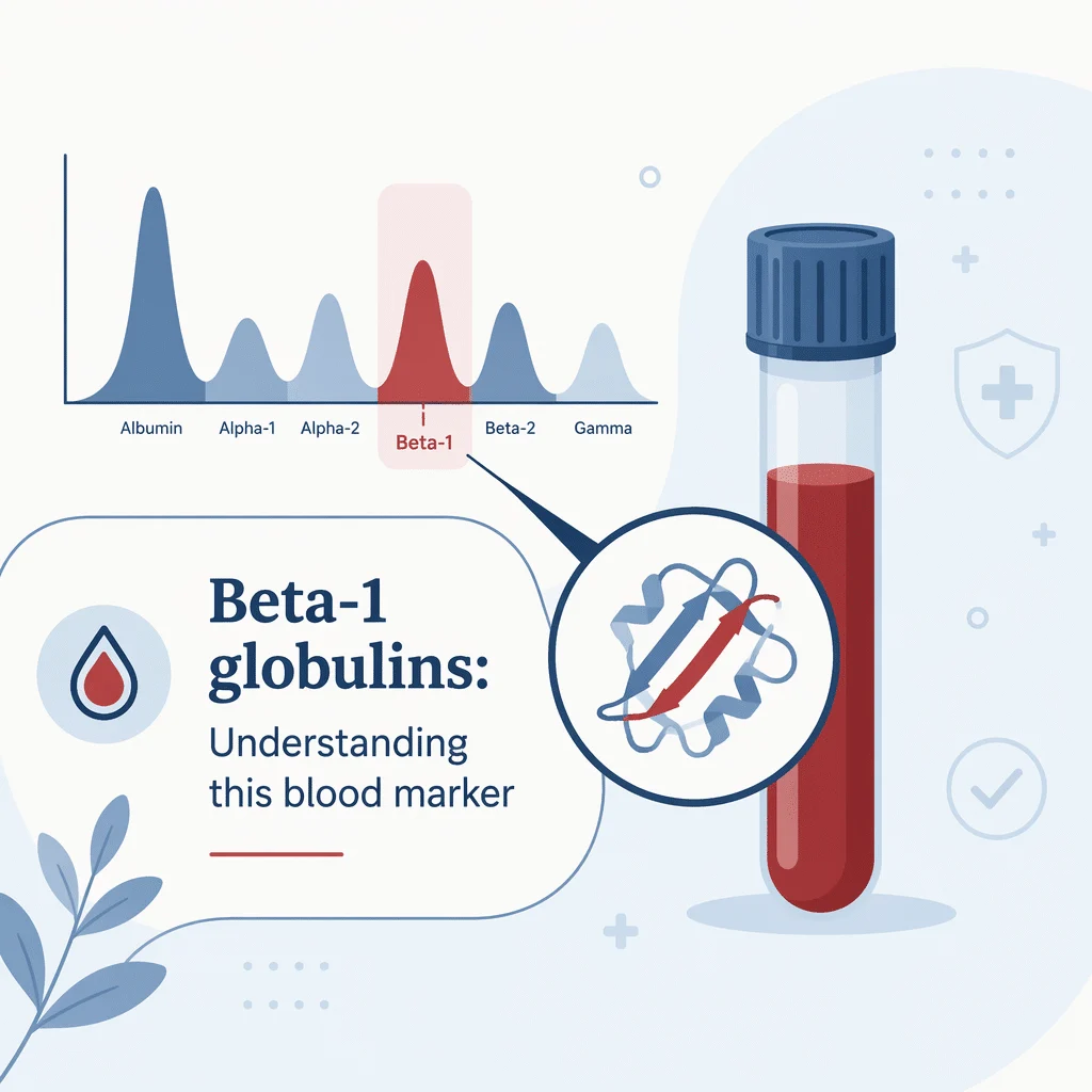
El plasma sanguíneo transporta miles de proteínas. Los laboratorios las agrupan en dos grandes familias: la albúmina, que forma un solo grupo grande, y las globulinas, que se dividen en fracciones más pequeñas. Las beta-1 globulinas son una de esas fracciones. Se ubican en la región beta del patrón de electroforesis, entre las globulinas alfa y las globulinas gamma, donde se encuentran los anticuerpos.
La banda beta-1 está dominada por una sola proteína llamada transferrina, la proteína transportadora que lleva el hierro por todo tu cuerpo. Como una sola proteína constituye la mayor parte de la banda, las beta-1 globulinas tienden a moverse en paralelo con tus niveles de hierro. Para ver cómo se comporta ese transportador por sí solo, puedes consultar el artículo dedicado al análisis de sangre de transferrina y transporte de hierro. Aquí también aparecen cantidades menores de otras proteínas, y la combinación exacta depende del método que use el laboratorio.
Dónde se ubica la beta-1 entre las fracciones de proteínas
La electroforesis generalmente reporta cinco o seis fracciones en un orden fijo: primero la albúmina, luego alfa-1, alfa-2, beta (a veces dividida en beta-1 y beta-2) y gamma. Las globulinas beta-1 forman el borde inicial de esa región beta. La banda vecina contiene proteínas diferentes, y puedes comparar ambas una vez que leas nuestro análisis de sangre de beta-2 globulinas. La primera fracción alfa tiene su propio artículo explicativo, así que también puedes leer nuestro globulinas alfa-1 en sangre.
Qué mide la electroforesis de proteínas séricas y cómo se toma la muestra
La electroforesis de proteínas séricas, frecuentemente abreviada como SPE o SPEP, coloca una pequeña cantidad de suero en un gel o en un tubo fino relleno de líquido y hace pasar una corriente eléctrica a través de él. Las proteínas se desplazan a distintas velocidades según su tamaño y carga eléctrica, por lo que se separan en bandas. Un escáner convierte esas bandas en una gráfica de picos, y el laboratorio mide el tamaño de cada pico, incluidas las globulinas beta-1. Para entender mejor la gráfica paso a paso, te recomendamos revisar nuestro guía para interpretar los resultados de la electroforesis de proteínas séricas.
Cómo se toma la muestra de sangre
El estudio utiliza una toma de sangre convencional. Un profesional de la salud limpia la piel, introduce una aguja en una vena del brazo y extrae un tubo de sangre. La mayoría de las personas no necesitan ayunar, pero sigue las indicaciones que imprima tu laboratorio, ya que otros estudios solicitados en la misma muestra pueden tener sus propias reglas. Luego, la sangre se centrifuga para separar el suero, que se carga en el analizador.
Las principales fracciones de la electroforesis de un vistazo
La tabla a continuación muestra las fracciones habituales, las principales proteínas que contiene cada una y lo que un cambio suele reflejar. La ubicación puede variar entre laboratorios, así que tómala como una guía general y no como una regla fija.
| Fracción proteica | Principales proteínas que contiene | Lo que un cambio suele reflejar |
|---|---|---|
| Albúmina | Albúmina | Nutrición, balance de líquidos y función hepática |
| Alfa-1 | Alfa-1 antitripsina y alfa-1 glicoproteína ácida | Inflamación aguda |
| Alfa-2 | Haptoglobina, alfa-2 macroglobulina y ceruloplasmina | Inflamación, destrucción de glóbulos rojos y pérdida de proteínas por el riñón |
| Beta-1 | Transferrina (componente principal) | Transporte de hierro y estado del hierro |
| Beta-2 | Complemento C3, beta-2 microglobulina y beta-lipoproteína (LDL) | Complemento y actividad inmunitaria |
| Gamma | Inmunoglobulinas (IgG, IgA e IgM) | Infección, inflamación y proteínas monoclonales |
Cómo se reportan las globulinas beta-1 y por qué el rango depende del laboratorio
Las beta-1 globulinas se reportan como concentración en gramos por litro (g/L) o gramos por decilitro (g/dL), o como porcentaje de la proteína total. Cada laboratorio imprime su propio rango de referencia junto a tu resultado, y esos rangos varían por razones válidas. Algunos laboratorios usan gel de agarosa y otros electroforesis capilar, y ambas técnicas dividen la región beta de forma ligeramente distinta. Varios laboratorios reportan una sola banda beta combinada en lugar de separar las beta-1 globulinas de las beta-2 globulinas.
Debido a esta variación, la comparación más confiable es siempre tu propio valor contra el rango impreso en tu reporte. Un resultado que parece alto en la escala de un laboratorio puede estar perfectamente dentro del rango en otro. Si quieres comparar los grupos de proteínas entre sí, también te puede ayudar revisar nuestra guía de la relación albúmina-globulina. La fracción beta-1 es solo una pequeña parte del total de proteínas, lo cual es otra razón por la que un valor único se interpreta mejor en contexto y no de forma aislada.
Qué eleva las beta-1 globulinas
Un resultado elevado de beta-1 generalmente apunta a la transferrina, la proteína principal de esta banda. Varias situaciones cotidianas pueden aumentarla, y la mayoría no son motivo de alarma por sí solas.
Anemia por deficiencia de hierro
Cuando el cuerpo tiene déficit de hierro, el hígado produce más transferrina para capturar el hierro disponible. Como la transferrina domina la banda, un mayor nivel de esta proteína puede elevar las beta-1 globulinas. La anemia por deficiencia de hierro es una de las causas más frecuentes de un resultado elevado de beta-1, y generalmente se confirma con estudios de hierro y no solo con la electroforesis.
Embarazo y estrógenos
El embarazo y los medicamentos que contienen estrógenos, como algunas pastillas anticonceptivas y la terapia hormonal, también elevan la transferrina. Por eso, las beta-1 globulinas pueden aumentar durante el embarazo o el uso de hormonas sin que esto indique ninguna enfermedad. Por eso tu médico interpreta el resultado tomando en cuenta tu situación general.
Una proteína monoclonal en la región beta
En ocasiones, un solo clon de células plasmáticas produce una gran cantidad de un anticuerpo idéntico, llamado proteína monoclonal o proteína M. Estas proteínas suelen aparecer en la zona gamma, pero algunas migran hacia la región beta y pueden elevar las beta-1 o las beta-2 globulinas. Cuando aparece un pico agudo y estrecho en lugar de un aumento amplio, los laboratorios agregan una prueba confirmatoria. Si se sospecha una proteína monoclonal, el laboratorio puede solicitar un análisis de cadenas ligeras libres kappa/lambda, y el médico puede solicitar un análisis de sangre de inmunoglobulina G (IgG).
Qué baja las beta-1 globulinas
Un resultado bajo de beta-1 es menos específico, pero algunos patrones aparecen con frecuencia.
Inflamación y enfermedades agudas
Durante una infección o inflamación activa, el cuerpo modifica su producción de proteínas. La transferrina se comporta como una proteína de fase aguda negativa, lo que significa que tiende a disminuir cuando la inflamación aumenta, por lo que las beta-1 globulinas pueden bajar. Para evaluar el grado de inflamación, los médicos suelen solicitar un proteína C reactiva, marcador de inflamación.
Ingesta baja de proteínas o pérdida de proteínas
Una nutrición deficiente, la malabsorción o condiciones que provocan pérdida de proteínas pueden reducir varias bandas al mismo tiempo, incluidas las beta-1 globulinas. En el síndrome nefrótico, los riñones liberan proteínas en la orina, lo que puede disminuir la transferrina y modificar todo el patrón.
Condiciones hepáticas
El hígado produce la transferrina, por lo que una enfermedad hepática significativa puede reducir su producción y disminuir la banda beta-1. Como el hígado también afecta la albúmina y otras fracciones, todo el trazado se interpreta en conjunto y no una sola banda de forma aislada.
| Dirección del cambio | Razones comunes | Siguiente paso habitual |
|---|---|---|
| Globulinas beta-1 elevadas | Anemia por deficiencia de hierro, embarazo o terapia con estrógenos, y en ocasiones una proteína monoclonal que migra hacia la región beta | Estudios de hierro, revisión del trazado completo e inmunofijación si aparece una banda bien definida |
| Globulinas beta-1 bajas | Inflamación aguda o infección, ingesta baja de proteínas o mala absorción, y pérdida de proteínas o enfermedad hepática | Marcadores de inflamación, revisión nutricional y estudios de riñón e hígado |
Cómo leer tu resultado de globulinas beta-1 y cuándo consultar a un médico
Una sola banda rara vez cuenta toda la historia. Los laboratorios y los médicos interpretan las globulinas beta-1 junto con las demás fracciones, tus síntomas y cualquier estudio realizado al mismo tiempo. Un cambio leve sin síntomas suele vigilarse en lugar de tratarse de inmediato, mientras que un cambio claro o un pico pronunciado generalmente lleva a más estudios.
Cuándo consultar a tu médico
- Tu reporte muestra una banda estrecha y bien definida, posiblemente una proteína monoclonal, en la región beta o gamma.
- Las globulinas beta-1 están elevadas y tienes signos de anemia por deficiencia de hierro, como cansancio, piel pálida o falta de aire.
- Varias fracciones están bajas al mismo tiempo, o notas hinchazón, orina espumosa o pérdida de peso sin explicación.
- El resultado no encaja con tu situación y simplemente quieres que te lo expliquen con claridad.
Ninguno de estos signos es un diagnóstico por sí solo. Son señales para revisar el panorama completo con un profesional de la salud, quien puede repetir el estudio o solicitar otros antes de llegar a alguna conclusión.
Cómo encaja la banda beta-1 en el contexto general de la electroforesis
Interpretar bien la electroforesis implica analizar todos los picos, no solo las globulinas beta-1. La albúmina refleja el estado nutricional y el equilibrio de líquidos; las fracciones alfa aumentan con la inflamación; la región beta contiene la transferrina y algunas proteínas del complemento; y la región gamma alberga los anticuerpos. Como el complemento puede aparecer cerca de esta zona, los médicos también pueden solicitar una análisis de sangre del complemento C3. Para entender cómo se comportan los anticuerpos cuando suben o bajan, puedes leer nuestro artículo sobre análisis de globulinas gamma en sangre.
Vistas en conjunto, las fracciones forman un patrón mucho más informativo que cualquier número por separado. Por eso, un resultado aislado de beta-1 se entiende mejor como una línea dentro de una historia más amplia, y por eso los médicos lo comparan con las bandas de los lados antes de llegar a una conclusión.
Avances científicos recientes
La investigación sobre la electroforesis de proteínas séricas y los estudios de hierro ha avanzado rápidamente en los últimos años. Esto es lo que sugieren los estudios recientes, en términos sencillos.
Las computadoras están aprendiendo a leer los trazados de electroforesis
Un estudio amplio entrenó un software con decenas de miles de resultados de electroforesis y encontró que el mejor modelo podía detectar una banda monoclonal anormal —un pico pronunciado producido por un solo clon de células— con una confiabilidad similar a la del personal de laboratorio con experiencia, e incluso de forma más consistente en algunas pruebas. Lo que esto significa para ti: las bandas inusuales, incluidas las pequeñas cerca de la región beta, pueden identificarse más rápido y de manera más uniforme, aunque un especialista sigue confirmando cada hallazgo antes de que llegue a tu reporte.
Los métodos más sensibles pueden detectar proteínas en cantidades muy pequeñas
Las revisiones de técnicas más recientes basadas en espectrometría de masas —un método que mide con gran precisión el peso de las moléculas de proteínas— reportan que pueden detectar proteínas anormales en cantidades pequeñas que la electroforesis estándar a veces no capta. Lo que esto significa para ti: si hay una banda tenue en la zona beta, estas herramientas reducen la posibilidad de que pase desapercibida y ayudan a los médicos a dar seguimiento a una condición ya conocida. Este enfoque todavía está incorporándose a los laboratorios de rutina, por lo que por ahora complementa la prueba habitual en lugar de reemplazarla.
Algunos medicamentos pueden imitar una banda anormal
Un estudio de laboratorio de 2025 mostró que varios medicamentos a base de anticuerpos aparecen en la electroforesis como un pico adicional, y al menos uno de ellos migra en la región beta, donde puede parecerse a una proteína monoclonal aunque en realidad sea solo el medicamento. Lo que esto significa para ti: si tomas uno de estos tratamientos, díselo a quien interprete tu estudio, porque un pico en la región beta o gamma puede reflejar el medicamento y no un problema nuevo. Los laboratorios usan un estudio complementario llamado inmunofijación, que identifica exactamente qué proteína está presente, para distinguirlos.
Las pruebas de hierro están siendo perfeccionadas
Dado que la transferrina es la principal proteína de la banda beta-1 y aumenta cuando el hierro está bajo, la forma en que los laboratorios evalúan el estado del hierro es importante para interpretar las globulinas beta-1. Revisiones recientes sugieren agregar marcadores como el receptor soluble de transferrina a medidas más antiguas como la ferritina y la saturación de transferrina, que indica qué proporción de la transferrina está transportando hierro en ese momento. Lo que esto significa para ti: un resultado alto de beta-1 o de transferrina se entiende mejor dentro de un pequeño panel de estudios de hierro y no de forma aislada, que es exactamente como la mayoría de los médicos ya lo evalúan.
Glosario
| Término | Definición |
|---|---|
| Beta-1 globulinas | Una fracción proteica en la electroforesis de proteínas séricas, compuesta principalmente por transferrina, la proteína encargada de transportar el hierro en el organismo. |
| Electroforesis de proteínas séricas (EPS) | Un análisis de sangre que separa las proteínas del suero en bandas según su tamaño y carga eléctrica para poder medir cada grupo. |
| Transferrina | La proteína que transporta el hierro en la sangre y el componente principal de la banda beta-1. |
| Globulinas | Una gran familia de proteínas en sangre que incluye las fracciones alfa, beta y gamma, y que trabaja junto con la albúmina. |
| Proteína monoclonal (proteína M) | Un anticuerpo único e idéntico producido en grandes cantidades por un solo clon de células, que puede aparecer como un pico agudo en la electroforesis. |
| Inmunofijación | Un estudio complementario que identifica el tipo exacto de una banda proteica sospechosa detectada en la electroforesis. |
| Electroforesis capilar | Un método moderno de electroforesis que separa las proteínas dentro de un tubo delgado relleno de líquido. |
| Síndrome nefrótico | Un trastorno renal en el que grandes cantidades de proteína se pierden en la orina, lo que puede reducir varias bandas proteicas. |
| Rango de referencia | El conjunto de valores que un laboratorio en particular considera típicos para una prueba; los rangos varían entre laboratorios. |
Preguntas frecuentes
¿Necesito estar en ayuno antes de un análisis de sangre de globulinas beta-1?
Por lo general, no. La electroforesis de proteínas en suero normalmente no requiere ayuno, así que puedes comer y beber con normalidad a menos que tu laboratorio indique lo contrario. Aun así, sigue las instrucciones impresas, ya que otras pruebas solicitadas en la misma muestra —como glucosa o un perfil de lípidos— pueden requerir ayuno. Si tienes dudas, pregunta al laboratorio cuando hagas tu cita. El estado de ayuno no cambia el significado de los valores de globulinas beta-1 en sí.
¿Los anticonceptivos o el embarazo pueden modificar las globulinas beta-1?
Sí. Los estrógenos elevan la transferrina, la principal proteína de la banda beta-1, por lo que el embarazo y los anticonceptivos o la terapia hormonal que contienen estrógenos pueden aumentar las globulinas beta-1. Este es un efecto fisiológico normal y no una señal de enfermedad. Dile a tu médico si estás embarazada o tomando hormonas para que el resultado se interprete en el contexto adecuado.
¿Un resultado alto de beta-1 significa que tengo cáncer?
No por sí solo. La razón más común de una banda beta-1 elevada es la anemia por deficiencia de hierro, en la que sube la transferrina. Con menos frecuencia, una proteína monoclonal relacionada con una enfermedad de la sangre migra hacia la región beta y eleva la banda. Lo que diferencia ambos casos es la forma: una elevación amplia suele ser inofensiva, mientras que un pico agudo y estrecho requiere una prueba confirmatoria como la inmunofijación. Solo un estudio completo puede distinguirlos.
¿Cuál es la diferencia entre las globulinas beta-1 y beta-2?
Son dos partes de la misma región beta. La beta-1 está dominada por la transferrina, mientras que la beta-2 generalmente contiene el complemento C3, la beta-2 microglobulina y, en muchos sistemas, la beta-lipoproteína (LDL). Algunos laboratorios reportan las dos por separado y otros las combinan en una sola banda beta, lo que explica en parte por qué los rangos de referencia varían de un laboratorio a otro.
¿Pueden ser normales las globulinas beta-1 aunque me sienta mal?
Sí. Un resultado normal de beta-1 no descarta una enfermedad, y muchas condiciones no modifican esta banda en absoluto. La electroforesis es una herramienta de detección entre muchas. Si tienes síntomas, tu médico revisará el trazado completo y puede pedir otros estudios en lugar de basarse únicamente en las globulinas beta-1.
¿En qué se diferencia el resultado de beta-1 de un examen de proteínas totales?
Una prueba de proteínas totales mide todas las proteínas de tu suero como un solo número, mientras que la electroforesis divide ese total en fracciones, incluidas las globulinas beta-1. Dos personas pueden tener el mismo nivel de proteínas totales y aun así presentar patrones muy distintos. Por eso, cuando un médico investiga un resultado anormal de proteínas totales, suele recurrir a la electroforesis para obtener más detalle.
Fuentes
- MedlinePlus, National Library of Medicine (NIH) — Total Protein and Albumin/Globulin (A/G) Ratio, 2024 — medlineplus.gov
- Cleveland Clinic — Serum Protein Electrophoresis (SPEP), 2023 — my.clevelandclinic.org
- Mayo Clinic — Multiple myeloma: Diagnosis and treatment, 2024 — mayoclinic.org
- MedlinePlus, National Library of Medicine (NIH) — Iron Tests (including transferrin), 2024 — medlineplus.gov
- Elfert E, et al. — Expert-level detection of M-proteins in serum protein electrophoresis using machine learning — Clinical Chemistry and Laboratory Medicine, 2024 — doi.org/10.1515/cclm-2024-0222
- Lee J, et al. — Paradigm shift in monoclonal protein detection: from electrophoresis-based to mass spectrometry-based methods — Annals of Laboratory Medicine, 2025 — doi.org/10.3343/alm.2025.0133
- Milandri J, et al. — Descifrando la interferencia: cómo los anticuerpos monoclonales terapéuticos desafían la electroforesis de proteínas séricas y la inmunofijación — Clinical Biochemistry, 2025 — doi.org/10.1016/j.clinbiochem.2025.111038
- Rusch JA, et al. — Diagnóstico de deficiencia de hierro: controversias y nuevas métricas — Best Practice & Research Clinical Anaesthesiology, 2023 — doi.org/10.1016/j.bpa.2023.11.001
Lecturas recomendadas
- Proteínas sanguíneas alfa-2 globulinas
- Guía completa del panel de sangre
- Guía de globulinas altas
- Guía de globulinas bajas
- Marcador sanguíneo complemento C4
Entiende tus resultados de laboratorio con AI DiagMe
Tus beta-1 globulinas son solo una línea en el reporte de electroforesis de proteínas séricas, y el panorama se vuelve más claro cuando puedes leerlo junto con estudios relacionados como la transferrina, un panel de hierro o la relación albúmina-globulina. AI DiagMe te explica en palabras sencillas qué significan estas proteínas en sangre, para que llegues a tu consulta ya preparado. Te ayuda a entender tus resultados, no hace diagnósticos y no reemplaza a tu médico.
Obtén la lectura de tus resultados en minutos