


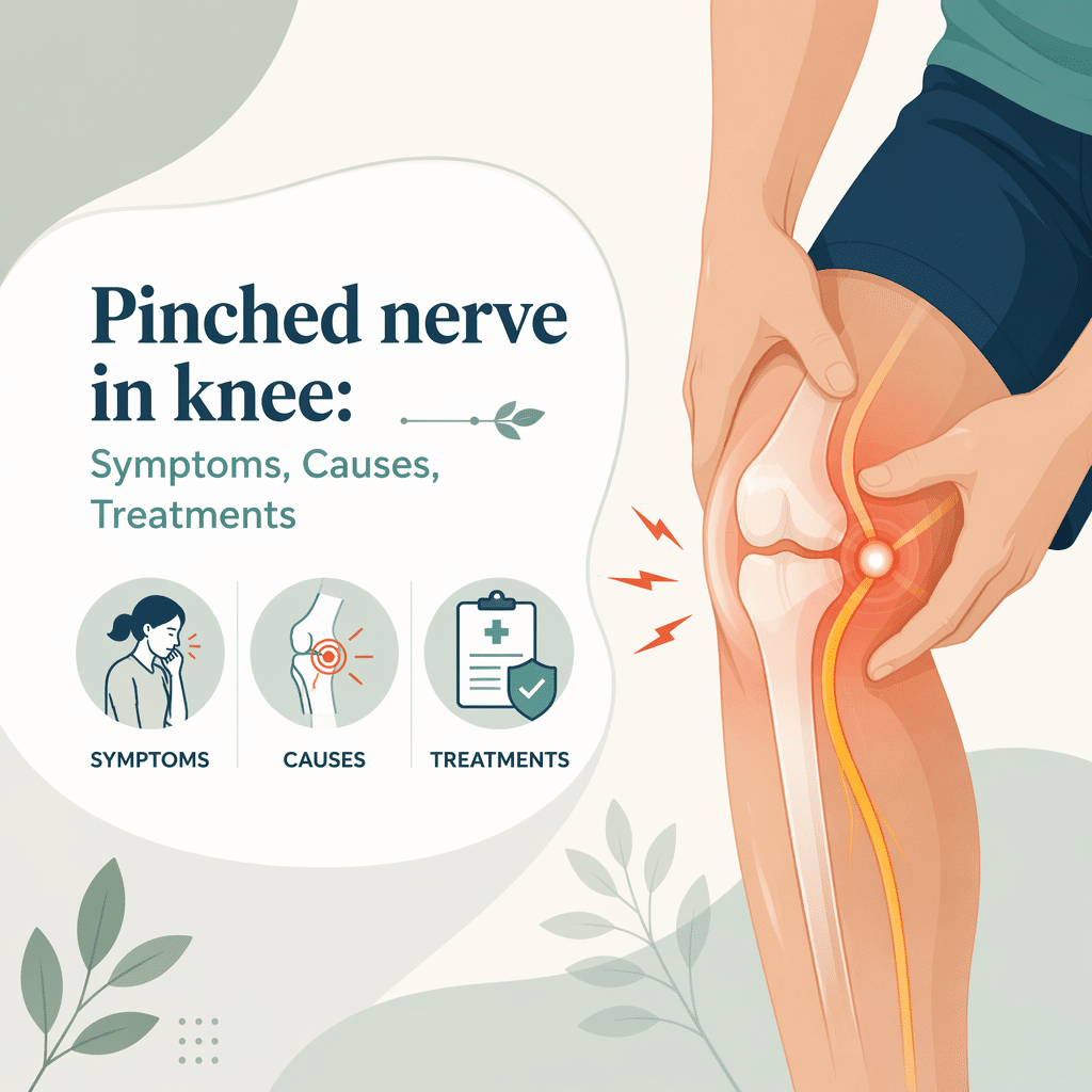
A pinched nerve in knee describes nerve compression or irritation around the knee that causes pain, numbness, tingling, or weakness. This article explains what a pinched nerve in knee means, how clinicians diagnose it, common causes and symptoms, treatment options, prevention strategies, and when to seek care. You will also find FAQs, a glossary of key terms, and a practical note about lab interpretation with AI DiagMe.
What is a pinched nerve in knee?
A pinched nerve in knee occurs when nearby tissues press on a sensory or motor nerve. Compression can come from swollen ligaments, nearby joint structures, a cyst, or repetitive strain. The pressure alters nerve signaling and produces distinct sensations. Symptoms vary with the affected nerve and the degree of compression. Most cases improve with noninvasive care, but some require procedural or surgical treatment.
Causes and risk factors
Nerve compression near the knee can arise from local problems and systemic processes. Common local causes include meniscal injury, Baker’s cyst, osteoarthritis with bone spurs, direct trauma, and scar tissue after surgery. Repetitive kneeling or prolonged pressure on the knee increases risk. Systemic factors such as diabetes or inflammatory disease can make nerves more vulnerable. Age and mechanical alignment issues, like knock-knees or bowlegs, also change load distribution and raise risk.
Common symptoms of a pinched nerve in knee
Pain at or around the knee commonly starts suddenly or worsens gradually. People often report sharp or burning pain that may radiate down the lower leg. Tingling, pins-and-needles, and numbness appear in the area served by the affected nerve. Muscle weakness or an unstable feeling can develop when motor fibers get involved. Symptoms often change with activity, position, or after prolonged sitting or standing.
How clinicians diagnose nerve compression
Clinicians begin with a focused history and physical exam. They test sensation, reflexes, and muscle strength. Providers perform specific maneuvers to reproduce symptoms and localize the problem. They also review prior injuries and systemic conditions that increase nerve vulnerability. A clear pattern of symptoms and exam findings often guides the next steps in testing and treatment.
Tests and imaging options
Providers order imaging when exam findings or symptom patterns suggest structural causes. Ultrasound detects fluid-filled cysts and guides injections. MRI shows soft tissues, cartilage, and meniscal injuries. X-rays reveal bone alignment and arthritis. Electrophysiology tests, such as nerve conduction studies and EMG, assess nerve function when diagnosis remains unclear. Lab tests check for diabetes or inflammatory markers when systemic disease is suspected.
Treatment options for a pinched nerve in knee
Treatment starts with conservative measures aimed at reducing pressure and improving function. Providers tailor care to the cause, severity, and the person’s goals. Most patients respond well to early nonoperative strategies. When conservative care fails or structural lesions cause persistent compression, procedural or surgical approaches may provide relief.
Conservative treatments for pinched nerve in knee
Rest and activity modification reduce repetitive stress. Physical therapy strengthens muscles, improves biomechanics, and teaches safer movement patterns. Ice and heat relieve pain and reduce swelling. Short courses of NSAIDs or acetaminophen control symptoms for many people. In some cases, clinicians inject corticosteroids around the compression site to reduce inflammation and improve mobility.
Interventional and surgical treatments
If a structural lesion causes ongoing compression, providers may recommend aspiration of a cyst or surgical removal of bone spurs. Arthroscopic procedures can repair meniscal tears or debride damaged tissue. Surgeons release scar tissue or revise previous repairs to free trapped nerves. Elective surgery depends on symptom severity, functional limitation, and response to conservative care.
Prevention and self-care
Maintain a healthy weight to reduce mechanical load across the knee. Strengthen the muscles around the hip and knee to improve alignment and absorb shock. Avoid prolonged kneeling and use knee pads when required. Use proper technique during sports and at work to limit repetitive strain. Manage blood sugar and systemic inflammation through medical care and lifestyle choices.
When to see a doctor
Seek medical attention if pain or numbness worsens, if weakness limits walking or standing, or if you notice loss of bladder or bowel control. Immediate evaluation is necessary for sudden severe symptoms after trauma. If conservative care does not improve symptoms within a few weeks, a clinician can reassess and order targeted tests. Early diagnosis helps prevent chronic nerve damage and preserves mobility.
Frequently Asked Questions (FAQ)
Q: How long does a pinched nerve in knee take to heal?
A: Recovery varies. Many people improve in weeks with conservative care. Structural causes may need longer treatment or surgery.
Q: Can exercises make the nerve compression worse?
A: Improper exercise can aggravate symptoms. A physical therapist will prescribe safe, progressive activities to strengthen without increasing compression.
Q: Will an MRI always show the cause?
A: MRI often reveals structural problems but not every case requires imaging. Clinicians use imaging alongside exam findings to confirm the diagnosis.
Q: Are injections safe for treating a pinched nerve in knee?
A: Injected corticosteroids can reduce inflammation and pain. Clinicians discuss risks, benefits, and alternatives before proceeding.
Q: Can diabetes cause a pinched nerve in knee?
A: Diabetes increases the risk of nerve injury and slows healing. Good blood sugar control reduces risk and aids recovery.
Q: When is surgery necessary?
A: Surgery becomes an option when conservative measures fail or when a clear structural lesion compresses the nerve and causes significant dysfunction.
Glossary of Key Terms
Compression: Pressure applied to a nerve that disrupts normal signaling.
Meniscus: A cartilage pad inside the knee that cushions and stabilizes the joint.
Baker’s cyst: A fluid-filled sac behind the knee that can press on nearby structures.
Electromyography (EMG): A test that records electrical activity of muscles to assess nerve function.
Arthroscopy: A minimally invasive surgical technique that uses a camera to treat joint problems.
Understand Your Lab Test Results with AI DiagMe
Interpreting tests and imaging helps you and your clinician make the best choices for a pinched nerve in knee. AI DiagMe can assist by translating lab and imaging results into clear explanations and by highlighting possible next steps. Use AI DiagMe to complement clinical advice and to understand how test results relate to symptoms and treatment options.
➡️ Analyze Your Lab Results with AI DiagMe Now

