
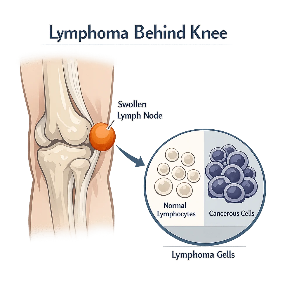
Lymphoma behind the knee is one of the least likely explanations for a lump in that spot, and understanding why can take a great deal of worry off your shoulders. The hollow at the back of the knee, called the popliteal fossa, does contain lymph nodes, but they sit deep in the tissue and rarely swell first when lymphoma develops. Far more often, a swelling there turns out to be a fluid-filled Baker’s cyst, a soft fatty lipoma, or a node reacting to a cut or an infection on the foot. In this article you’ll learn what usually causes a lump behind the knee, which features genuinely warrant a doctor’s attention, when swelling is an emergency, and how clinicians separate benign from serious using examination, ultrasound, biopsy and blood tests.
Why lymphoma behind the knee is a rare explanation for a lump
Lymphoma is a cancer that starts in white blood cells called lymphocytes, and it usually announces itself in the lymph nodes you can feel most easily: the neck, the armpits and the groin. The popliteal fossa is a much quieter neighborhood. It holds only a handful of lymph nodes, and they sit deep, tucked around the popliteal vein rather than just under the skin. That depth matters. A node there has to grow considerably before anyone notices it, and by that point lymphoma would almost always have shown itself somewhere more accessible first.
What the popliteal lymph nodes drain
The popliteal nodes collect lymph fluid from the back of the calf, the heel, the outer edge of the foot and the deep tissues of the lower leg. They are a filtering station for that territory. When they swell, the cause is usually sitting downstream: an infected blister, an ingrown toenail, athlete’s foot, a puncture wound, cellulitis, or a healing sports injury. Doctors call this reactive lymphadenopathy, meaning the node has enlarged because it is doing its job.
There is one exception worth knowing. Because these nodes drain the lower leg and foot, they can be the first stop for a skin cancer that started there. Surgeons mapping lymph drainage in limb melanoma found that roughly one patient in seventeen drained to a popliteal or elbow node instead of the usual groin or armpit basin, and in some of those patients the popliteal node was the only one holding cancer cells. Our team explains the warning signs of melanoma.
Why a swollen gland does not mean cancer
Infection is by far the most common reason any lymph node enlarges. MedlinePlus places colds, dental abscesses, ear infections, mononucleosis and skin infections well ahead of cancer on that list. A reactive node is typically tender, mobile under the fingers, smaller than a grape, and shrinks over two to four weeks once the trigger settles. Autoimmune conditions enlarge nodes too, and our team details the symptoms and blood tests of rheumatoid arthritis.
What a lump behind the knee usually turns out to be
Most swellings in the popliteal fossa are not lymph nodes at all. Radiologists reviewing masses in this region list synovial, meniscal and ganglion cysts, lipomas, vascular problems and nerve tumors as the usual suspects, with cancers a distant last. The table below sets out what each one typically feels like and what generally happens next.
| Likely cause | How it typically feels | What usually happens next |
|---|---|---|
| Baker’s cyst (popliteal cyst) | Soft, rounded fullness in the crease, clearer when the knee is straight, tighter after standing or activity | Ultrasound confirms fluid; treatment targets the knee problem that produced it |
| Lipoma | Soft, doughy, slides under the skin, painless, unchanged for years | Usually left alone; removed only if it presses on something or bothers you |
| Ganglion cyst | Firm, smooth, attached to deeper tissue, may change size from week to week | Ultrasound or MRI; often watched, sometimes drained or removed |
| Reactive lymph node | Small, tender, mobile, appears after a foot or leg infection, settles within weeks | Treat the infection, then recheck the node once it has cleared |
| Popliteal artery aneurysm | Pulsating swelling, sometimes with calf cramping or cold toes | Prompt ultrasound and referral to a vascular specialist |
| Nerve sheath tumor (schwannoma) | Firm and painless at first, later pins and needles or numbness down the leg or foot | MRI, then surgical removal if symptoms progress |
| Node enlarged by lymphoma | Hard, painless, anchored in place, keeps growing over weeks, often with swollen nodes elsewhere | Ultrasound, then a lymph node biopsy to confirm the diagnosis |
A Baker’s cyst is the single most common answer. Mayo Clinic describes it as swelling behind the knee caused by excess joint fluid, most often triggered by arthritis in the knee or a torn cartilage. Our guide explains the main forms of arthritis and how they are diagnosed, which matters here because treating the joint problem usually settles the cyst.
When swelling behind the knee is an emergency
One scenario should never wait. If your calf or the back of your knee becomes swollen, painful, warm or discolored over a few hours to a couple of days, that pattern can mean a deep vein thrombosis, a blood clot in a deep leg vein. The CDC lists swelling, pain or tenderness, warmth, and redness or discoloration as the signs to act on, and notes that about half of people with a clot have no symptoms at all. Seek same-day medical care rather than waiting to see whether it settles.
Two things make this confusing. A Baker’s cyst that bursts spills fluid down into the calf and produces almost identical pain and swelling, which is exactly why Mayo Clinic advises seeking care right away for pain and swelling behind the knee. And a clot left untreated can travel to the lungs. Our team describes the warning signs of a pulmonary embolism, and our guide explains the coagulation panel, including PT, PTT, INR and D-dimer.
How lymphoma behind the knee actually presents
Lymphoma rarely starts in the popliteal fossa. When a hematologist does find lymphoma in a node there, it is usually one part of a wider pattern rather than an isolated lump: nodes enlarged in more than one area of the body, sometimes an enlarged spleen, and whole-body symptoms alongside. Primary lymphoma of bone or soft tissue in the leg exists, but it is uncommon and tends to produce deep aching pain rather than a lump you can roll under your fingers.
The two broad families are Hodgkin and non-Hodgkin lymphoma, each covering dozens of subtypes that behave differently and are treated differently. Sorting them out is a job for a pathologist, not for a symptom checklist. Our team covers the full picture of lymphoma symptoms, causes and treatments, including how the subtypes are staged and managed. Blood cancers that start in the bone marrow can also enlarge nodes, and our guide explains the blood tests used to investigate leukemia.
The B symptoms that change the picture
Hematologists pay close attention to three whole-body symptoms, known together as B symptoms:
- Drenching night sweats that soak your nightclothes or sheets, not simply feeling warm
- Unexplained weight loss with no change in diet or activity
- Persistent or recurring fever with no infection to explain it
Itching without a rash and unusual fatigue often keep them company. None of these prove lymphoma on their own. Combined with a hard, growing, painless node, they shift the conversation from watchful waiting to prompt investigation.
Warning signs that a lump needs checking
Benign and worrying lumps tend to feel different. MedlinePlus advises contacting a clinician when nodes fail to shrink after several weeks or keep getting larger, when they feel hard or fixed in place, or when fever, night sweats or unexplained weight loss come with them. Applied to the back of the knee, these are the features that justify an appointment:
- A lump still present after four weeks, or one that keeps getting bigger
- A hard or rubbery texture rather than soft and squashy
- A lump anchored to deeper tissue that will not slide under the skin
- No pain at all, which sounds reassuring but is more typical of malignant nodes than reactive ones
- Swollen nodes in the neck, armpit or groin at the same time
- Any of the B symptoms described above
- New numbness, tingling or weakness in the leg or foot
One or two of these does not make a diagnosis. It makes an examination worthwhile, which is the entire point.
How doctors work up a lump behind the knee
The sequence is predictable, and knowing it removes some of the anxiety of a first appointment.
Examination first, then ultrasound
Your clinician will feel the lump for size, firmness, mobility and tenderness, check the other node areas, inspect your foot and lower leg for a source of infection, and ask about fevers, sweats and weight. Ultrasound is the usual first imaging test: quick, painless, free of radiation, and able to separate a fluid-filled cyst from a solid mass within minutes. It also shows the features radiologists use to judge a node, such as its shape, whether the fatty center called the hilum is still visible, and how blood flows through it. MRI is added when the mass is solid, deep, or sitting close to nerves and vessels.
Why a whole node beats a needle sample
If lymphoma is genuinely suspected, the diagnosis is made on tissue, not on imaging or blood work. The National Cancer Institute describes removing an entire node (an excisional biopsy), part of a node (an incisional biopsy), or a fragment taken with a wide needle (a core biopsy). Whenever it is practical, surgeons prefer to take the whole node. Lymphoma is identified partly by how the cells are arranged inside the node, and a fine needle aspirate collects loose cells while destroying that architecture. Removing the node intact lets the pathologist see the pattern, run the immune-marker and genetic tests that name the subtype, and avoid sending you back for a second procedure.
What blood tests can and cannot tell you
No blood test diagnoses lymphoma, and none can rule it out. Several are still ordered early because they map the terrain and point toward infection, inflammation or a blood disorder.
- A complete blood count reveals anemia, low platelets or an unusual white cell pattern. Our guide walks through how to read a complete blood count.
- Lactate dehydrogenase (LDH) is an enzyme released when cells break down quickly. It can rise in lymphoma, but also after hard exercise, a heart problem or liver injury. Our team explains what an LDH blood test measures.
- The erythrocyte sedimentation rate and C-reactive protein both flag inflammation without naming its cause. Our guide covers the erythrocyte sedimentation rate and what raises it, and our team also explains C-reactive protein as an inflammation marker.
- Lymphocyte counts move with infections, medications and blood conditions alike. Our guide covers the causes of a high lymphocyte count.
Taken together, a normal blood count with normal inflammatory markers alongside a soft, mobile, shrinking lump is reassuring. Abnormal numbers alongside a hard, growing node argue for imaging and a biopsy. Neither combination replaces the examination itself.
Latest scientific advances
Research published between 2023 and 2026 has focused less on rare leg lymphomas and more on something patients meet much sooner: making the ultrasound scan of a lump more informative, so that fewer people go through a biopsy they did not need.
Ultrasound is learning to read nodes better
A 2025 review of lymph node ultrasound set out the features that separate a reactive node from a cancerous one, including its shape, the loss of the fatty center, thickening of the outer rim, and unusual blood flow. What this means for you: the scan is no longer simply solid or fluid. A skilled operator can often place a node in a reassuring or a suspicious category on the spot, which shapes how urgently anything else needs to happen.
Two add-on techniques are maturing alongside it. Elastography measures how stiff a node is, since cancerous tissue tends to be firmer than inflamed tissue; a 2025 study of just over 200 patients found stiffness to be a strong independent signal of malignancy, particularly for Hodgkin lymphoma. Contrast-enhanced ultrasound, which uses tiny gas bubbles to display a node’s blood supply in real time, improved accuracy further in a 2025 comparison. What this means for you: if a node behind your knee looks borderline on a standard scan, these tools can add information without another appointment or a needle. Both are still being validated, and neither replaces a biopsy.
A shared scoring language for nodes
Radiologists are also testing Node-RADS, a standardized system that turns node size and shape into a score from 1 to 5. In a 2026 study of more than 500 nodes, the scores tracked closely with whether a node proved cancerous, and two radiologists reading the same scans agreed most of the time. What this means for you: a score in a report is meant to translate into a clear next step instead of a vague cannot-exclude. This remains an early result, and the system performs best combined with your clinical history rather than used on its own.
Case reports keep confirming the ordinary answer
Recent published reports of popliteal masses continue to land on benign diagnoses: Baker’s cysts dissecting into the calf muscle and mimicking a clot, a giant inflammatory mass filling a cyst in rheumatoid arthritis, a benign nerve tumor pressing on the nerve that lifts the foot. What this means for you: when specialists consider an unusual lump behind the knee worth publishing, it is far more often an unusual benign lump than a cancer.
Glossary
| Term | Definition |
|---|---|
| Popliteal fossa | The soft hollow at the back of the knee. It contains nerves, blood vessels and a small number of deep lymph nodes. |
| Lymphadenopathy | The medical term for enlarged lymph nodes, whatever the cause. It describes a finding, not a diagnosis. |
| Reactive node | A lymph node that has enlarged while responding to a nearby infection or inflammation, then shrinks again once the trigger clears. |
| Baker’s cyst | A fluid-filled swelling behind the knee, also called a popliteal cyst, caused by excess joint fluid escaping backward. |
| B symptoms | The combination of drenching night sweats, unexplained weight loss and persistent fever that hematologists look for when assessing lymphoma. |
| Excisional biopsy | Surgical removal of an entire lymph node so a pathologist can examine its internal structure as well as its cells. |
| Hilum | The fatty center of a lymph node, visible on ultrasound. Losing it is one of the signs that a node may be abnormal. |
| LDH (lactate dehydrogenase) | An enzyme released when cells break down quickly. It is a supporting clue in lymphoma, never a diagnosis on its own. |
| Deep vein thrombosis (DVT) | A blood clot in a deep vein, usually in the leg, causing swelling, pain and warmth. It needs same-day medical care. |
| Elastography | An ultrasound technique that measures how stiff a tissue is, used as an extra clue when a lymph node looks borderline. |
Frequently asked questions
Can you feel the lymph nodes behind your knee?
Usually not. Healthy popliteal nodes are small and sit deep in the fatty tissue around the popliteal vein, well below the surface, so most people cannot feel them at all. That is different from the nodes in the neck or groin, which many people can find with light pressure even when they are perfectly normal. If you can clearly feel a lump in the crease behind your knee, it is more likely a cyst, a lipoma or a markedly enlarged node than a node of normal size.
How do you check the lymph nodes behind your knee?
Sit down and bend the knee slightly so the muscles and the hollow relax, then press gently into the crease with the pads of your fingers, moving in small circles from one side to the other. Compare the two knees, since a difference between sides is more informative than an absolute size. Note whether anything you find is tender, whether it moves under your fingers, and whether it changes over the following weeks. Self-examination has real limits, and it does not replace a clinical examination.
What causes popliteal lymph nodes to swell?
The usual cause is an infection or an injury in the area those nodes drain: the foot, the heel, the outer edge of the foot, the back of the calf and the deep tissues of the lower leg. Athlete’s foot, an infected blister, an ingrown toenail, a puncture wound, cellulitis and post-surgical healing are all common triggers. Inflammatory joint disease around the knee can also enlarge them. Cancer, whether lymphoma or a melanoma that has spread from the foot or lower leg, sits far down that list.
Can a swollen lymph node behind the knee be cancer?
It can, but this is uncommon, and the pattern matters more than the mere presence of a lump. Nodes that raise concern are hard rather than springy, anchored rather than mobile, painless, steadily growing over weeks, and often accompanied by swollen nodes in other parts of the body or by night sweats, fever and weight loss. A single tender node that appeared after a foot infection and is already shrinking behaves very differently. Only a biopsy can settle the question.
How long should I wait before getting a lump behind my knee checked?
Four weeks is a reasonable yardstick for a lump that is stable, soft and painless, especially if it appeared after an infection or an injury. Do not wait at all if the lump is growing, if it is hard and fixed, if you have night sweats, fever or unexplained weight loss, or if you develop new numbness or weakness in the leg. Swelling that comes on quickly with pain, warmth or discoloration in the calf needs same-day care, because a blood clot has to be excluded first.
Does a painful lump behind the knee mean it is less serious?
Pain is a weak signal in either direction, though it is more often associated with benign causes. Infected and inflamed nodes are typically tender, whereas nodes enlarged by lymphoma are classically painless, which is precisely why a painless lump should not be dismissed. Pain can equally come from a cyst pressing on tissue, a nerve tumor, or a blood clot. What a clinician weighs is the whole picture: texture, mobility, growth rate, other swollen nodes and whole-body symptoms.
Sources
- Mayo Clinic — Baker cyst: symptoms and causes, 2026 — mayoclinic.org
- MedlinePlus Medical Encyclopedia (National Library of Medicine) — Swollen lymph nodes, 2024 — medlineplus.gov
- Centers for Disease Control and Prevention — About blood clots: signs and symptoms of deep vein thrombosis, 2025 — cdc.gov
- National Cancer Institute — Adult Non-Hodgkin Lymphoma Treatment (PDQ), Patient Version, 2026 — cancer.gov
- Tavarozzi R, Lombardi A, Scarano F, et al. — Lymph node ultrasound in lymphoproliferative disorders: clinical characteristics and applications — Journal of Basic and Clinical Physiology and Pharmacology, 2025 — doi.org/10.1515/jbcpp-2025-0089
- Pugliese N, Picardi M, Giordano C, et al. — Elastography enhances the diagnostic performance of conventional ultrasonography in differentiating benign from malignant superficial lymphadenopathies — Cancers, 2025 — doi.org/10.3390/cancers17091480
- Liang S, Han P, Fei X, et al. — High-frequency contrast-enhanced ultrasound in discriminating benign and malignant superficial lymph nodes: a diagnostic comparison — BMC Cancer, 2025 — doi.org/10.1186/s12885-025-14238-1
- Zhang H, Li R, He D, et al. — Ultrasonography-based application of Node-RADS in differentiating malignant and benign superficial lymph nodes: a retrospective study — Ultrasound in Medicine and Biology, 2026 — doi.org/10.1016/j.ultrasmedbio.2025.11.669
- West NJ, Wadhwa S, Ayars C, et al. — Interval sentinel lymph nodes with the use of routine lymphoscintigraphy in extremity melanoma — Journal of Surgical Research, 2023 — doi.org/10.1016/j.jss.2023.08.055
- Augustin D, Augustin DH, Pharol A, et al. — Intramuscular dissecting Baker’s cysts: a case series highlighting ultrasound diagnosis and differential considerations — Cureus, 2024 — doi.org/10.7759/cureus.53658
- Gupta S, Kanna H, Saran S — A large calf mass in a patient with rheumatoid arthritis: giant pannus filling Baker’s cyst — Journal of Medical Ultrasound, 2024 — doi.org/10.4103/jmu.jmu_40_24
- Lykos S, Pallis D, Chatzikyriakos A, et al. — Common peroneal nerve schwannoma presenting as a popliteal fossa mass: a case report — Cureus, 2026 — doi.org/10.7759/cureus.111000
Further reading
- How to read your blood test results
- Abnormal blood test results and what they mean
- Tumor markers and their limits
- Low lymphocytes: causes, symptoms and risks
- Lymphoma: symptoms, causes and treatments
Understand your lab results with AI DiagMe
A lump behind the knee is assessed by examination and imaging, but the blood tests ordered alongside it are often what leave people puzzled. AI DiagMe turns those numbers into plain language, so you can see what a complete blood count, an LDH level, a sedimentation rate or a C-reactive protein result actually says about inflammation, infection and your blood cells. It helps you understand your results and prepare better questions for your appointment. It does not diagnose, and it does not replace your doctor.
Get your results interpreted in minutes