




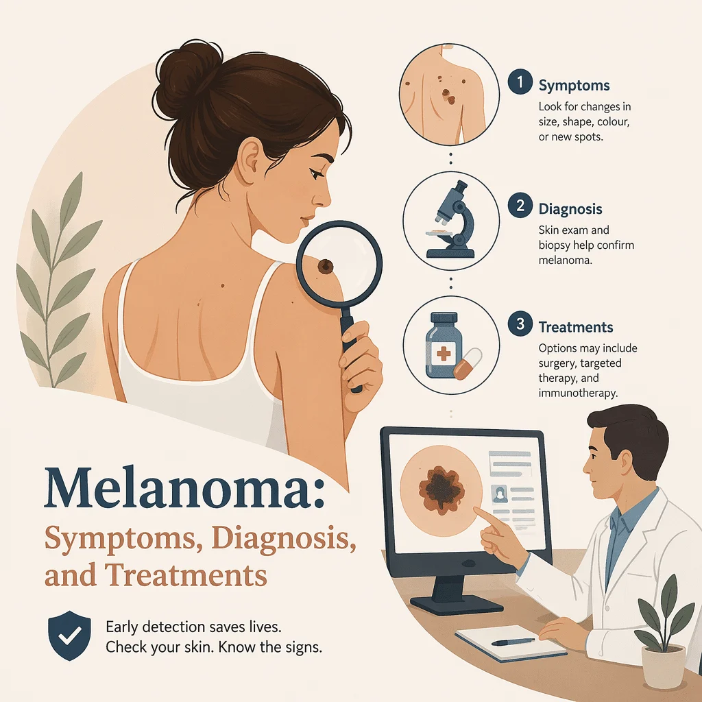
Mengenali gejala melanoma sejak dini bisa menjadi perbedaan antara pengobatan yang cepat dan sederhana dengan penanganan yang jauh lebih rumit. Melanoma adalah bentuk kanker kulit paling serius karena, tidak seperti kanker kulit yang lebih umum, melanoma dapat menyebar ke bagian tubuh lain jika tidak terdeteksi tepat waktu. Kabar baiknya adalah sebagian besar melanoma muncul di permukaan kulit sehingga bisa terlihat, dan banyak yang ditemukan sejak dini — seringkali oleh orang yang sekadar memperhatikan tahi lalat yang tampak berbeda dari biasanya. Dalam artikel ini Anda akan belajar cara mengenali tanda-tanda peringatan dengan aturan ABCDE, bagaimana dokter mendiagnosis dan menentukan stadium penyakit, tes darah dan biomarker apa yang penting dan kapan, serta bagaimana pengobatan melanoma telah berkembang dalam beberapa tahun terakhir. Tujuannya adalah memberikan informasi yang jelas dan tenang, bukan menimbulkan kekhawatiran.
Apa itu melanoma?
Melanoma adalah kanker yang bermula dari melanosit, yaitu sel-sel yang memproduksi melanin — pigmen yang memberi warna pada kulit. Karena melanosit terutama ditemukan di kulit, sebagian besar melanoma muncul di sana, namun melanoma juga dapat berkembang di mata, di bawah kuku, atau pada lapisan mulut maupun permukaan internal lainnya. Melanoma hanya menyumbang sebagian kecil dari seluruh kasus kanker kulit, namun menjadi penyebab sebagian besar kematian akibat kanker kulit karena kemampuannya untuk menyebar, atau bermetastasis, jika tidak ditangani.
Tidak semua melanoma tampak sama. Memahami pola-pola utamanya membantu menjelaskan mengapa tanda-tanda peringatannya bisa berbeda dari satu orang ke orang lain.
Jenis-jenis utama melanoma
- Melanoma penyebaran superfisial, bentuk yang paling umum, yang cenderung tumbuh melebar di permukaan kulit sebelum tumbuh ke dalam.
- Melanoma nodular, lesi yang tumbuh lebih cepat, sering menonjol dan keras, yang bisa berwarna gelap atau, lebih jarang, sama sekali tidak berpigmen.
- Melanoma lentigo maligna, yang biasanya muncul pada kulit yang rusak akibat paparan sinar matahari pada orang dewasa yang lebih tua, seperti di area wajah.
- Melanoma lentiginous akral, yang berkembang di telapak tangan, telapak kaki, atau di bawah kuku, dan dapat terjadi pada orang dengan warna kulit apa pun.
Gejala melanoma: tanda peringatan ABCDE
Alat yang paling berguna untuk mengenali gejala melanoma di rumah adalah aturan ABCDE. Setiap huruf menunjukkan ciri yang lebih umum ditemukan pada melanoma dibandingkan tahi lalat biasa. Sebuah bintik tidak harus menunjukkan semua ciri untuk layak mendapat perhatian, dan setiap perubahan tunggal yang baru atau berkembang patut dikonsultasikan ke dokter.
| Tanda | Apa yang perlu diperhatikan |
|---|---|
| A — Asimetri | Satu sisi bintik tidak sama dengan sisi lainnya. |
| B — Border (Tepi) | Tepinya tidak rata, bergerigi, berlekuk, atau kabur, bukan halus. |
| C — Color (Warna) | Beberapa warna muncul dalam satu bintik: cokelat, hitam, kecokelatan, dan terkadang merah, putih, atau biru. |
| D — Diameter | Lebih besar dari sekitar 6 milimeter, kira-kira selebar penghapus pensil, meskipun melanoma bisa lebih kecil. |
| E — Evolving (Berkembang) | Perubahan apa pun dalam beberapa minggu atau bulan pada ukuran, bentuk, warna, atau ketinggian, maupun rasa gatal, perdarahan, atau keropeng yang baru muncul. |
Tanda peringatan lainnya
Dokter kulit juga memperhatikan tanda “bebek buruk rupa”: tahi lalat yang tampak berbeda dari tahi lalat lainnya pada seseorang. Bintik baru di usia dewasa, luka yang tidak kunjung sembuh, garis gelap di bawah kuku jari tangan atau kaki, atau perubahan pada tahi lalat yang sudah lama ada semuanya perlu diperiksa. Pertumbuhan yang tidak berbahaya pun bisa terlihat mengkhawatirkan. Untuk memahami bagaimana bintik jinak yang umum bisa disalahartikan sebagai kanker, baca perbandingan keratosis seboroik dan melanoma oleh kami.
Kapan harus menemui dokter?
Buat janji dengan dokter atau dokter kulit jika Anda memperhatikan salah satu dari berikut ini:
- Tahi lalat atau bintik yang berubah ukuran, bentuk, atau warnanya.
- Bintik baru yang tampak berbeda dari bintik lainnya, terutama setelah usia 30 tahun.
- Bintik yang terasa gatal, berdarah, mengeluarkan cairan, atau tidak sembuh-sembuh.
- Garis gelap di bawah kuku yang tidak dapat Anda jelaskan akibat cedera.
- Lesi apa pun yang membuat Anda khawatir, meskipun tidak sepenuhnya sesuai dengan pola ABCDE.
Apa penyebab melanoma, dan siapa yang berisiko lebih tinggi?
Melanoma berkembang ketika melanosit mengalami kerusakan genetik yang membuatnya tumbuh tidak terkendali. Penyebab tunggal terbesar yang dapat diubah adalah radiasi ultraviolet, atau UV, dari sinar matahari dan dari alat penyamakan kulit dalam ruangan. Menurut Centers for Disease Control and Prevention dan American Academy of Dermatology, melindungi kulit dari UV adalah cara paling efektif untuk menurunkan risiko.
Faktor risiko yang perlu diketahui
- Kulit terbakar matahari berulang kali, terutama yang melepuh saat masa kanak-kanak.
- Penggunaan alat penyamakan kulit dalam ruangan.
- Kulit cerah yang mudah terbakar, mata berwarna terang, serta rambut pirang atau merah.
- Jumlah tahi lalat yang banyak (50 atau lebih) atau beberapa tahi lalat atipikal.
- Riwayat melanoma pada diri sendiri atau anggota keluarga.
- Sistem kekebalan tubuh yang melemah, misalnya setelah transplantasi organ.
Orang dewasa yang lebih tua paling sering didiagnosis, tetapi melanoma juga termasuk salah satu kanker yang cukup umum pada orang dewasa muda, itulah mengapa tanda-tanda peringatannya penting di setiap usia. Memiliki faktor risiko tidak berarti melanoma pasti akan terjadi, dan banyak penderita melanoma hanya memiliki satu atau dua faktor risiko.
Cara melanoma didiagnosis dan ditentukan stadiumnya
Diagnosis melanoma adalah proses bertahap yang dimulai dari pemeriksaan mata dan berakhir di bawah mikroskop. Tidak ada tes darah yang dapat memastikan atau menyingkirkan melanoma; diagnosis dibuat berdasarkan sampel jaringan yang dicurigai.
Pemeriksaan kulit dan dermoskopi
Dokter pertama-tama memeriksa bercak tersebut dan sering kali seluruh permukaan kulit. Banyak yang menggunakan dermoskopi, yaitu kaca pembesar genggam dengan cahaya terpolarisasi yang mengungkap pola yang tidak terlihat oleh mata biasa, sehingga meningkatkan akurasi dan mengurangi prosedur yang tidak perlu.
Biopsi dan histopatologi
Jika suatu lesi dicurigai, langkah standar berikutnya adalah biopsi, biasanya dengan mengangkat seluruh bercak beserta sedikit kulit normal di sekitarnya. Seorang ahli patologi kemudian memeriksanya di bawah mikroskop untuk memastikan melanoma dan mengukur ciri-ciri penting, termasuk ketebalan Breslow (seberapa dalam melanoma menembus kulit) dan apakah permukaannya mengalami ulserasi. Detail-detail ini menentukan stadium dan rencana penanganan.
Biopsi kelenjar getah bening sentinel dan pencitraan
Untuk melanoma yang melampaui ketebalan tertentu, dokter mungkin akan memeriksa kelenjar getah bening sentinel, yaitu kelenjar pertama yang menerima aliran dari area tersebut, untuk melihat apakah sel-sel telah mulai menyebar. Pencitraan seperti USG, CT, PET-CT, atau MRI umumnya diperuntukkan bagi melanoma yang lebih tebal atau ketika penyebaran dicurigai, bukan untuk lesi tipis yang masih dini. Penentuan stadium menggabungkan ketebalan tumor, ulserasi, status kelenjar getah bening, dan, bila penyakit sudah lanjut, hasil pemeriksaan darah.
Tes darah dan biomarker pada melanoma
Banyak orang bertanya tes darah apa yang dapat mendeteksi melanoma. Jawaban jujurnya adalah tidak ada yang bisa melakukannya sendiri, dan melanoma tidak memiliki tes darah skrining rutin seperti beberapa jenis kanker yang dipantau dengan penanda protein. Untuk memahami apa yang bisa dan tidak bisa dilakukan oleh penanda-penanda ini, lihat penjelasan kami tentang penanda tumor. Sebaliknya, tes darah dan jaringan tertentu memiliki perannya masing-masing pada momen-momen tertentu, terutama untuk menilai prognosis, memandu pengobatan, atau memantau penyakit stadium lanjut.
| Tes | Apa yang dilihatnya | Kapan biasanya diperlukan |
|---|---|---|
| LDH (laktat dehidrogenase) | Enzim umum dalam darah yang dapat meningkat saat jaringan mengalami stres. | Melanoma lanjut (stadium IV), sebagai penanda prognostik yang sudah masuk dalam sistem penentuan stadium. |
| S100B | Protein yang cenderung mencerminkan jumlah melanoma dalam tubuh. | Prognosis dan pemantauan di beberapa pusat kesehatan, terutama di Eropa. |
| Tes mutasi BRAF | Perubahan genetik pada jaringan tumor, paling sering terjadi pada titik yang disebut V600. | Penyakit stadium lanjut atau risiko tinggi, untuk menentukan apakah terapi bertarget dapat digunakan. |
| DNA tumor yang bersirkulasi (ctDNA) | Fragmen kecil DNA tumor yang dilepaskan ke dalam aliran darah. | Penggunaan yang berkembang untuk memantau melanoma stadium lanjut; bukan tes skrining. |
LDH: penanda prognosis pada penyakit stadium lanjut
Laktat dehidrogenase adalah enzim yang terdapat di banyak jaringan, sehingga tidak spesifik untuk melanoma. Namun, pada melanoma stadium lanjut, kadar LDH yang tinggi dikaitkan dengan prognosis yang lebih buruk dan secara resmi menjadi bagian dari cara mengklasifikasikan penyakit stadium IV. Penanda ini digunakan untuk membantu menilai prognosis dan memantau perkembangan penyakit, bukan untuk mendeteksi melanoma sejak awal. Untuk mengetahui cara enzim ini diukur dan dilaporkan, baca panduan kami tentang tes darah LDH.
S100B: memantau beban tumor
S100B adalah protein yang sering meningkat seiring bertambahnya jumlah melanoma dalam tubuh. Di beberapa negara, penanda ini diukur untuk membantu memperkirakan prognosis dan memantau kondisi pasien dari waktu ke waktu. S100B merupakan penanda pendukung, bukan penanda diagnostik, dan penggunaannya bervariasi dari satu pusat layanan ke pusat lainnya.
Uji mutasi BRAF: kunci pemilihan terapi
Sekitar setengah dari melanoma kutaneus membawa mutasi pada gen yang disebut BRAF, paling sering berupa perubahan yang dikenal sebagai V600. Uji ini dilakukan pada jaringan tumor, bukan pada darah, dan penting karena membuka peluang untuk terapi bertarget. Mengetahui status BRAF pada melanoma stadium lanjut membantu tim medis memilih jalur pengobatan yang tepat.
DNA tumor yang bersirkulasi: biopsi cair yang terus berkembang
DNA tumor yang bersirkulasi terdiri dari potongan-potongan kecil DNA yang dilepaskan oleh tumor ke dalam darah. Pengukurannya, yang kadang disebut biopsi cair, merupakan bidang penelitian yang berkembang pesat dengan tujuan mendeteksi melanoma yang kambuh lebih awal dan menilai seberapa baik pengobatan bekerja. Biopsi cair ini menggunakan teknologi yang sama dengan alat skrining terbaru; untuk gambaran yang lebih lengkap, baca ikhtisar kami tentang tes darah untuk deteksi dini berbagai jenis kanker. Beberapa jenis kanker dipantau menggunakan penanda protein dalam darah, meskipun melanoma saat ini tidak bergantung pada satu penanda pun; untuk memahami cara pendekatan tersebut bekerja, baca artikel kami tentang tes darah CEA.
Cara melanoma diobati
Pengobatan sangat bergantung pada stadiumnya. Melanoma stadium awal yang belum menyebar biasanya ditangani dan disembuhkan hanya dengan operasi, sementara melanoma stadium lanjut memerlukan berbagai pilihan pengobatan yang telah berkembang pesat selama satu dekade terakhir.
Operasi
Pembedahan adalah pengobatan utama untuk melanoma yang terdeteksi sejak dini. Dokter bedah mengangkat melanoma beserta sebagian kulit sehat di sekitarnya, prosedur ini disebut eksisi lokal luas. Jika diperlukan, biopsi kelenjar getah bening sentinel dilakukan bersamaan untuk memeriksa apakah kanker sudah menyebar.
Imunoterapi
Imunoterapi membantu sistem imun mengenali dan menyerang sel melanoma. Kelas utamanya, yang disebut penghambat pos pemeriksaan imun, mencakup obat-obatan yang memblokir PD-1 (seperti pembrolizumab dan nivolumab) serta obat yang memblokir CTLA-4 (ipilimumab), yang terkadang diberikan secara kombinasi. Menurut National Cancer Institute, obat-obatan ini telah mengubah harapan hidup banyak penderita melanoma stadium lanjut.
Terapi target
Untuk melanoma yang membawa mutasi BRAF V600, terapi bertarget dapat mematikan sinyal pertumbuhan yang bermasalah. Pengobatan ini memadukan penghambat BRAF (seperti dabrafenib atau vemurafenib) dengan penghambat MEK (seperti trametinib) agar bekerja lebih efektif dan lebih lama. Itulah mengapa pengujian BRAF dilakukan sebelum memilih jalur pengobatan ini.
Radioterapi dan perawatan suportif
Radioterapi dapat digunakan dalam situasi tertentu, misalnya untuk menangani melanoma yang telah menyebar ke otak atau untuk meringankan gejala. Selama imunoterapi atau terapi bertarget, tim medis memantau hasil laboratorium rutin; untuk memahami angka-angka tersebut, baca penjelasan kami tentang pemeriksaan darah lengkap. Perawatan juga mencakup pengelolaan efek samping dan dukungan untuk kesehatan secara keseluruhan.
Jika laporan patologi Anda menggambarkan sel imun yang ditemukan di dalam tumor sebagai brisk atau non-brisk, lihat panduan kami tentang limfosit yang menginfiltrasi tumor.
Mengurangi risiko: pencegahan dan tindak lanjut
Karena paparan sinar UV adalah penyebab utama yang dapat dimodifikasi, perlindungan dari sinar matahari menjadi inti dari pencegahan. American Academy of Dermatology menyarankan untuk mencari tempat teduh, mengenakan pakaian pelindung dan topi bertepi lebar, serta menggunakan tabir surya spektrum luas dengan SPF 30 atau lebih tinggi, sekaligus menghindari penggunaan alat tanning dalam ruangan sama sekali.
Pemeriksaan kulit mandiri secara rutin membantu Anda mengenali kondisi normal kulit Anda, sehingga perubahan apa pun lebih mudah terdeteksi. Banyak dokter menyarankan untuk memeriksa kulit setiap bulan dan menemui profesional untuk pemeriksaan kulit sesuai risiko pribadi Anda. Perlindungan matahari yang baik jarang menyebabkan masalah vitamin D, tetapi jika Anda khawatir tentang kadar vitamin D Anda, baca panduan kami tentang tes darah vitamin D. Jika ditemukan dan ditangani sejak dini, melanoma sangat sering dapat disembuhkan; prognosisnya menjadi lebih serius setelah menyebar, itulah mengapa mendeteksi perubahan sejak awal sangat penting.
Kemajuan ilmiah terbaru dalam melanoma
Penelitian melanoma berkembang pesat, dan beberapa temuan terbaru sedang mengubah cara penanganannya. Poin-poin di bawah ini disederhanakan untuk pembaca umum, dan tidak satu pun dari poin tersebut menggantikan saran dari tim perawatan Anda sendiri.
Imunoterapi sebelum operasi
Untuk melanoma yang telah menyebar ke kelenjar getah bening terdekat namun masih bisa diangkat, sebuah uji klinis tahun 2024 menemukan bahwa pemberian imunoterapi sebelum operasi — pendekatan yang disebut terapi neoadjuvan — membuat lebih banyak pasien bebas kanker lebih lama dibandingkan pemberian imunoterapi hanya setelah operasi. Artinya bagi Anda: waktu pemberian imunoterapi, bukan hanya jenis obatnya, kini mulai menjadi bagian dari diskusi penanganan melanoma stadium III.
Tes darah untuk memantau kemungkinan kekambuhan
Para peneliti terus menyempurnakan tes DNA tumor yang beredar di darah (circulating tumor DNA) untuk mendeteksi kekambuhan melanoma lebih awal daripada yang bisa terlihat melalui pemindaian, serta untuk mengetahui lebih cepat apakah suatu pengobatan berhasil. Artinya bagi Anda: biopsi cair ini masih digunakan terutama dalam penelitian dan pusat kesehatan khusus, bukan untuk skrining rutin, namun menunjukkan masa depan di mana pengambilan darah sederhana dapat membantu memantau melanoma stadium lanjut.
Cara yang lebih baik untuk membaca prognosis
Selain tes LDH yang sudah lama digunakan, para ilmuwan sedang mempelajari penanda darah tambahan — termasuk protein dan sinyal imun — untuk memprediksi lebih tepat bagaimana melanoma pada seseorang akan berkembang. Artinya bagi Anda: saat ini tes LDH dan pemeriksaan mutasi BRAF berbasis jaringan tetap menjadi andalan utama, sementara penanda yang lebih baru masih dalam tahap validasi dan belum menjadi bagian dari perawatan sehari-hari.
Glosarium
| Ketentuan | Definisi |
|---|---|
| Melanosit | Sel kulit yang menghasilkan melanin, yaitu pigmen yang memberi warna pada kulit, rambut, dan mata. Melanoma bermula dari sel-sel ini. |
| Aturan ABCDE | Daftar periksa sederhana (Asimetri, Batas, Warna/Color, Diameter, Evolusi) untuk mengenali tahi lalat yang mungkin merupakan melanoma. |
| Dermoskopi | Pemeriksaan kulit menggunakan kaca pembesar dan cahaya khusus untuk melihat pola yang tidak terlihat oleh mata biasa. |
| Biopsi | Pengambilan sampel jaringan, atau seluruh area yang mencurigakan, untuk diperiksa di bawah mikroskop. |
| Ketebalan Breslow | Pengukuran seberapa dalam melanoma telah tumbuh ke dalam kulit, digunakan untuk membantu menentukan stadium. |
| Biopsi kelenjar getah bening sentinel | Pemeriksaan pada kelenjar getah bening pertama yang menerima aliran dari area tersebut, untuk memeriksa apakah kanker sudah menyebar lebih awal. |
| Metastasis | Penyebaran kanker dari tempat asalnya ke bagian tubuh lainnya. |
| Laktat dehidrogenase (LDH) | Enzim darah yang digunakan sebagai penanda prognostik pada melanoma stadium lanjut, bukan sebagai tes skrining. |
| Mutasi BRAF | Perubahan genetik pada tumor yang dapat ditargetkan dengan obat-obatan tertentu; ditemukan pada sekitar separuh kasus melanoma kulit. |
| DNA tumor yang bersirkulasi (ctDNA) | Fragmen DNA tumor yang beredar dalam darah, diukur melalui biopsi cair untuk membantu memantau perkembangan penyakit. |
Pertanyaan yang sering diajukan
Seperti apa tampilan melanoma stadium awal?
Melanoma dini sering berupa bercak datar atau sedikit menonjol yang melanggar aturan ABCDE: bisa asimetris, memiliki tepi yang tidak rata, menampilkan lebih dari satu warna, atau sekadar berubah. Ukurannya bisa kecil, sehingga ukuran saja tidak bisa menjadi patokan. Banyak melanoma dini tidak terasa sakit, itulah mengapa penampilan dan perubahan lebih penting daripada rasa tidak nyaman. Jika ada bercak yang terlihat berbeda dari tahi lalat Anda yang lain atau terus berkembang, segera periksakan daripada menunggu gejala muncul.
Apakah tes darah bisa mendeteksi melanoma?
Tidak ada satu pun tes darah yang dapat mendiagnosis melanoma, dan tidak ada pemeriksaan darah rutin untuk kondisi ini. Diagnosis ditegakkan melalui biopsi pada kulit yang dicurigai. Tes darah memiliki peran pendukung: LDH membantu menilai prognosis pada penyakit stadium lanjut, S100B digunakan di beberapa pusat kesehatan untuk memantau beban tumor, dan DNA tumor yang bersirkulasi merupakan metode baru untuk melacak melanoma stadium lanjut. Tes-tes ini berperan dalam penanganan setelah melanoma sudah diketahui, bukan untuk menemukannya pertama kali.
Apa perbedaan tahi lalat biasa dengan melanoma?
Tahi lalat biasa umumnya berukuran kecil, berwarna seragam, berbentuk bulat atau oval, dengan tepi yang mulus, dan tidak berubah selama bertahun-tahun. Melanoma cenderung asimetris, memiliki banyak warna, tepi tidak beraturan, ukuran lebih besar, atau berubah seiring waktu. Bercak yang tampak berbeda dari tahi lalat Anda yang lain — seperti “bebek buruk rupa” — merupakan petunjuk yang berguna. Karena beberapa pertumbuhan kulit yang tidak berbahaya bisa menyerupai melanoma, dokter kulit memberikan jawaban pasti, biasanya dengan dermoskopi dan, jika diperlukan, biopsi.
Apakah setiap pasien melanoma perlu menjalani tes BRAF?
Tidak selalu. Tes BRAF umumnya dilakukan untuk melanoma stadium lanjut atau berisiko tinggi, karena tujuan utamanya adalah menentukan apakah terapi bertarget merupakan pilihan yang tepat. Tes ini dilakukan pada jaringan tumor dari hasil biopsi, bukan dari sampel darah. Banyak orang dengan melanoma tipis stadium dini yang telah diangkat sepenuhnya melalui operasi tidak memerlukan tes BRAF sama sekali. Dokter onkologi Anda akan merekomendasikannya jika hasilnya akan mengubah rencana pengobatan.
Apakah melanoma selalu mengancam jiwa?
Tidak. Jika melanoma ditemukan dan diangkat sejak dini, kondisi ini sangat sering dapat disembuhkan, dan sebagian besar orang yang didiagnosis pada stadium awal memiliki prognosis yang baik. Risiko meningkat ketika melanoma tumbuh lebih dalam atau menyebar ke kelenjar getah bening atau organ lain, itulah mengapa deteksi dini sangat penting. Bahkan untuk melanoma stadium lanjut, imunoterapi modern dan terapi bertarget telah meningkatkan hasil pengobatan secara signifikan dibandingkan satu dekade lalu.
Seberapa sering saya harus memeriksa kulit saya?
Banyak dokter kulit menyarankan pemeriksaan mandiri setiap bulan agar Anda lebih mengenal tanda lahir Anda sendiri dan dapat mendeteksi perubahan. Gunakan cermin atau minta bantuan untuk memeriksa area yang sulit dilihat, seperti punggung, kulit kepala, dan telapak kaki. Seberapa sering Anda perlu menjalani pemeriksaan kulit oleh dokter bergantung pada faktor risiko Anda — orang dengan banyak tahi lalat, kulit cerah, atau riwayat melanoma pada diri sendiri maupun keluarga mungkin disarankan untuk lebih rutin memeriksakan diri.
Sumber
- National Cancer Institute — Melanoma Treatment (PDQ), Patient Version — cancer.gov
- American Academy of Dermatology — Melanoma: overview and warning signs — aad.org
- American Cancer Society — Signs and Symptoms of Melanoma Skin Cancer — cancer.org
- Tasdogan et al. — Cutaneous melanoma — Nature Reviews Disease Primers, 2025 — doi.org/10.1038/s41572-025-00603-8
- Boutros et al. — The treatment of advanced melanoma: current approaches and new challenges — Critical Reviews in Oncology/Hematology, 2024 — doi.org/10.1016/j.critrevonc.2024.104276
- Castellani et al. — BRAF mutations in melanoma: biological aspects, therapeutic implications, and circulating biomarkers — Cancers, 2023 — doi.org/10.3390/cancers15164026
- Blank et al. — Neoadjuvant nivolumab and ipilimumab in resectable stage III melanoma — New England Journal of Medicine, 2024 — doi.org/10.1056/NEJMoa2402604
- Eroglu et al. — Circulating tumor DNA-based molecular residual disease detection for treatment monitoring in advanced melanoma — Cancer, 2023 — doi.org/10.1002/cncr.34716
- Kaleem et al. — Imaging and laboratory workup for melanoma — Oral and Maxillofacial Surgery Clinics of North America, 2022 — doi.org/10.1016/j.coms.2021.11.004
- Ding et al. — Prognostic biomarkers of cutaneous melanoma — Photodermatology, Photoimmunology & Photomedicine, 2022 — doi.org/10.1111/phpp.12770
Bacaan lebih lanjut
- Cara pengambilan darah, langkah demi langkah
- Berapa lama hasil tes darah keluar
- Ruam kulit: penyebab, gejala, dan pengobatan
- Eksim: memahami kondisi kulit ini
- Psoriasis: gejala, jenis, penyebab, dan pemicunya
Pahami hasil lab Anda dengan AI DiagMe.
Dapatkan interpretasi hasil Anda dalam hitungan menit.
Melanoma mengingatkan kita betapa pentingnya satu perubahan kecil pada kulit, atau satu angka dalam hasil laboratorium. Jika pemeriksaan melanoma mencakup tes darah seperti LDH, hitung darah lengkap, atau tes DNA tumor yang beredar (circulating tumor DNA), hasil tersebut bisa sulit dipahami hanya dari kertas. Untuk memahami angka-angka tersebut, Anda dapat berkonsultasi panduan kami untuk membaca hasil tes darah, dan AI DiagMe dapat membantu Anda memahami arti nilai-nilai tersebut dalam bahasa yang mudah dimengerti. Ini adalah alat bantu untuk memahami hasil Anda, bukan untuk mendiagnosis melanoma, dan tidak menggantikan dokter atau dokter kulit Anda.