




A troponin blood test measures a protein that leaks into your bloodstream when heart muscle cells are damaged, and it is the test doctors rely on most to detect injury to the heart. If you or someone near you has chest pain or pressure, pain spreading to an arm, the jaw, neck or back, sudden breathlessness, cold sweats, nausea or lightheadedness, call 999 now. Do not wait for a blood test, do not finish reading this page, and do not drive yourself to hospital.
This article is written for a different moment. You are most likely reviewing a result after the fact, or you are a patient or a relative trying to make sense of an emergency department visit that has already happened. In this article you'll learn what troponin is, how high-sensitivity testing and repeat draws work, why a raised result means heart injury rather than automatically a heart attack, which conditions outside the heart can lift the number, and what a normal result does and does not settle.
Emergency: call 999 first, ask questions later
Call 999 immediately, and do not wait to see whether symptoms pass, if you have any of the following:
- Chest pain, pressure, tightness or heaviness, in the centre or the left side
- Pain or discomfort spreading to one or both arms, the back, shoulders, neck, jaw or upper stomach
- Sudden shortness of breath, at rest or with light activity
- Cold sweats, nausea or vomiting, unusual exhaustion, dizziness or fainting
An ambulance is safer than a car. Paramedics can record a heart tracing, start treatment on the way, and hospitals often treat people who arrive by ambulance faster. The National Heart, Lung, and Blood Institute is explicit on this point: never delay calling 999 to do anything else first.
What is troponin, and why is it the reference marker for heart injury?
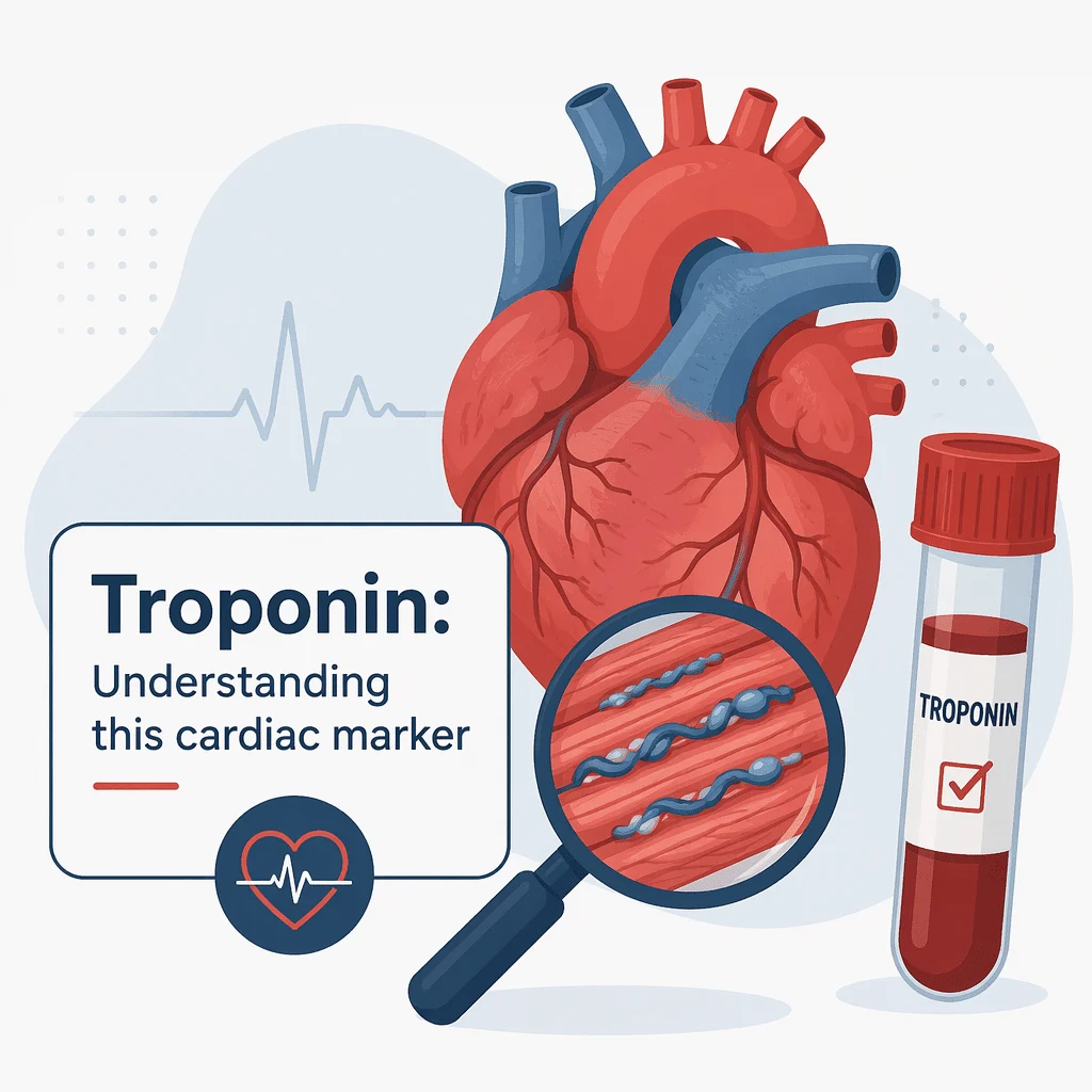
Troponin is a group of proteins inside muscle cells that controls each contraction. Your heart muscle cells, called cardiomyocytes, carry their own distinct versions: cardiac troponin I and cardiac troponin T. These forms are structurally different from the troponin in your arm and leg muscles, and that difference is the whole point. A laboratory can measure them and know the signal came from heart tissue.
While heart cells are intact, troponin stays inside them, and blood levels sit extremely low. When heart muscle cells are stressed or damaged, troponin escapes into the circulation and the level rises. That is why troponin became the reference marker for myocardial injury, displacing older tests that were less specific to the heart. You can still see some of those older markers on a report, and you can read our guides to the CK-MB cardiac marker and to the myoglobin muscle and blood marker. Both rise in heart injury, but both also rise from ordinary skeletal muscle damage, which is exactly the ambiguity troponin removed.
Why the troponin blood test is done and how repeat testing works
The test has one dominant purpose: to find out whether heart muscle has been injured. It is ordered in emergency departments for suspected heart attack, and sometimes around major surgery or in intensive care to monitor a heart under strain. It is not a routine screening test for people without symptoms.
High-sensitivity assays changed the timeline
Modern laboratories use high-sensitivity troponin assays, written as hs-cTnI or hs-cTnT. An assay is simply the laboratory method used to measure a substance. These detect far smaller amounts than older versions could, measuring a level in most healthy people rather than reporting “undetectable.” That brought two consequences: heart attacks can be confirmed or excluded within hours instead of overnight, and many small elevations now show up that would once have passed unnoticed.
The 0/1-hour and 0/2-hour algorithms
Because the number moves over time, hospitals do not judge a single value in isolation. They use structured protocols built around two draws taken close together. The European Society of Cardiology recommends a 0/1-hour algorithm, with blood taken at arrival and again an hour later; a 0/2-hour version exists for assays validated at that interval. The American College of Cardiology and the American Heart Association, in their joint chest pain guideline, similarly endorse serial high-sensitivity troponin measurement combined with the heart tracing and the clinical picture.
These algorithms sort people into three groups: rule-out, meaning a heart attack is very unlikely; rule-in, meaning it is very likely and treatment proceeds; and an observation group needing more testing. No algorithm relies on troponin alone — the electrocardiogram, the story of the symptoms, and the examination all carry weight. Troponin is one input, not a verdict.
A raised troponin means myocardial injury, not automatically a heart attack
This is the single most misread part of a troponin result. A level above the reference threshold tells you that heart muscle cells have been damaged. It does not tell you why. Doctors call the finding itself myocardial injury, and then work out the mechanism behind it. Three broad explanations account for most cases.
Type 1 myocardial infarction
This is the classic heart attack. A fatty plaque in a coronary artery ruptures, a clot forms on top of it, blood flow to a section of heart muscle is choked off, and those cells die. Troponin rises steeply. This is the scenario that emergency treatment, such as opening the artery, is designed for.
Type 2 myocardial infarction
Here the coronary arteries are not blocked by a fresh clot. Instead the heart's demand for oxygen outstrips the supply reaching it. A severe infection, a dangerously fast heart rhythm, major bleeding, very low or very high blood pressure, or a serious lung problem can all create this mismatch. The muscle suffers a shortfall rather than a blockage, and the treatment is to fix the underlying cause — a different path from a type 1 heart attack.
Non-ischaemic myocardial injury
In this group the damage has nothing to do with blood supply. Inflammation of the heart muscle, a bruised heart after chest trauma, or the strain of failing kidneys can injure cardiomyocytes directly. Troponin rises, but no artery is starving. This category is not harmless: research following patients for years has found that people with myocardial injury carry a risk of later heart problems comparable to people who had an outright heart attack, which is why it warrants follow-up rather than dismissal.
Other causes of a raised troponin, inside and outside the heart
Once you accept that troponin marks injury rather than a specific diagnosis, the long list of causes makes sense. Anything that stresses or damages heart muscle can raise it. Chronic kidney disease deserves special mention: troponin is cleared partly by the kidneys, and people with reduced kidney function often run a persistently raised baseline that is not an emergency. Reviewing kidney numbers alongside the result matters, and our guide to the kidney function panel explains those markers.
| Cause of a raised troponin | Cardiac or non-cardiac | What usually happens next |
|---|---|---|
| Heart attack from a blocked artery (type 1) | Cardiac | Emergency treatment to restore blood flow; heart tracing and coronary imaging |
| Myocarditis, inflammation of the heart muscle | Cardiac | Cardiac MRI and monitoring; often follows a recent viral illness |
| Heart failure that has suddenly worsened | Cardiac | Echocardiogram and BNP testing; treatment of the decompensation |
| Fast or irregular heart rhythm | Cardiac | Rhythm control; troponin usually settles once the rate is controlled |
| Chronic kidney disease | Non-cardiac | Comparison with the person’s own usual baseline rather than a population cutoff |
| Sepsis or severe infection | Non-cardiac | Treatment of the infection; troponin read as a severity signal |
| Pulmonary embolism, a clot in the lung | Non-cardiac | CT scan of the lung arteries; troponin helps grade severity |
| Strenuous endurance exercise, such as a marathon | Non-cardiac | Usually a modest rise that settles within a day or two, with no symptoms |
Several of these situations are assessed with other blood markers at the same time. Inflammation and infection are tracked with our guide to the CRP inflammation marker and our article on the procalcitonin infection marker. Strain on a failing heart is measured with the BNP cardiac marker. If muscle damage away from the heart is the question, laboratories may add the CPK creatine phosphokinase blood test or the LDH lactate dehydrogenase blood test. For an overview of how these fit together, see our guide to the cardiac markers panel.
Why the change across draws, and sex-specific cutoffs, matter most
The delta carries more information than one number
Doctors care less about a single value than about the delta, the change between two draws taken a defined interval apart. The reason is mechanical. In an evolving heart attack, troponin climbs sharply over an hour or two. In chronic kidney disease or long-standing heart disease, the level may sit above the threshold but stay flat, draw after draw. A flat raised level suggests a chronic condition; a rising or falling level suggests something acute is in progress. This is why a lone number, read off a portal without its companion draw, is hard to interpret, and why your own previous results are so useful.
Sex-specific 99th percentiles
Reference thresholds are set at the 99th percentile: the level below which 99 out of 100 apparently healthy people fall. Healthy women generally have lower circulating troponin than healthy men, largely reflecting differences in heart muscle mass. Applying one shared cutoff can therefore make a real elevation in a woman look unremarkable. Many laboratories now report separate thresholds by sex, and large studies have used values roughly twice as high for men.
This connects to a pattern worth knowing. Women’s heart attack symptoms are more often atypical: nausea, unusual fatigue lasting days, jaw, neck or back pain, and breathlessness, without the crushing central chest pain of the stereotype. That combination, atypical symptoms plus thresholds historically calibrated on men, has contributed to heart attacks in women being recognised later and less often. If your symptoms don’t match the textbook picture, that is a reason to call 999, not a reason to talk yourself out of it.
What a normal troponin does and does not rule out
A low or normal troponin is the desirable result. Unlike markers where a low value signals a deficiency, there is no such thing as troponin being too low. It means no significant recent damage to heart muscle has been detected.
But the timing of the draw governs how much that reassures. Troponin takes time to appear after injury begins, typically a couple of hours. A single sample taken very soon after symptoms started can therefore be normal even whilst a heart attack is underway. This is why emergency departments repeat the test rather than discharging people on one result, and why a result sitting under the standard threshold very early is not an all-clear. It is also why interpreting your own troponin at home, mid-symptoms, is unsafe: the number is only meaningful alongside the timing, the heart tracing, and an examination.
If you are working through a report with several flagged values, our guides to abnormal blood test results and to reading your blood test results explain how reference ranges are built and why a flag is a prompt for a conversation rather than a diagnosis.
Emergency red flags: call 999 now, do not wait for a blood test
This section is not about booking an appointment. Call 999 immediately, from wherever you are, if you or someone with you has chest pain or pressure lasting more than a few minutes or coming and going; pain radiating to an arm, the jaw, neck, back or upper stomach; sudden breathlessness; cold sweats with nausea; or sudden dizziness or fainting. Call even if you are unsure. Call even if the symptoms seem mild or vague. Call even if you are young, fit, or have no risk factors.
Do not drive yourself. Do not ask a relative to drive you. Do not wait to see whether it passes. Do not look for a blood test first, and do not use any online tool, including this one, to decide whether your symptoms are serious. A troponin test is performed after you reach medical care, not before.
Separately, and only when you are not acutely unwell: if a past result was raised, or a doctor mentioned myocardial injury on a discharge summary, arrange follow-up rather than filing it away. That finding predicts future risk, and it is worth a proper conversation about your heart and your risk factors, including your lipid panel.
Latest scientific advances in troponin testing
Research identified through PubMed has sharpened several of the points above. The findings below are translated into plain language, without raw statistics.
A large study of more than 40,000 people led by Matthew Lowry and colleagues, published in the European Heart Journal in 2023, examined patients tested at different times after their symptoms began. In those arriving within three hours, a single result below the assay’s limit of detection, meaning the smallest amount the machine can register at all, reliably ruled out a heart attack. The standard 99th-percentile threshold performed poorly for ruling out, missing a substantial share of heart attacks in early arrivals. What this means for you: how soon the blood was drawn after symptoms started changes what a normal result proves, and “within the reference range” very early is not an all-clear.
Ziwen Li and colleagues, writing in the Journal of the American College of Cardiology in 2024, compared one shared rule-out threshold against separate thresholds for women and men across roughly 17,000 patients. A single uniform low threshold proved safe for both sexes, though sex-specific thresholds would shift who gets labelled low risk, identifying somewhat more women and fewer men. What this means for you: separate cutoffs for women are an active question in cardiology, not a settled one, and the threshold printed on your report may differ between laboratories.
Kuan Ken Lee and colleagues reported in the BMJ in 2023 on a trial across ten Scottish hospitals involving more than 48,000 patients, in which sites switched to a high-sensitivity troponin assay with sex-specific thresholds at randomly assigned times. Among patients whose classification changed because of the more sensitive test, the risk of a heart attack or death over the following five years fell. The clearest gains were in people with non-ischaemic myocardial injury, not those with heart attacks. What this means for you: detecting heart injury that is not a heart attack still improves care, which argues against brushing such a result aside.
Finally, Paul Haller and colleagues followed roughly 2,700 patients for a median of about five years, publishing in Clinical Research in Cardiology in 2023. This was a cohort study, meaning a group of people followed forward over time. Patients with myocardial injury, a raised troponin without a heart attack, faced a risk of death and cardiovascular events broadly similar to patients who had a genuine heart attack. What this means for you: being told “it wasn’t a heart attack” is reassuring about the mechanism but is not a clean bill of health. It is a reason to follow up, not to relax.
A reliability note: these are large, well-conducted studies, but most were run in European hospital systems using particular assays. Thresholds and algorithms do not transfer perfectly between laboratories or countries, so your local reference values are the ones that apply to your result.
Glossary
| Term | Definition |
|---|---|
| Troponin | A group of proteins inside muscle cells that controls contraction. The heart’s versions leak into the blood when heart muscle is damaged. |
| Cardiac troponin I and T (cTnI, cTnT) | The two heart-specific forms measured in blood. Both are reliable markers of heart muscle injury; which one is used depends on the laboratory. |
| High-sensitivity assay (hs-cTn) | A laboratory method able to measure very small troponin amounts, allowing faster decisions and detection of smaller injuries. |
| 99th percentile | The reference threshold, set at the level below which 99 out of 100 apparently healthy people fall. Often reported separately for women and men. |
| Delta | The change in troponin between two draws taken a set interval apart. A rising delta suggests an acute event; a flat one suggests a chronic cause. |
| Myocardial injury | Damage to heart muscle cells shown by a raised troponin, whatever the cause. It is a finding, not a diagnosis. |
| Type 1 myocardial infarction | A heart attack caused by a plaque rupturing in a coronary artery and a clot blocking blood flow. |
| Type 2 myocardial infarction | Heart muscle damage caused by demand for oxygen exceeding supply, without a fresh blocking clot. |
| Limit of detection | The smallest amount of troponin an assay can register at all, lower than the reference threshold. |
| Acute coronary syndrome | An umbrella term for sudden reduced blood flow to the heart, covering heart attacks and unstable angina. |
Frequently asked questions
I have chest pain right now — should I wait for a troponin blood test?
No. Call 999 immediately. A troponin test happens after you reach medical care, and waiting for one costs time that heart muscle does not have. Emergency services can record a heart tracing on the spot and begin treatment on the way to hospital. Do not drive yourself, do not wait to see if it eases, and do not use this article or any online tool to judge whether your symptoms qualify. If you are unsure whether it is serious, that uncertainty is itself the reason to call.
What is a normal range for a troponin blood test?
There is no single universal number. Each laboratory sets its own threshold, usually at the 99th percentile for its specific assay, and many report separate values for women and men. Units differ too, commonly ng/L or pg/mL. This is why comparing your figure against one you found online is unreliable: it may come from a different method entirely. The threshold printed next to your result on your own report is the one that applies to you.
How long does a troponin blood test take to come back?
In a hospital laboratory a high-sensitivity troponin result is typically available within about an hour of the sample arriving. The longer part is the protocol rather than the analysis, because a decision usually needs two draws, taken one or two hours apart, to show which way the level is moving. That is why an emergency department assessment for chest pain often takes several hours even when the first result is reassuring.
Is there a difference between a troponin I and a troponin T blood test?
They are two different proteins from the same complex, and both are excellent markers of heart muscle injury. In practice the choice depends on your laboratory’s equipment. There are subtle differences: troponin T tends to be more affected by reduced kidney function, while troponin I is sometimes considered slightly more heart-specific in that setting. Neither is meaningfully better for most patients, and results from the two are not interchangeable, so they cannot be compared directly against each other.
How long does troponin stay high after a heart attack?
Levels typically begin rising within a couple of hours, peak within roughly the first day, then decline gradually. Troponin I can remain detectable for about a week to ten days afterwards, and troponin T for up to around two weeks. This long tail is useful for diagnosing a heart attack someone presented late for, but it complicates matters if new symptoms appear soon after, since a still-falling level from the first event has to be distinguished from a fresh rise.
Can exercise raise my troponin blood test result?
Yes. Prolonged strenuous endurance exercise, such as a marathon, can produce a measurable rise in otherwise healthy people. It is usually modest, appears without symptoms, and settles within a day or two. That said, this is a distinction made by a doctor with the full picture, not one to make yourself. Exercise is never a safe explanation to fall back on if you have chest pain or other heart attack symptoms — those require emergency care regardless of what you were doing beforehand.
Sources
- Troponin Test — MedlinePlus, National Library of Medicine
- Heart Attack Symptoms — National Heart, Lung, and Blood Institute (NHLBI), NIH
- Analytical and Clinical Aspects of Troponin Testing — StatPearls, NCBI Bookshelf
- Lowry MTH, Doudesis D, Boeddinghaus J, et al. — Troponin in early presenters to rule out myocardial infarction — European Heart Journal, 2023 — https://doi.org/10.1093/eurheartj/ehad376
- Li Z, Wereski R, Anand A, et al. — Uniform or Sex-Specific Cardiac Troponin Thresholds to Rule Out Myocardial Infarction at Presentation — Journal of the American College of Cardiology, 2024 — https://doi.org/10.1016/j.jacc.2024.03.365
- Lee KK, Doudesis D, Ferry AV, et al. — Implementation of a high sensitivity cardiac troponin I assay and risk of myocardial infarction or death at five years — BMJ, 2023 — https://doi.org/10.1136/bmj-2023-075009
- Haller PM, Kellner C, Sörensen NA, et al. — Long-term outcome of patients presenting with myocardial injury or myocardial infarction — Clinical Research in Cardiology, 2023 — https://doi.org/10.1007/s00392-023-02334-w
Further reading
- Get an overview of how heart tests fit together with our guide to the cardiac markers panel
- Understand the marker of strain on a struggling heart in our article on the BNP cardiac blood marker
- Compare troponin with an older heart test by reading our guide to the CK-MB cardiac marker
- Check the kidney numbers that shape troponin interpretation in our guide to the kidney function panel
- Learn how reference ranges are built by reading our guide to abnormal blood test results
Understand your lab results with AI DiagMe
Once you are safely through the acute episode and reviewing paperwork at your own pace, AI DiagMe can help you make sense of what the numbers mean. It puts markers such as troponin, BNP, kidney function and your lipid panel into plain language so you arrive at your appointment with better questions. It helps you understand your results; it does not diagnose you, and it does not replace your doctor. It is not for emergencies: if you have symptoms that might be a heart attack, call 999 instead.
Get your results interpreted in minutes