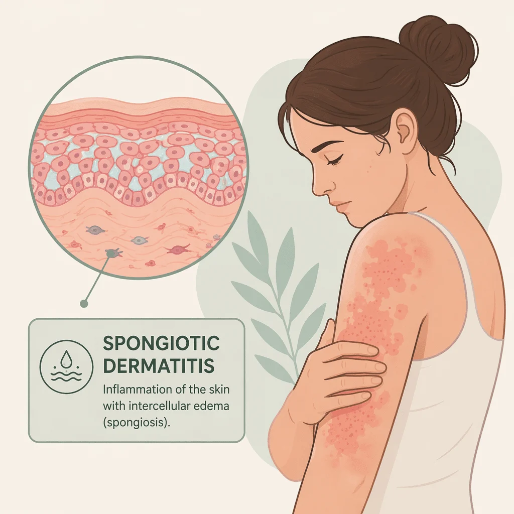
Jika Anda sedang memegang laporan biopsi yang bertuliskan dermatitis spongiotic, mulailah dari sini: dua kata tersebut bukan nama sebuah penyakit. Keduanya menggambarkan sebuah pola — tampilan kulit Anda ketika seorang ahli patologi meletakkan irisan kulit tersebut di bawah mikroskop.
Poin kedua perlu disampaikan dengan jelas, karena itulah yang paling banyak dicari orang. Dermatitis spongiotic bukan kanker. Bukan prakanker, dan tidak akan berkembang menjadi kanker. Ini adalah pola peradangan, dan merupakan tanda mikroskopis dari eksim.
Dalam artikel ini Anda akan mempelajari apa sebenarnya spongiosis itu, mengapa ahli patologi menggambarkan sebuah pola alih-alih menyebutkan nama kondisi, masalah kulit apa saja yang menghasilkan pola tersebut, cara membaca kata-kata lain yang tercetak di sampingnya, dan apa yang biasanya terjadi selanjutnya — termasuk satu tes yang paling sering terlewatkan.
Apa yang sebenarnya dilihat oleh ahli patologi
Lapisan terluar kulit Anda, epidermis, tersusun dari sel-sel yang disebut keratinosit yang tersusun rapat seperti bata dalam sebuah dinding. Pada kulit yang sehat, sel-sel ini berdampingan satu sama lain.
Ketika kulit tersebut mengalami peradangan dengan cara tertentu, cairan meresap di antara sel-sel tersebut dan mendorongnya saling menjauh. Di bawah mikroskop, dinding yang rapat itu terbuka menjadi sesuatu yang tampak seperti spons dapur, dengan ruang-ruang jernih tempat cairan berkumpul. Tampilan itulah yang disebut spongiosis, dan dari situlah kata spongiotic berasal.
Jika cukup banyak cairan terkumpul, ruang-ruang tersebut bergabung dan membentuk lepuhan kecil di dalam epidermis. Itulah mengapa eksim yang sedang aktif mengeluarkan cairan, dan mengapa beberapa ruam menunjukkan lepuhan sekecil titik yang terlihat dengan mata telanjang.
Spongiosis adalah ciri mikroskopis utama dari peradangan eksim. Jadi ketika sebuah laporan menyebutkan dermatitis spongiotik, ahli patologi pada dasarnya sedang mengatakan: pola dalam sampel ini terlihat seperti eksim.
Mengapa ahli patologi mendeskripsikan, bukan mendiagnosis
Seorang ahli patologi biasanya menerima potongan kulit berukuran beberapa milimeter, dimasukkan dalam wadah berisi cairan pengawet, disertai catatan klinis satu baris. Mereka dapat mendeskripsikan potongan tersebut dengan sangat tepat. Yang tidak dapat mereka lihat adalah pekerjaan Anda, sabun yang Anda gunakan, atau sudah berapa minggu ruam itu muncul.
Apa yang tidak dimaksud oleh dermatitis spongiotik
Ini bukan berarti kanker. Spongiosis adalah cairan di antara sel-sel normal, bukan sel-sel abnormal yang berkembang biak tanpa kendali. Institut Kanker Nasional AS mendefinisikan kanker sebagai penyakit di mana sel-sel tumbuh tanpa terkendali dan menyerang jaringan di sekitarnya. Peradangan tidak melakukan keduanya.
Ini juga bukan kondisi prakanker. Perubahan prakanker memiliki istilahnya sendiri dalam laporan patologi — kata-kata seperti displasia atau karsinoma in situ — dan dermatitis spongiotik tidak termasuk di antaranya. Eksim yang sudah berlangsung lama tidak berubah menjadi kanker kulit.
Ada satu catatan penting yang perlu disampaikan. Biopsi terkadang dilakukan justru untuk menyingkirkan kemungkinan tertentu. Jika dokter Anda mengambil sampel kulit karena suatu area terlihat tidak biasa, atau karena ruam tidak kunjung membaik, hasil spongiotik benar-benar memberikan ketenangan terkait kemungkinan yang mereka pikirkan. Meski begitu, ini bukan alasan untuk membatalkan janji tindak lanjut.
Biopsi juga memiliki keterbatasan. Biopsi dapat memastikan bahwa suatu proses bersifat inflamasi, bukan ganas atau infeksius. Namun biasanya tidak dapat memberi tahu Anda kondisi inflamasi mana yang Anda alami, dan tidak dapat memberi tahu Anda apa yang memicunya. Laporan Anda harus selalu diinterpretasikan oleh dokter yang memintanya, bersama dengan semua informasi lain yang mereka ketahui tentang Anda.
Mengapa laporan Anda tidak menyebutkan nama penyakit
Para dokter menyebut langkah yang hilang ini sebagai korelasi kliniko-patologis. Ini adalah istilah panjang untuk gagasan sederhana: temuan mikroskopis dan gambaran klinis harus dipadukan terlebih dahulu sebelum sesuatu dapat diberi nama.
Banyak kondisi berbeda yang menghasilkan pola spongiotik yang sama. Seorang ahli patologi yang hanya melihat preparat sering kali tidak dapat membedakan eksim atopik dari alergi nikel atau reaksi terhadap obat baru, karena pada tingkat sel semuanya bisa terlihat hampir identik.
Dokter bisa melakukannya. Mereka tahu di mana ruam itu berada, sudah berapa lama ada, apakah simetris, apa yang Anda tangani di tempat kerja, apa yang Anda oleskan ke kulit, dan apa yang berubah sesaat sebelum ruam muncul. Gabungkan kedua sudut pandang itu, dan diagnosis pun terbentuk. Hilangkan salah satunya, dan diagnosis tidak akan muncul.
Jadi laporan yang terasa membingungkan seringkali memang bekerja persis seperti yang dimaksudkan. Ahli patologi telah menyelesaikan separuh tugasnya. Separuh lainnya menjadi tanggung jawab orang yang ada di ruang konsultasi.
Kondisi-kondisi yang menghasilkan pola spongiotik
Daftarnya panjang, dan itulah intinya. Semua kondisi berikut ini dapat menghasilkan spongiosis pada biopsi.
- Dermatitis atopik, yaitu eksem gatal jangka panjang yang sering dimulai sejak masa kanak-kanak dan diturunkan dalam keluarga yang juga memiliki riwayat asma dan demam hay.
- Dermatitis kontak alergi, yaitu reaksi imun yang tertunda terhadap sesuatu yang menyentuh kulit — nikel, wewangian, pewarna rambut, pengawet, bahan kimia karet.
- Dermatitis kontak iritan, yaitu kerusakan langsung akibat pekerjaan basah yang berulang, detergen, pelarut, atau gesekan, tanpa melibatkan alergi sama sekali.
- Eksem numuler atau diskoid, yang membentuk plak berbentuk koin, sering ditemukan di anggota tubuh.
- Eksem dishidrotik, dengan lepuhan yang dalam di telapak tangan, sisi-sisi jari, dan telapak kaki.
- Dermatitis stasis, yaitu peradangan pada tungkai bawah yang disebabkan oleh sirkulasi vena yang buruk.
- Reaksi id, yang juga disebut autoeksematisasi, yaitu ruam yang menyebar dan muncul di tempat yang jauh dari ruam aktif di bagian tubuh lainnya.
- Erupsi obat, yaitu ruam yang dipicu oleh suatu obat — kisah yang sudah tidak asing lagi dengan antibiotik seperti amoksisilin, dan salah satu alasan mengapa dugaan alergi penisilin perlu dikonfirmasi dengan benar.
- Reaksi gigitan serangga dan tungau, termasuk ruam berbenjol dan gatal dari dermatitis akibat kumbang karpet.
Gatal adalah gejala yang hampir semuanya miliki, dan bagi banyak orang gejalanya paling parah setelah gelap. Jika gatal di malam hari adalah keluhan utama Anda, bukan laporan itu sendiri, panduan kami tentang kaki gatal di malam hari membahas penyebab-penyebab tersebut secara mendalam. Untuk ruam di bagian tubuh lainnya, ikhtisar kami tentang penyebab ruam kulit adalah titik awal yang lebih tepat.
Dermatitis stasis dan kekeliruan dengan selulitis
Satu entri dalam daftar itu layak mendapat perhatian tersendiri. Dermatitis stasis pada tungkai bawah sering kali keliru didiagnosis sebagai selulitis, yaitu infeksi kulit bakteri — dan selulitis terkadang keliru didiagnosis sebagai dermatitis stasis.
Keduanya dapat membuat kaki tampak merah, terasa hangat, bengkak, dan nyeri. Dermatitis stasis biasanya menyerang kedua kaki, berkembang selama berbulan-bulan, dan disertai noda kecokelatan serta perubahan varises akibat gangguan vena yang sudah lama. Selulitis umumnya hanya menyerang satu kaki, muncul dalam satu atau dua hari, dan sering disertai demam serta rasa tidak enak badan. Artikel kami tentang kaki merah membahas perbedaan keduanya secara lengkap.
Perbedaan ini penting karena keduanya memerlukan penanganan yang berlawanan. Salah diagnosis berarti pemberian antibiotik yang sebenarnya tidak diperlukan, atau sebaliknya, infeksi yang dibiarkan tanpa pengobatan.
Istilah lain dalam laporan Anda, dijelaskan
Laporan patologi jarang hanya memuat dua kata. Tabel di bawah ini mencakup frasa yang paling sering muncul berdampingan dengan dermatitis spongiosis.
| Frasa dalam laporan Anda | Maksud dari ahli patologi | Kemungkinan artinya |
|---|---|---|
| Dermatitis spongiosis akut | Banyak cairan di antara sel-sel, kadang membentuk lepuhan kecil; prosesnya tampak baru terjadi | Flare yang dimulai beberapa hari lalu, bukan berbulan-bulan — bisa berupa reaksi kontak, erupsi obat, atau flare eksim yang tiba-tiba |
| Dermatitis spongiosis subakut | Masih ada sedikit cairan, dengan lapisan luar yang mulai menebal; ini adalah fase pertengahan | Ruam yang sudah ada selama beberapa minggu — tidak benar-benar baru, tetapi juga belum berlangsung lama |
| Dermatitis spongiosis kronis | Cairan hampir tidak ada, tetapi epidermis menebal dan bersisik; kulit telah beradaptasi terhadap peradangan yang berlangsung lama | Eksim yang sudah lama, biasanya dengan berbulan-bulan kebiasaan menggosok dan menggaruk di atasnya |
| Dermatitis spongiosis dengan eosinofil | Sel darah putih yang berkaitan dengan alergi ditemukan dalam jaringan | Mengarah pada kemungkinan reaksi obat, alergi kontak, atau reaksi gigitan serangga — ini merupakan petunjuk, bukan kesimpulan akhir |
| Dermatitis spongiosis dengan parakeratosis | Lapisan luar yang sudah mati terbentuk terlalu cepat dan masih mengandung inti sel | Kulit yang mengalami pergantian sel dengan cepat, khas pada ruam yang aktif atau sering kambuh |
| Dermatitis perivaskular superfisial | Sel-sel radang mengelompok di sekitar pembuluh darah kecil tepat di bawah permukaan kulit | Temuan yang sangat umum dan tidak spesifik, yang menyertai sebagian besar ruam peradangan |
Beberapa istilah lain juga muncul tersendiri. Hiperkeratosis berarti lapisan luar yang sudah mati mengalami penebalan. Parakeratosis berarti lapisan tersebut terbentuk terlalu cepat, dengan inti sel yang masih terperangkap di dalamnya. Akantosis berarti epidermis yang masih hidup itu sendiri telah menebal. Likenifikasi adalah gambaran klinis dari kondisi yang sama: kulit yang terasa seperti kulit, berkerut, akibat berbulan-bulan digaruk dan digosok.
Tidak ada kata-kata tersebut yang mengubah inti permasalahan. Kata-kata itu hanya menggambarkan sudah berapa lama proses berlangsung dan bagaimana kulit meresponsnya, bukan apa yang menjadi penyebabnya.
Apa yang sebenarnya terjadi selanjutnya
Setelah biopsi mengonfirmasi pola ekzematosa, pertanyaan yang lebih berguna adalah: ekzema jenis apa ini? Menjawab pertanyaan ini yang akan mengubah hasil pengobatan, karena beberapa kemungkinan penyebabnya bisa hilang sepenuhnya begitu pemicunya dihilangkan.
Uji tempel, langkah yang paling sering terlewatkan
Jika dermatitis kontak alergi masuk dalam daftar kemungkinan, uji tempel adalah langkah definitif berikutnya. Sayangnya, langkah ini juga sangat jarang dilakukan.
Uji tempel tidak sama dengan uji tusuk kulit atau tes darah alergi yang mengukur IgE spesifik. Kedua tes tersebut mencari alergi yang dimediasi antibodi secara langsung — jenis yang menimbulkan biduran atau demam hay dalam hitungan menit. Alergi kontak adalah reaksi yang tertunda dan digerakkan oleh sel, yang membutuhkan satu hingga dua hari untuk muncul, sehingga memerlukan metode yang sama sekali berbeda.
Dalam praktiknya, kamar-kamar kecil berisi alergen terstandar ditempelkan di punggung Anda dan dibiarkan selama sekitar dua hari. Kemudian dilepas dan dibaca, lalu dibaca lagi beberapa hari kemudian untuk menangkap reaksi yang muncul belakangan. Itulah mengapa uji tempel memerlukan beberapa kali kunjungan yang tersebar dalam satu minggu dan tidak bisa diselesaikan dalam satu kali kunjungan saja. Logam terkadang bereaksi bahkan lebih lambat.
Meninjau apa saja yang telah bersentuhan dengan kulit Anda
Selain pengujian, Anda akan diminta membuat daftar lengkap: apa yang Anda tangani di tempat kerja, sabun atau produk yang Anda gunakan untuk mandi, kosmetik, produk rambut, perhiasan, alas kaki, sarung tangan, dan — yang penting — krim serta salep yang selama ini Anda oleskan pada ruam itu sendiri.
Produk topikal itu sendiri bisa menjadi alergen, sehingga ruam yang semakin parah semakin rajin Anda obati adalah petunjuk yang perlu disampaikan kepada dokter. Jangan menghentikan pengobatan yang diresepkan atas inisiatif sendiri; mintalah dokter Anda untuk meninjau daftarnya bersama Anda.
Cara pengobatan dermatitis spongiotik, secara umum
Tidak ada satu pengobatan tunggal untuk dermatitis spongiotik, karena ini bukan satu penyakit tunggal. Yang diobati adalah kondisi yang mendasarinya, dan caranya sepenuhnya bergantung pada kondisi mana yang terjadi, di bagian tubuh mana letaknya, seberapa parah, dan siapa Anda.
Secara garis besar, perawatan berjalan dalam tiga jalur: menghilangkan apa pun yang memicu peradangan, memulihkan pelindung kulit dengan pelembap rutin, dan meredakan peradangan dengan pengobatan anti-inflamasi yang diresepkan. Dermatitis stasis menambahkan jalur keempat yang ditujukan pada pembuluh vena dan pembengkakan.
Baris ketiga itu adalah keputusan dokter yang meresepkan. Obat anti-inflamasi mana, dengan kekuatan berapa, untuk berapa lama, dan pada bagian tubuh mana — semua itu bergantung pada pemeriksaan langsung dan tidak bisa ditentukan hanya dari sebuah artikel. Jika infeksi muncul di atas ruam, rencana penanganannya pun akan berubah.
Kapan harus kembali ke dokter
Hasil biopsi yang jinak memang melegakan, tetapi hasil itu hanya menggambarkan satu momen dan satu potongan kecil kulit. Kulit terus berubah. Hasil dari bulan lalu belum tentu masih berlaku sekarang.
Segera cari pertolongan medis jika Anda mengalami
- Ruam disertai demam, kemerahan yang menyebar, rasa hangat, dan nyeri — kemungkinan selulitis, yang memerlukan penilaian medis pada hari yang sama.
- Kulit melepuh atau mengelupas, terutama jika disertai luka di mulut atau mata — tangani ini sebagai kedaruratan medis.
- Ruam yang menyebar dengan cepat setelah mulai mengonsumsi obat baru.
- Ruam yang mengeluarkan cairan dan berkerak dengan kerak berwarna kuning atau seperti madu, yang mengindikasikan adanya infeksi.
- Bintik tunggal yang berdarah, membentuk luka terbuka, atau terus membesar — ini perlu dievaluasi ulang meskipun hasil biopsi sebelumnya jinak.
- Ruam yang tidak kunjung membaik meskipun sudah diobati.
Selain kondisi-kondisi di atas, jadwalkan kunjungan ulang jika ruam terus muncul kembali di tempat yang sama, jika periode pengobatan yang disepakati telah berlalu tanpa kemajuan, atau jika Anda tidak memahami apa yang tertulis dalam laporan Anda. Meminta dokter yang meminta biopsi untuk menjelaskannya kepada Anda adalah hal paling berguna yang bisa Anda lakukan dengan dokumen yang ada di tangan Anda.
Kemajuan ilmiah terkini dalam diagnosis dermatitis spongiosis
Eosinofil adalah petunjuk yang lebih lemah dari yang selama ini diajarkan
Apa yang ditemukan: sebuah tim dari Amerika Serikat membandingkan biopsi kulit dari orang-orang yang dermatitis kontak alerginya telah dikonfirmasi melalui uji tempel dengan biopsi dari ruam spongiosis lainnya. Bertentangan dengan ajaran yang sudah lama berlaku, eosinofil dermal — sel darah putih yang berkaitan dengan alergi dan sering dicantumkan dalam laporan — ternyata tidak mengarah pada alergi kontak, dan infiltrasi eosinofil yang berat justru lebih umum ditemukan pada diagnosis lainnya. Kelompok sel Langerhans, yaitu sel penjaga imun yang berada di epidermis, adalah satu-satunya ciri yang cenderung mengarah pada alergi kontak.
Apa artinya bagi Anda: jika laporan Anda menyebutkan adanya eosinofil, anggaplah itu sebagai petunjuk bahwa obat-obatan, alergen kontak, atau reaksi gigitan serangga perlu dipertimbangkan — bukan sebagai bukti dari salah satunya. Kesimpulan para peneliti cukup tegas: dermatitis kontak alergi tidak dapat dibedakan secara andal dari eksim lainnya hanya berdasarkan gambaran di bawah mikroskop. Uji tempel yang menentukan jawabannya, bukan biopsi.
Apa yang sebenarnya ditemukan oleh uji tempel di Amerika Utara
Apa yang ditemukan: North American Contact Dermatitis Group menguji beberapa ribu pasien di lebih dari selusin pusat kesehatan menggunakan serangkaian skrining standar. Sebagian besar menunjukkan setidaknya satu reaksi positif, dan hampir setengahnya didiagnosis utama dengan dermatitis kontak alergi. Nikel menempati urutan teratas, diikuti oleh pengawet methylisothiazolinone, linalool teroksidasi dari wewangian, dan kobalt. Lebih dari seperlima bereaksi terhadap sesuatu yang relevan secara klinis yang sama sekali tidak termasuk dalam panel standar.
Apa artinya bagi Anda: logam, pengawet, dan wewangian adalah penyebab yang paling umum dijumpai sehari-hari, bukan bahan-bahan yang langka. Hal ini juga menjelaskan mengapa dokter akan meminta Anda membawa produk yang biasa Anda gunakan — panel standar hanyalah titik awal, bukan jawaban yang lengkap.
Kaki merah lebih sering salah didiagnosis daripada yang kebanyakan orang sadari
Apa yang ditemukan: sebuah tinjauan sistematis dan meta-analisis menggabungkan berbagai studi di mana orang yang didiagnosis dengan selulitis tanpa komplikasi dievaluasi ulang dalam dua minggu. Sebagian besar ternyata tidak mengalami infeksi sama sekali, dan tiga kondisi menyumbang sebagian besar diagnosis alternatif: dermatitis stasis, dermatitis eksematosa, dan pembengkakan sederhana atau limfedema.
Apa artinya bagi Anda: jika Anda telah berulang kali diobati untuk selulitis di kaki yang sama tanpa perbaikan yang bertahan lama, hasil biopsi spongiotik mungkin menjadi informasi yang mengubah gambaran keseluruhan, dan hal ini wajar untuk disampaikan kepada dokter. Pemeriksaan darah seperti hitung darah lengkap dan CRP mendukung penilaian tersebut, tetapi tidak dapat memberikan kesimpulan sendiri.
Sebagian dari apa yang terlihat di bawah mikroskop kini dapat dibaca dari permukaan kulit
Apa yang ditemukan: sebuah studi observasional terhadap lebih dari dua ratus orang dengan eksem membandingkan apa yang dilihat dokter dengan mata telanjang, apa yang ditunjukkan oleh dermoskopi — alat pembesar genggam dengan cahaya terpolarisasi — dan apa yang ditunjukkan oleh biopsi. Pola dermoskopik sangat sesuai dengan stadium mikroskopis, dengan ciri-ciri pembuluh darah, sisik, dan pigmen yang khas yang membedakan penyakit akut, subakut, dan kronis.
Apa artinya bagi Anda: sebagian informasi yang diberikan oleh biopsi dapat diperoleh tanpa melakukan sayatan. Jika ruam Anda sedang dipantau dan bukan sedang diselidiki untuk pertama kalinya, dokter spesialis kulit mungkin akan menggunakan dermatoskop, bukan pisau bedah.
Spongiosis tidak hanya terjadi pada eksem
Apa yang ditemukan: dalam serangkaian kasus di rumah sakit pada orang dengan psoriasis telapak tangan dan telapak kaki, biopsi dibandingkan dengan diagnosis klinis. Spongiosis muncul pada sebagian besar sampel psoriasis ini, bersama dengan gambaran yang lebih khas dari psoriasis. Diagnosis klinis dan mikroskopis sesuai pada sebagian besar kasus, tetapi hanya jika keduanya dipertimbangkan bersama-sama.
Apa artinya ini bagi Anda: spongiosis dapat muncul pada kondisi yang sama sekali bukan eksim, termasuk psoriasis. Ini adalah satu alasan lagi mengapa laporan yang menggambarkan suatu pola tidak sama dengan diagnosis, dan mengapa gambaran klinis yang menjadi penentu akhir.
Pertanyaan yang sering diajukan
Apakah dermatitis spongiosis itu kanker?
Tidak. Dermatitis spongiosis adalah pola peradangan, bukan pertumbuhan sel abnormal. Ini bukan kanker, bukan prakanker, dan tidak berkembang menjadi kanker seiring waktu. Yang dilihat oleh ahli patologi adalah cairan yang terkumpul di antara sel-sel kulit biasa — tanda khas eksim di bawah mikroskop. Meski demikian, jika biopsi dilakukan karena dokter Anda ingin menyingkirkan sesuatu yang spesifik, diskusikan hasilnya dengan mereka daripada berasumsi bahwa masalahnya sudah selesai. Dan periksakan kembali setiap bercak tunggal yang berdarah, mengalami ulserasi, atau terus membesar, apa pun yang dikatakan laporan sebelumnya.
Apakah dermatitis spongiosis bisa sembuh?
Polanya akan hilang ketika peradangan yang mendasarinya mereda, sehingga jawaban yang jujur bergantung pada penyebabnya. Dermatitis kontak iritan dan alergi dapat sembuh sepenuhnya dan permanen setelah pemicunya diidentifikasi dan dihilangkan — itulah mengapa mengidentifikasinya sangat penting. Dermatitis atopik cenderung datang dan pergi selama bertahun-tahun, dengan periode kambuh dan periode tenang. Dermatitis stasis biasanya membaik ketika masalah pembuluh vena dan pembengkakan yang mendasarinya ditangani, tetapi memerlukan perhatian berkelanjutan. Tanyakan mana yang berlaku untuk Anda, karena jawabannya akan mengubah prospek secara keseluruhan.
Mengapa biopsi saya tidak memberikan diagnosis?
Karena banyak kondisi yang berbeda terlihat sama di bawah mikroskop. Ahli patologi dapat melaporkan dengan tepat bahwa jaringan menunjukkan pola eksematosa, tetapi mereka tidak dapat melihat di mana ruam berada, sudah berapa lama ada, apa yang Anda terpapar, atau apa pekerjaan Anda. Memberikan konteks tersebut adalah tugas dokter klinis, dan kedua bagian itulah yang bersama-sama menghasilkan diagnosis. Laporan deskriptif bukan laporan yang gagal. Itu adalah satu bagian dari proses dua bagian, dan bagian yang hilang adalah sebuah percakapan.
Apakah dermatitis spongiosis sama dengan dermatitis atopik?
Tidak sepenuhnya tepat. Dermatitis atopik adalah nama suatu kondisi; dermatitis spongiotik adalah deskripsi tentang bagaimana dermatitis atopik — dan beberapa kondisi lainnya — terlihat di bawah mikroskop. Jadi, dermatitis atopik bersifat spongiotik saat dilakukan biopsi, tetapi tidak semua dermatitis spongiotik bersifat atopik. Dermatitis kontak alergi dan iritan, eksem numular, eksem dishidrotik, dermatitis stasis, reaksi obat, dan reaksi gigitan serangga semuanya dapat menghasilkan gambaran yang sama. Menentukan kondisi mana yang Anda miliki adalah keputusan klinis, bukan keputusan berdasarkan mikroskop.
Apakah ada penyakit autoimun di balik dermatitis spongiotik?
Biasanya tidak. Dermatitis spongiotik adalah pola peradangan yang paling sering dipicu oleh alergi, iritasi, atau kerusakan pelindung kulit — bukan oleh tubuh yang menyerang jaringannya sendiri. Dermatitis atopik melibatkan respons imun yang terlalu aktif, tetapi tidak diklasifikasikan sebagai penyakit autoimun. Jika dokter Anda mencurigai adanya masalah sistemik, mereka akan menyampaikannya dan melakukan pemeriksaan secara langsung. Laporan spongiotik saja bukan alasan untuk mulai mencari kondisi autoimun.
Apakah ada makanan yang perlu dihindari pada dermatitis spongiotik?
Tidak ada daftar makanan yang menyebabkan atau menyembuhkan dermatitis spongiotik, dan tidak ada diet berbasis bukti untuk kondisi ini. Alergi makanan kadang-kadang berkontribusi pada kambuhnya eksem pada anak kecil, dan sebagian kecil orang dengan alergi kontak terhadap zat tertentu — seperti balsam Peru atau rempah-rempah tertentu — juga bereaksi terhadap sumber makanan tersebut. Kedua situasi ini memerlukan penilaian yang tepat, bukan tebak-tebakan. Menghilangkan seluruh kelompok makanan tanpa saran medis berisiko menimbulkan masalah gizi dan jarang membantu kondisi kulit.
Glosarium
| Ketentuan | Definisi |
|---|---|
| Spongiosis | Cairan yang terkumpul di antara sel-sel lapisan kulit terluar dan mendorongnya terpisah, sehingga terlihat seperti spons di bawah mikroskop |
| Epidermis | Lapisan tipis terluar kulit, bagian tempat terjadinya spongiosis |
| Keratinosit | Jenis sel utama epidermis, tersusun berlapis-lapis seperti bata dalam dinding |
| Hiperkeratosis | Penebalan lapisan terluar kulit yang sudah mati, sering ditemukan pada ruam yang sudah berlangsung lama |
| Parakeratosis | Inti sel yang tertahan di lapisan terluar kulit yang sudah mati, sebagai tanda bahwa kulit telah memperbarui dirinya terlalu cepat |
| Akantosis | Penebalan bagian epidermis yang masih hidup |
| Likenifikasi | Kulit yang menebal, keras, dan berkerut akibat gesekan atau garukan yang berkepanjangan |
| Eosinofil | Sel darah putih yang berkaitan dengan alergi, parasit, dan beberapa reaksi obat, yang kadang-kadang dilaporkan dalam sampel kulit |
| Uji tempel | Tes untuk alergi kontak tipe lambat, di mana zat-zat standar ditempelkan pada kulit dan hasilnya dibaca selama beberapa hari |
| Korelasi kliniko-patologis | Menggabungkan temuan di bawah mikroskop dengan pengamatan klinis dokter untuk mencapai diagnosis |
Sumber
- Dermatitis Atopik: Dasar-Dasar, Gejala, dan Penyebab — National Institute of Arthritis and Musculoskeletal and Skin Diseases (NIAMS), US National Institutes of Health.
- Dermatitis kontak — MedlinePlus Medical Encyclopedia, US National Library of Medicine.
- Laporan Patologi — National Cancer Institute, US National Institutes of Health.
- Apa Itu Kanker? — National Cancer Institute, US National Institutes of Health.
- Wu PA, Wu J, Liu R, et al. Epidermal spongiotic Langerhans cell collections, but not eosinophils, are a clue to the diagnosis of allergic contact dermatitis: a series of 170 clinically- and patch test-confirmed cases. Journal of the American Academy of Dermatology. 2025;92(4):853-860 — data diambil melalui PubMed.
- Houle MC, DeKoven JG, Atwater AR, et al. North American Contact Dermatitis Group Patch Test Results: 2021-2022. Dermatitis. 2025;36(5):464-476 — data diambil melalui PubMed.
- Nightingale R, Yadav K, Hamill L, et al. Misdiagnosis of Uncomplicated Cellulitis: a Systematic Review and Meta-analysis. Journal of General Internal Medicine. 2023;38(10):2396-2404 — data diambil melalui PubMed.
- Bhatt MM, Jamale V, Hussain AA, et al. An Observational Study of Dermoscopic and Histopathological Correlation in Spongiotic Disorders: A Hospital Based Cross Sectional Study. Indian Journal of Dermatology. 2024;68(6):634-641 — data diambil melalui PubMed.
- Das G, Mathur M, Shrestha A, et al. Palmoplantar psoriasis: A clinicopathological correlation in a tertiary care hospital. Skin Research and Technology. 2024;30(8):e13882 — data diambil melalui PubMed.
Bacaan lebih lanjut
- Eksim: memahami kondisi kulit ini
- Ruam kulit: penyebab, gejala, dan pengobatan
- Ruam kulit yang terasa nyeri: penyebab dan cara mengatasinya
- Kaki gatal di malam hari: penyebab dan cara mengatasinya
- Keratosis seboroik vs melanoma: panduan mengenali gejalanya
Pahami hasil lab Anda dengan AI DiagMe.
Laporan biopsi bukan hasil laboratorium yang diinterpretasikan oleh AI DiagMe. AI DiagMe tidak membaca slide patologi dan tidak mendiagnosis kondisi kulit — hal itu merupakan wewenang dokter yang meminta biopsi tersebut.
Yang sering datang bersamaan adalah hasil tes darah. Sebuah hitung darah lengkap, sebuah hitung eosinofil, CRP atau sebuah tes alergi IgE spesifik mungkin semuanya diminta saat ruam Anda sedang diperiksa.
Itulah angka-angka yang dapat dibantu dipahami oleh AI DiagMe untuk Anda, dalam bahasa yang mudah dimengerti, sebelum janji temu Anda berikutnya.
Dapatkan interpretasi hasil Anda dalam hitungan menit.