




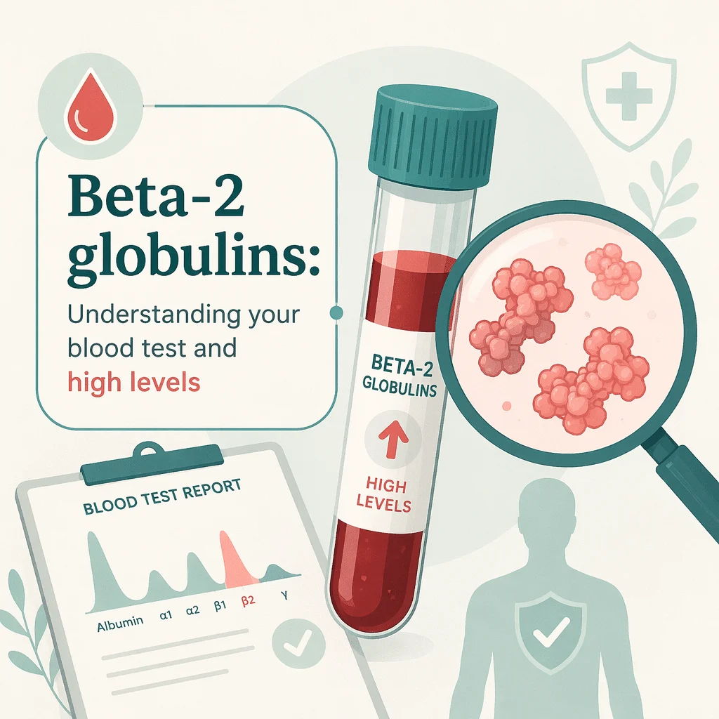
Beta-2 globulin is one of the fractions reported on a serum protein electrophoresis, the laboratory test that sorts the proteins in your blood into groups. A raised beta-2 globulin is not a cancer marker. Most of the time it reflects something ordinary: iron deficiency, higher blood fats, or everyday inflammation. One point clears up most of the worry on this page, so it comes first: beta-2 globulin is not the same thing as beta-2 microglobulin. The names look almost identical, the two are measured in completely different ways, and only the second is used in myeloma staging. In this article you’ll learn what actually sits inside the beta-2 fraction, why laboratories report it so differently, what pushes it up or down, and when a doctor decides to look further.
What serum protein electrophoresis measures, and where beta-2 sits
Your blood serum contains thousands of different proteins, and measuring each one individually would be slow and expensive. So laboratories use a shortcut dating from the 1930s. The sample is placed in an electric field, where proteins drift at different speeds depending on their charge, size and shape. Proteins that behave alike travel together and settle into bands, and the laboratory measures how much protein sits in each one.
The result is a curve of peaks and valleys. Reading from left to right, the usual zones are albumin, then alpha-1, alpha-2, beta and gamma. Some systems split the beta zone into two smaller ones, beta-1 and beta-2. Your report may give each zone as a percentage of total protein, as a concentration in grams per litre or grams per decilitre, or both. For the whole curve rather than one band, our guide walks through the complete serum protein electrophoresis results.
This matters for how you read your own result. Beta-2 globulin is not a single protein with a single job. It is a postcode, not a person. It tells you how much protein happens to be travelling at a particular speed, and several unrelated proteins share that speed.
Beta-2 globulin is not beta-2 microglobulin
This is the confusion that brings most people to this page, and it is worth untangling carefully, because the two are genuinely different tests that happen to share a name.
Beta-2 globulin is a zone on the electrophoresis curve: a bulk measurement of whatever protein lands in that part of the strip, reported alongside albumin, alpha-1, alpha-2, beta-1 and gamma. No one orders it on its own. It appears because the whole electrophoresis was run.
Beta-2 microglobulin is one specific, small protein. It sits on the surface of nearly every cell in the body that has a nucleus, and it is shed into the blood as cells are made and replaced. It is measured individually, by a targeted immunoassay rather than by electrophoresis, and it can be measured in blood, urine or spinal fluid. According to the National Cancer Institute, it is used in multiple myeloma, chronic lymphocytic leukaemia and some lymphomas, to judge prognosis and to follow the response to treatment.
The name overlap is a historical accident. Beta-2 microglobulin was christened decades ago precisely because it travels in the beta-2 part of an electrophoresis strip. But it circulates in milligrams per litre, whilst the beta-2 zone is measured in grams per litre. It is roughly a thousand times too dilute to shift the fraction it was named after. In practice, beta-2 microglobulin contributes essentially nothing to your beta-2 globulin number.
So if you have been told your beta-2 globulin is slightly high and you have found alarming pages about myeloma staging, you have almost certainly landed on material about a different test.
| Beta-2 globulin | Beta-2 microglobulin | |
|---|---|---|
| What it is | A zone on the electrophoresis curve containing several different proteins | One specific small protein found on the surface of nucleated cells |
| How it is measured | By serum protein electrophoresis, as part of the whole curve | By a separate, targeted immunoassay ordered on its own |
| How it is reported | As a percentage of total protein and/or in g/L or g/dL | In mg/L, from blood, urine or spinal fluid |
| What it is used for | Part of the overall pattern a doctor reads across all fractions | Prognosis and treatment monitoring in myeloma, CLL and some lymphomas |
| Is it a cancer marker? | No | Not for diagnosis; it is used once a diagnosis is already established |
What is actually in the beta-2 fraction
When laboratories do split the beta zone in two, the contents divide roughly like this. The beta-2 zone is dominated by complement C3, an immune protein that helps tag bacteria for destruction. Beta-lipoproteins, the particles carrying LDL cholesterol around the body, also travel in the beta region. Depending on the system, some transferrin may reach beta-2 as well, although most stays in beta-1.
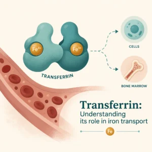
The beta-1 zone, by contrast, carries the bulk of the transferrin, the protein that ferries iron through the bloodstream, along with hemopexin. We have a separate piece on beta-1 globulins.
Why your report may not even have a beta-2 line
Here is something most pages skip. Many laboratories never split beta at all. They report a single combined beta fraction, and that is a perfectly standard way to run the test. The MedlinePlus encyclopaedia, for instance, lists reference values for albumin, alpha-1, alpha-2, beta and gamma, with beta as one number.
So if you are hunting for a beta-2 line and cannot find one, nothing has gone wrong. Equally, if two reports disagree, they may not be measuring the same thing in the same way. Reference ranges differ between laboratories, and so does the decision to subdivide. Always compare your value against the range printed on your own report, not against a number from the internet.
What causes a raised beta-2 globulin
Because several unrelated proteins share the beta zone, anything that raises any of them can lift the fraction. The common explanations are reassuringly mundane.
| Cause | What is happening |
|---|---|
| Iron deficiency | The body makes more transferrin to scavenge scarce iron, lifting the beta region |
| High blood fats | Beta-lipoproteins carrying LDL cholesterol travel in the beta zone |
| Acute inflammation or infection | Complement C3 rises as part of the body’s response |
| Pregnancy or oestrogen | Oestrogen increases transferrin production, a normal physiological shift |
| A monoclonal protein | An abnormal antibody can happen to migrate in beta and hide inside the peak |
Iron deficiency
This is one of the most common reasons for a beta reading to drift upwards, and it surprises people because iron and globulins sound unrelated. When iron runs short, the liver produces more transferrin to capture whatever is available, and more transferrin means more protein in the beta region. Since transferrin sits mostly in beta-1, the effect is clearest there, but on a report giving beta as one number the whole fraction rises. A doctor would check your iron status directly, using an iron studies panel, rather than drawing conclusions from the electrophoresis alone. A low ferritin result often accompanies this picture.
Blood fats
Beta-lipoproteins carry LDL cholesterol, and they migrate in the beta zone. If your cholesterol is high, or if you did not fast before the blood draw when your laboratory asked you to, the beta fraction can look elevated for reasons that have nothing to do with your immune system. This is usually clarified by a straightforward lipid panel.
Inflammation, pregnancy and oestrogen
Complement C3 rises when the body is fighting something, so a recent infection or a flare of an inflammatory condition can lift beta-2. This is normally read alongside other markers of inflammation such as C-reactive protein, and alongside the alpha-2 globulins, which also react to inflammation. Pregnancy and oestrogen-containing medication raise transferrin, which is why a beta rise in a pregnant woman is generally expected rather than concerning.
When a beta abnormality leads to more testing
There is one situation where a beta finding genuinely prompts further investigation, and it deserves a calm, accurate explanation rather than either silence or alarm.
Some people produce a monoclonal protein: a large quantity of a single, identical antibody made by one clone of cells. On electrophoresis this typically shows up as a narrow, sharp spike, most often in the gamma zone. But not always. Certain antibody types, IgA in particular, naturally travel in the beta region. The spike then lands on top of the beta peak instead of standing alone in an empty part of the curve, partly camouflaged by the C3 and lipoproteins already there.
This is why a laboratory or doctor may follow up an odd-looking beta peak with a test called immunofixation, which uses antibodies to identify whether a band is one single antibody type or a normal mixture of many. It answers a question the basic electrophoresis cannot.
It is important to keep this in proportion. Being sent for immunofixation is not a diagnosis and not a prediction. It is the laboratory doing its job properly: the beta zone is a place where an abnormal band could hide, so when the shape looks unusual, it gets checked. The great majority of raised beta results are explained by iron, lipids or inflammation, and never need any of this. If the pathway is followed further, related tests may include kappa and lambda free light chains. Those tests, and any discussion of conditions such as multiple myeloma, belong with a doctor who can see your whole clinical picture.
What a low beta-2 globulin can mean
A low beta fraction attracts far less attention than a high one, and is less common. The usual reasons are straightforward.
Losing protein is the first. If protein escapes through the kidneys, as in nephrotic syndrome, or through an inflamed digestive tract, many serum proteins fall together. Poor nutrition is the second: without the raw materials, the liver cannot manufacture proteins at a normal rate. Since the liver makes most of them, liver disease is the third, and a doctor would generally look at liver function tests alongside the electrophoresis.
There is also a mechanism specific to beta-2. Complement C3 can be consumed faster than it is replaced when the immune system is actively attacking something, including in some autoimmune and kidney diseases, and the beta-2 zone can then dip. Because low LDL cholesterol also empties the beta zone, an unusually low result sometimes simply reflects low blood fats.
In nearly all these cases the beta number is a supporting detail. The albumin level usually says more, which is why low albumin tends to drive the conversation instead.
Beta-gamma bridging and the liver
One pattern on the curve is worth knowing by name, because it is one of the few things the shape says on its own.
Normally there is a clear valley between the beta peak and the gamma peak. In advanced liver disease, particularly cirrhosis, that valley fills in and the two peaks merge into a continuous plateau. Laboratory staff call this beta-gamma bridging. The reason is that chronic liver disease drives up production of a class of antibody called IgA, which happens to travel in exactly the gap between beta and gamma. As it accumulates, it fills the trough, and the bridge appears.
This illustrates the honest limit of a single number. Beta-gamma bridging is not detected by looking at a beta-2 value; it is detected by looking at the shape of the whole curve, and someone reading only the numbers would miss it entirely. The pattern is closely tied to the gamma globulins next door, where the extra antibody largely shows up.
When to see a doctor
An isolated beta-2 globulin result, on its own, is rarely a reason to act. It is one number in a pattern, and the pattern is what gets read.
Bring your report to your doctor, and let them interpret it in context, if any of the following apply.
- Your laboratory has flagged the result, or the report mentions an unusual peak, a band, or a suggestion of immunofixation.
- The beta change comes with symptoms you cannot explain, such as persistent tiredness, unintended weight loss, bone pain, fever that will not settle, or frequent infections.
- Several fractions have moved together, or your albumin has fallen.
- You are already being followed for a kidney, liver, or blood condition.
- You are taking an antibody-based medication, since some of these are visible on electrophoresis and your doctor needs to know.
What you should not do is try to score yourself. There is no percentage above the reference range that reliably separates “harmless” from “serious”, and any page offering you one is inventing it. The meaning of a beta value depends entirely on the rest of the curve, your history and your symptoms. That interpretation belongs to your doctor.
Latest scientific advances in protein electrophoresis
Research on the beta region in recent years has focused less on what a high number means and more on making sure that what laboratories see there is real. Here is what that work has found, in plain terms.
Antibody medicines can imitate an abnormal protein
A systematic review published in 2025 in Clinical Chemistry and Laboratory Medicine pooled 30 studies covering 30 therapeutic monoclonal antibodies, the growing family of drugs used in cancer and autoimmune disease, and mapped where each shows up on an electrophoresis strip. Most appeared in the gamma zone, but one migrated in the beta-2 globulin region and another in alpha-2.
What this means for you: if you are receiving one of these treatments, the drug itself can produce a small peak that looks like a monoclonal protein, including in the beta area. It is the medication showing up, not a new disease. Make sure your doctor and your laboratory know every treatment you are on.
One drug lands squarely in beta-2
A 2025 study in Clinical Biochemistry tested seven antibody treatments to see how little of each could still be spotted. One, ravulizumab, migrated specifically in the beta-2 region and remained detectable at low concentrations. The authors argued for routinely confirming suspicious peaks with immunofixation rather than judging on the electrophoresis alone.
What this means for you: it explains why a doctor confirms rather than assumes when something appears in beta-2.
A beta-2 pattern that mimics something worse
Researchers in Italy reviewed more than 127,000 electrophoresis tests run at their hospital over two years. They described a recognisable pattern of a raised beta-2 zone with beta-gamma bridging that can be mistaken for a monoclonal protein, but actually reflects a high polyclonal level of an antibody subtype called IgG4. It turned up in about 3 of every 1,000 tests, and most of their new IgG4 cases were first noticed on the electrophoresis rather than by looking for them.
What this means for you: an unusual beta-2 and beta-gamma picture is not automatically a blood cancer. It can point to an entirely different, treatable group of conditions, and the shape of the curve is what reveals it.
Software is learning to read the curve
A 2024 study in PLoS One applied machine-learning methods to 67,073 capillary electrophoresis samples to see whether software could flag monoclonal proteins. One finding is directly relevant here: when the researchers asked which parts of the curve the algorithms actually relied on, the gamma fraction and part of the beta fraction carried most of the information.
What this means for you: it confirms the theme of this article. The beta region carries real information, but it carries it as part of a whole curve. Even a computer needs the full picture, not one fraction.
How much weight to give these findings
These are laboratory and method studies, not studies of patients’ outcomes, which limits how far they stretch. Several tested drugs spiked into donated serum rather than measured in treated patients: a clean way to establish where a drug migrates, but not the same as real-world conditions. The IgG4 work came from a single hospital, so the frequency elsewhere may differ. The machine-learning study looked back at stored results rather than testing software in live practice. Together they say something modest but useful: the beta region is a place where things can be misread, and confirmation exists for good reason.
Glossary
| Term | Definition |
|---|---|
| Serum protein electrophoresis (SPEP) | A laboratory test that separates the proteins in blood serum into zones using an electric field |
| Beta-2 globulin | A zone on that curve, containing mainly complement C3 and beta-lipoproteins; not a single protein |
| Beta-2 microglobulin | A separate small protein measured by its own immunoassay, used for prognosis in some blood cancers |
| Complement C3 | An immune protein that helps mark bacteria for destruction, and the main occupant of beta-2 |
| Transferrin | The protein that carries iron in the blood; it travels mainly in the beta-1 zone |
| Beta-lipoprotein | The particle that transports LDL cholesterol, which migrates in the beta region |
| Monoclonal protein | A large amount of one identical antibody made by a single clone of cells, seen as a narrow spike |
| Polyclonal | A rise in many different antibodies at once, producing a broad hump rather than a sharp spike |
| Immunofixation | A follow-up test that identifies whether a band is one single antibody type or a normal mixture |
| Beta-gamma bridging | A filled-in valley between the beta and gamma peaks, classically seen in cirrhosis |
Frequently asked questions
Is beta-2 globulin the same as beta-2 microglobulin?
No, and this is the single most common mix-up on this subject. Beta-2 globulin is a zone on a serum protein electrophoresis curve that holds several different proteins, reported alongside albumin and the other fractions in grams per litre. Beta-2 microglobulin is one specific small protein, measured on its own by a different method, reported in milligrams per litre, and used in myeloma and some lymphomas to judge prognosis and follow treatment. They share a name because beta-2 microglobulin was originally named after where it travels on an electrophoresis strip, but it is far too dilute to affect the beta-2 fraction itself. If your report shows a beta-2 globulin, you have not had a beta-2 microglobulin test.
Does a high beta globulin mean cancer?
No. A raised beta globulin is not a cancer marker, and on its own it does not indicate cancer. The overwhelming majority of raised beta results come from iron deficiency, high blood fats, inflammation, pregnancy or oestrogen-containing medication. There is one nuance worth knowing calmly: because certain abnormal antibodies can migrate in the beta region, a beta peak with an unusual shape is one of the things a laboratory checks with a confirmatory test. That check is routine caution, not a finding. Your doctor reads the whole curve, your symptoms and your history together before anything is concluded.
What is a normal beta-2 globulin level?
There is no single universal number, and you should be sceptical of any page that gives you one. Reference ranges vary between laboratories because they depend on the method, the equipment and the population the laboratory serves. Some laboratories report beta as one combined fraction and never produce a beta-2 value at all. The only range that applies to you is the one printed next to your result on your own report. Comparing your value to a figure found online is one of the easiest ways to worry yourself unnecessarily.
Why does my report show beta globulin but not beta-1 and beta-2?
Because your laboratory reports beta as a single fraction, which is entirely standard. Whether the beta zone gets subdivided into beta-1 and beta-2 depends on the technique and the equipment used. Neither approach is better or more thorough than the other; they are simply different conventions. If you have two reports from two laboratories that look different, this is often the explanation. It also means a beta-1 or beta-2 figure from one laboratory cannot be compared directly against a combined beta figure from another.
Can iron deficiency raise beta globulins?
Yes, and it is one of the most common explanations. When your body is short of iron, the liver produces more transferrin, the protein that transports iron in the blood, to make the most of what is available. Transferrin travels in the beta region, so more of it means more protein in that zone. On systems that split beta, the effect shows up mainly in beta-1; on systems reporting a single beta, the whole fraction rises. A doctor investigating this would look at your iron studies directly rather than relying on the electrophoresis, which was never designed to measure iron status.
What does it mean if beta-2 globulin and gamma globulin are both high?
It depends entirely on the shape of the curve, which is why this needs a doctor rather than a lookup table. A broad rise across both zones usually suggests a polyclonal picture, meaning many antibodies increasing together, as happens in chronic inflammation, infection or liver disease. If the two zones have merged with no valley between them, that is beta-gamma bridging, which points towards the liver. A narrow spike in either zone is a different matter and is what immunofixation exists to clarify. The combination itself does not identify a cause; the pattern does.
Sources
- MedlinePlus Medical Encyclopedia (US National Library of Medicine) — Protein electrophoresis, serum
- Sonagra AD, Zubair M, Dholariya SJ. Electrophoresis. StatPearls, NCBI Bookshelf (updated 2025)
- National Cancer Institute — Tumour Marker Tests in Common Use, including beta-2-microglobulin
- Pelletier S, Florent L, Gillery P, Oudart JB. Interference of therapeutic monoclonal antibodies with electrophoresis and immunofixation of serum proteins: state of knowledge and systematic review. Clin Chem Lab Med. 2025 (via PubMed)
- Milandri J, Jaillard M, Laffineur M, Dechomet M, Kolopp-Sarda MN. Decoding the interference: how therapeutic monoclonal antibodies challenge serum protein electrophoresis and immunofixation. Clin Biochem. 2025 (via PubMed)
- Michetti L, Antelmi E, Ravasio R, Ghirardi A. Serum protein electrophoresis pattern associated with elevated polyclonal IgG4 concentrations. Clin Chem Lab Med. 2026 (via PubMed)
- Sopasakis A, Nilsson M, Askenmo M, Nyholm F, Mattsson Hultén L, Rotter Sopasakis V. Machine learning evaluation for identification of M-proteins in human serum. PLoS One. 2024 (via PubMed)
Further reading
- How to read your serum protein electrophoresis results
- What beta-1 globulins show on a blood test
- Understanding high and low gamma globulins
- Transferrin and how the body transports iron
- The kappa lambda ratio and free light chains
Understand your lab results with AI DiagMe
Reports that list protein electrophoresis, beta globulins, transferrin and total protein side by side are hard to read when nobody explains how the fractions relate to each other. AI DiagMe turns that page of numbers into plain language, so you can see what each line refers to and arrive at your appointment with better questions. It helps you understand your results. It does not diagnose anything, and it does not replace your doctor.
Get your results interpreted in minutes